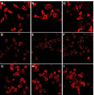
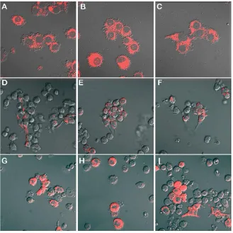
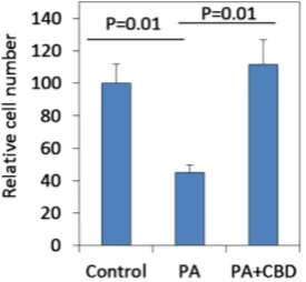
Cannabidiol (CBD) Prevents Palmitic Acid Induced Drop in Mitochondrial Membrane Potential
Full text
Figure
Related documents
TheUGC (Submission of Metadata and Full–text of Doctoral Theses in Electronic Format) Regulation, 2005,mandate for all Universities of India to creation of metadata
The surgery performed on the upper eyelid was a conventional blepharoplasty with removal of excess skin, treatment for the bags when they were present and a transpalpebral
Her analysis of the apparent paradox of the coexistence of a certain idealization of the past when workers lived on the traditional sugar cane properties, and a demand for
Building on the relevant survey work that has been done in other jurisdictions [28,29] and in other related areas of research [30], we conducted a series of in- depth
Apoptosis cannot occur in cell lines in vitro, because cell lines are immortalized by reprogramming the death program of the parental cells, because in culture there lack scavengers
In our case, although the size of the carcinoid tumor was small and the tumor was hormonally inactive, the concomitant pancreas divisum led to an early diagnosis because obstruction
Table 11 Significant difference of outcomes between- groups of 3 studies that compared a spinal manipulation to some type of physical therapy included in a systematic review on
However, this individual had many of the functional deficits and congenital anomalies commonly associated with deletion of the distal 1p36 critical region including developmental
