*Dr. Kelly Pearl Silva Lobo Norton, Dr. Surendra Kumar G. P., Dr. Bharath Raj, Dr.
Nalinakshamma
Department of Prosthodontics, VokkaligaraSangha Dental College and Hospital, KR Road,
Vishweshwarapura, Bangalore, Karnataka, India
ARTICLE INFO ABSTRACT
The disfigurement after the loss of an eye can cause significant physical and psychological disturbance to a patient. The rehabilitation of an ocular defect patient is a challenging task both clinically and technically. A Custom made ocular prosthesis aids in replicating the exact location of the iris and gaze of the patient along with an accurate reproduction of the color, contour, and size of the contra
esthetic and well
normal appearance of an anophthalmic patient.
Copyright © 2018, Kelly Pearl Silva Lobo Norton et al
unrestricted use, distribution, and reproduction in any medium,
INTRODUCTION
The disfigurement caused after the loss of an eye causes significant physical and psychological disturbance to the patient. Psychological distress can be reduced by timely replacement with a suitable ocular prosthesis (stock or custom). It is often tempting for clinicians to use a stock eye which can be economic, however, such a prosthesis may not perfectly adaptable to the tissue bed of the eye socket and it usually causes significant discomfort and fails to satisfy the patient’s esthetic expectations. Improved esthetics is achieved only when the iris color and position is perfectly matched with the normal eye, and this is often possible only by a custom-made ocular prosthesis (Goiato MC
Prosthetic replacement of lost eye presents with many challenges one of which being accurate positioning of iris (
UY et al, 2011). Any asymmetry in positioning causes a
squinted eye appearance which leads to unaesthetic results. This article describes a simple and innovative technique along with a case report for the fabrication of a custom
prosthesis in an attempt to avoid time consuming and expensive procedures that are required in other methods. Article History:
Received 04th February, 2018
Received in revised form
24th March, 2018
Accepted 27th April, 2018
Published online 31st May, 2018
Citation: Dr. Kelly Pearl Silva Lobo Norton, Dr. Surendra Kumar G. P., Dr. Bharath Raj, Dr. Nalinakshamma Muniswamy Reddy and Dr. Tanuja
N Murthy, 2018. “Customised ocular prosthesis- a simplified approach
Key words:
Ocular Defect, Ocular Prosthesis, Graph Grid, Iris Positioning, Custom Ocular Prosthesis, Artificial eye.
*Corresponding author:
*Dr. Kelly Pearl Silva Lobo Norton, Dr. Surendra Kumar G. P., Dr. Bharath Raj, Dr.
Nalinakshamma Muniswamy Reddy and Dr. Tanuja N Murthy
Department of Prosthodontics, VokkaligaraSangha Dental College and Hospital, KR Road,
Vishweshwarapura, Bangalore, Karnataka, India - 560004
ARTICLE INFO ABSTRACT
The disfigurement after the loss of an eye can cause significant physical and psychological disturbance to a patient. The rehabilitation of an ocular defect patient is a challenging task both clinically and technically. A Custom made ocular prosthesis aids in replicating the exact location of the iris and gaze of the patient along with an accurate reproduction of the color, contour, and size of the contra lateral natural eye. This article describes a simplified approach for fabricating an accurate, esthetic and well-fitting custom-made ocular prosthesis in an attempt to restore facial symmetry and normal appearance of an anophthalmic patient.
Norton et al. This is an open access article distributed under the Creative Commons medium, provided the original work is properly cited.
The disfigurement caused after the loss of an eye causes physical and psychological disturbance to the patient. Psychological distress can be reduced by timely replacement with a suitable ocular prosthesis (stock or custom). It is often tempting for clinicians to use a stock eye uch a prosthesis may not perfectly adaptable to the tissue bed of the eye socket and it usually causes significant discomfort and fails to satisfy the patient’s esthetic expectations. Improved esthetics is achieved perfectly matched with the normal eye, and this is often possible only by a Goiato MC et al, 2013). Prosthetic replacement of lost eye presents with many challenges one of which being accurate positioning of iris (Pai Any asymmetry in positioning causes a squinted eye appearance which leads to unaesthetic results. This article describes a simple and innovative technique along with a case report for the fabrication of a custom-made ocular pt to avoid time consuming and expensive procedures that are required in other methods.
Clinical Report
The patient reported to the Department of Prosthodontics with the chief complaint of discomfort using stock eye which had been given after enucleation of the right eye
procedure was explained to the patient and written consent was obtained.
To take an impression of the eye socket the patient was seated in an erect position to allow the tissues involved in the defect to be recorded in their natural drape. An impression of the orbital socket was made with medium body addition silicone impressio
DENTSPLY) using a custom perforated tray attached to the mixing tip of the dispensing gun
Figure. 3a, b).
An index was made using putty addition silicone impression material (Aquasil, DENTSPLY). To obtain a wax conformer, molten wax (Modelling Wax, Deepti Dental Products of India Pvt. Ltd.) was flown in the index (Figure. 4a, b).
Pearl Silva Lobo Norton, Dr. Surendra Kumar G. P., Dr. Bharath Raj, Dr. Nalinakshamma Muniswamy Reddy and Dr. Tanuja
a simplified approach”, International Journal of Current Research, 10
*Dr. Kelly Pearl Silva Lobo Norton, Dr. Surendra Kumar G. P., Dr. Bharath Raj, Dr.
N Murthy
Department of Prosthodontics, VokkaligaraSangha Dental College and Hospital, KR Road,
560004
The disfigurement after the loss of an eye can cause significant physical and psychological disturbance to a patient. The rehabilitation of an ocular defect patient is a challenging task both clinically and technically. A Custom made ocular prosthesis aids in replicating the exact location of the iris and gaze of the patient along with an accurate reproduction of the color, contour, and size of bes a simplified approach for fabricating an accurate, made ocular prosthesis in an attempt to restore facial symmetry and
Commons Attribution License, which permits
The patient reported to the Department of Prosthodontics with the chief complaint of discomfort using stock eye which had ion of the right eye (Figure.1b). The procedure was explained to the patient and written consent was
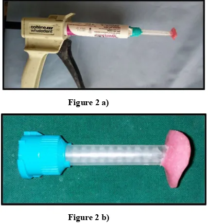
To take an impression of the eye socket the patient was seated in an erect position to allow the tissues involved in the defect to be recorded in their natural drape. An impression of the orbital socket was made with medium body addition silicone impression material (Aquasil, DENTSPLY) using a custom perforated tray attached to the mixing tip of the dispensing gun (Figure. 2a, b and
An index was made using putty addition silicone impression material (Aquasil, DENTSPLY). To obtain a wax conformer, molten wax (Modelling Wax, Deepti Dental Products of India Pvt. Ltd.) was flown in the Pearl Silva Lobo Norton, Dr. Surendra Kumar G. P., Dr. Bharath Raj, Dr. Nalinakshamma Muniswamy Reddy and Dr. Tanuja
Figure 1 a)
Figure 1 b)
Preoperative Photograph: a) Orbital Socket and b) Stock eye Prosthesis
Figure 2 a)
Figure 2 b)
a) Modified Custom tray on mixing tip of the dispensing gun b) Close up of Modified Tip
Figure 3 a)
Figure 3 b)
(a, b)Final Impression of the ocular defect
Figure 4 a)
Figure 4 b)
a) Putty adapted over the impression b) Putty Index separated with the wax conformer
[image:2.595.309.541.53.723.2] [image:2.595.111.246.59.504.2] [image:2.595.327.540.75.434.2] [image:2.595.347.551.461.727.2] [image:2.595.71.285.547.776.2]Figure 5 .Wax Conformer Trial
Figure 6. Graph Grid used to mark and simulate the location of the iris of the natural eye
The positioning of corneal/iris buttons is assessed using a graph grid so that its location would exactly simulate the other eye. A graphic grid was custom made using OHP sheet on which graph paper was printed. This graphic grid was relieved in a triangular form in the nose area. The patient was asked to gaze straight at an object kept 4 feet away. Vertical lines coinciding with the medial and distal extremities of the iris of the natural eye and horizontal lines referring to the center, inferior and superior limits of the iris were marked. The facial markings were transferred to the grid template by placing it on the patient's face. These markings were transported to the side of the defect and onto the sculptured wax pattern and the iris button was attached to the wax pattern. The iris cornea piece has a stem that indicated the direction of gaze to orient the iris in coordination with the other eye (Fig. 6 and Fig. 7) The selected shade of the sclera was matched with the
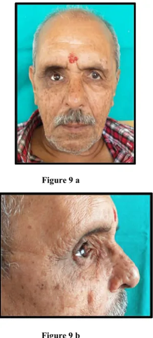
tooth colored heat cure resin (DPI) which was then packed in a two-piece flask and cured. After curing, the prosthesis was retrieved from the mold (Fig. 8 a-d). The prosthesis was then tried in the patient’s eye socket and adjusted for desired volume, retention and comfort. (Figure 9 a,b) Scleral characterization was done by extrinsic staining (acrylic paint) to match the contralateral natural eye (Fig. 10 a, b).
[image:3.595.71.288.54.195.2] The prosthesis was covered with a thin layer of modeling wax and further acrylized using clear polymethyl methacrylate.
Figure 7 a.
Figure 7 b.
a) Wax conformer with Corneal button b) Trial
Figure 8 a
Figure 8 b
[image:3.595.76.280.230.423.2]Figure 9 a
Figure 9 b
[image:4.595.381.525.53.262.2]Trial of the Ocular prosthesis prior to Characterisation a) Frontal View b) Profile view
Figure 10 a
Figure 10 b.
a) Characterisation of the sclera using oil paints b) Final characterized Ocular Prosthesis
Figure 11a.
[image:4.595.375.523.429.766.2]Figure 11 b.
Figure 11 c.
[image:4.595.59.285.439.768.2]DISCUSSION
The ocular prosthesis is an artificial replacement for the bulb of the eye. After the surgeon enucleates the eye, the maxillofacial prosthodontist provides the patient with an artificial eye to overcome the agony of losing the eye and helps him psychologically and socially (Laney WR et al, 1979). Ocular prosthesis can be either prefabricated stock eyes or custom-made. A custom-made ocular prosthesis replicates the orientation, natural color, contour, and size of the pupil and iris, improving the aesthetics and symmetry of the patient’s face (Raizada K et al, 2007; Frank RN et al 2000). It also allows uniform distribution of volume and weight, appropriate contour, providing excellent esthetics, and easy adaptation of the patient in wearing the prosthesis.(Goiato MC et al, 2013).Custom-made ocular prosthesis achieves intimate contact with the tissue bed enabling ideal fit and distributes pressure equally on the tissue bed (Ow KKR et al, 1997) The procedures for a custom-made ocular prosthesis includes an impression of the socket, wax pattern trial, iris positioning and acrylisation. The use of the putty index to fabricate the wax pattern eliminates the additional steps of fabricating a master cast which is not utilized for further fabrication of the ocular prosthesis. Different methods have been suggested to determine the size and position of the iris by visual judgment (Benson P., 1977), usingpupillometer (Roberts AC, 1969), or other calipers. Benson advocated a method of determining the size and position of the iris by visual judgment (Benson P., 1977).Since iris positioning is a technique-sensitive procedure, visual assessment alone may not be reliable and may incorporate parallax errors. Roberts suggested the use of a pupillometer for exact alignment of the pupil in the ocular prosthesis ( Roberts AC, 1969).Pupillometer is an instrument consisting of two parallel cylinders with positive lens for locating the pupil. However, the technical difficulty of making a pupillometer available in every clinical setup is a limitation for using this method (McArthur DR, 1977). A transparent graph template was used to accurately locate and position the iris rather than counting on the visual assessment alone, which can introduce interobserver variability because of binocular vision and parallax errors. The method described here is straightforward and can be carried out in any clinical set-up. However, the limitation of the method described here is that in the case of facial asymmetry, accurate determination of facial midline and iris positioning will be challenging (McArthur DR, 1977). The use of the corneal button aids in replicating the gaze of the patient. It allows for coordinating movements with the contralateral natural eye. The close adaptation of the custom made prosthesis to the tissue bed allows for some movement of the eye prosthesis aided by intact extraocular muscles. Various methods have been suggested for customizing the iris such as conventional painting on the artificial iris, reverse painting using prefabricated caps and using digital and hard copy images of the patient’s healthy eye(Raizada K et al, 2007; Sykes LM, 1996; Artopoulou II,
refabrication of the prosthesis as aging occurs (César R, 2008; Canadas MD, 2010; Fernandes AU, 2009; Haug SP, 2000).
Conclusion
Restoration of an ocular defect is indicated not only to prevent changes of the supporting tissue but also to avoid distress to the patient. Symmetry is important for the aesthetic appearance of the maxillofacial prosthesis, with ocular and orbital prosthesis being no exception. An accurate orientation of iris disk assembly largely contributes to the success of ocular and orbital prosthesis. The technique described here has attempted at positioning the iris, virtually eliminating the subjective errors arising out of visual illusion by using a transparent graph template. The patient satisfaction and acceptance were further improved using manual paint on technique for characterization of iris and sclera.
REFERENCES
Artopoulou, I.I., Montgomery, P.C., Wesley, P.J., Lemon, J.C. 2006. Digital imaging in the fabrication of ocular prostheses. J Prosthet Dent., 95:327‑30
Benson, P. 1977. The fitting and fabrication of a custom resin artificial eye. J Prosthet Dent., 38:532‑8.
Canadas, M.D., Garcia, L.F., Consani, S., Pires‑de‑Souza, F.C. 2010. Color stability, surface roughness, and surface porosity of acrylic resinsfor eye sclera polymerized by different heat sources. J Prosthodont, 19:52‑7.
César, R. 2008. Evaluation of iris color stability in ocular prosthesis. Braz Dent J., 19:370‑4.
Fernandes, A.U., Goiato, M.C., dos Santos, D.M. 2009. Effect of weathering and thickness on the superficial microhardness of acrylic resin and ocularbutton. Cont Lens
Anterior Eye, 32:283‑7
Frank, R.N., Puklin, J.E., Stock, C., Canter, L.A. 2009. Race, iris color, and age‑related macular degeneration. Trans Am
OphthalmolSoc., 98:109‑15.
Goiato, M.C, Micheline, D., Dds, H., Helga, K., Turcio, L., Fili, M. 2013. An alternate impression technique for ocular prostheses. J Prosthodont, 22:338‑40.
Haug, S.P., Andres, C.J. 2000. Fabrication of custom ocular prosthesis. In: Taylor TD, editor. Clinical maxillofacial prosthetics. 1st ed. Chicago: Quintessence Publishing; p. 265-76.
Laney, W.R., Gardner, A.F. 1979. Maxillofacial prosthetics. PSG, Littleton
McArthur, D.R. 1977. Aids for positioning prosthetic eyes in orbital prostheses. J Prosthet Dent., 37:320‑6
Ow, K.K.R., Amrith, S. 1997. Ocular prosthetics: Use of a tissue conditioner material to modify a stock ocular prosthesis. J Prosthet Dent., 78:218-22.
Ocular and Orbital Prostheses. J Prosthodont., 20(3): 244-246.
Raizada, K., Rani, D. 2007. Ocular prosthesis. Cont Lens Anterior Eye, 30:152‑62.
Roberts, A.C. 1969. An instrument to achieve pupil alignment in eye prosthesis. J Prosthet Dent., 22:487‑9.
Sykes, L.M. 1996. Custom made ocular prostheses: A clinical report. J Prosthet Dent., 75:1‑3.