doi: 10.1093/femspd/ftv067
Advance Access Publication Date: 7 September 2015 Minireview
M I N I R E V I E W
Roads to the development of improved pertussis
vaccines paved by immunology
Jolanda Brummelman
1
, Mieszko M. Wilk
2
, Wanda G.H. Han
1
,
C ´ecile A.C.M. van Els
1
,
∗
and Kingston H.G. Mills
2
1
Centre for Infectious Disease Control, National Institute for Public Health and The Environment,
Bilthoven, the Netherlands and
2Immune Regulation Research Group, School of Biochemistry and
Immunology, Trinity Biomedical Sciences Institute, Trinity College Dublin, Dublin 2, Ireland
∗Corresponding author:Centre for Infectious Disease Control, National Institute for Public Health and The Environment, Postbus 1, 3720 BA Bilthoven, the Netherlands. Tel:+31-30-2743665; E-mail:[email protected]
One sentence summary:To develop improved pertussis vaccines capable of inducing long-lived protective immunity, lessons have to be learned from immunology ofBordetella pertussisinfection and current vaccination.
Editor:Nicholas Carbonetti
ABSTRACT
Current acellular pertussis vaccines have various shortcomings, which may contribute to their suboptimal efficacy and waning immunity in vaccinated populations. This calls for the development of new pertussis vaccines capable of inducing long-lived protective immunity. Immunization with whole cell pertussis vaccines and natural infection withBordetella pertussisinduce distinct and more protective immune responses when compared with immunization with acellular pertussis vaccines. Therefore, the immune responses induced with whole cell vaccine or after infection can be used as a benchmark for the development of third-generation vaccines against pertussis. Here, we review the literature on the immunology ofB. pertussisinfection and vaccination and discuss the lessons learned that will help in the design of improved pertussis vaccines.
Keywords:Bordetella pertussis; vaccine; T cell; Toll-like receptor agonist; immunology
Introduction
Bordetella pertussisis a respiratory tract pathogen that causes the severe disease whooping cough (pertussis) in infants and young children. It can also infect adolescents and adults. The disease was largely controlled in developed countries follow-ing the introduction of whole cell pertussis (wP) vaccines in the 1940s/1950s. However, pertussis still kills over 200 000 infants yearly worldwide, mostly in developing countries, but the in-cidence of disease is also increasing in many developed coun-ties (Blacket al.2010). This has been attributed to the intro-duction of acellular pertussis (aP) vaccines in the 1990s, which were developed in response to concerns around the safety of wP vaccines. Unlike wP vaccines, which are composed of killed
bacteria with a wide range of antigens and pathogen-associated molecular patterns (PAMPs) that bind to pattern recognition re-ceptors (PRRs) and activate innate immune cells, aP vaccines are composed of three to five B. pertussisantigens absorbed to alum as the adjuvant. A monocomponent vaccine based on hydrogen peroxide-detoxified pertussis toxin (PT) was also developed (Trollfors et al.1995), but had lower efficacy than the three and five component aP vaccines. Although initial reports from phase 3 clinical trials carried out in the 1990s suggested that aP vaccines were at least as effective as wP vaccines (Grecoet al.1996; Gustafssonet al.1996), recent stud-ies have demonstrated a rapid and alarming drop in protec-tion over time, suggesting a failure to sustain immunity (Klein et al.2012).
Received:18 June 2015;Accepted:17 August 2015
C
FEMS 2015. This is an Open Access article distributed under the terms of the Creative Commons Attribution Non-Commercial License (http://creativecommons.org/licenses/by-nc/4.0/), which permits non-commercial re-use, distribution, and reproduction in anymedium, provided the original work is properly cited. For commercial re-use, please [email protected]
Table 1.The putative roles of different immune cells in protective immunity toB. pertussis.
Cell Type Infectiona wP aP Species Function Reference
Th1 cell +++ ++ +/– Man IFNγproduction, macrophage
activation, opsonizing Ab, prevents dissemination of Bp
(Ryanet al.1997,1998b; Ausielloet al. 1999; Mascartet al.2003; Mascart
et al.2007; Schureet al.2012)
+++ + + Baboon (Warfel and Merkel2013)
+++ ++ +/– Mouse (Millset al.1993; Rosset al.2013;
Raevenet al.2014; Brummelman
et al.2015)
Th2 cell +/– +/– +++ Man No identified role in mice;
unknown role in baboons and humans
(Ryanet al.1997,1998b; Ausiello
et al.1999; Mascartet al.2003; Mascartet al.2007; Schureet al. 2012), van Twillert unpublished
– – +++ Baboon (Warfel and Merkel2013)
– – +++ Mouse (Millset al.1993; Rosset al.2013;
Raevenet al.2014; Brummelman
et al.2015)
Th17 cell +++ ++ +/– Man IL-17 production, neutrophil
recruitment and activation
(Schureet al.2012), van Twillert, unpublished
+++ ++ – Baboon (Warfel, Zimmerman and Merkel
2014)
+++ ++ +/– Mouse (Rosset al.2013; Raevenet al.2014;
Brummelmanet al.2015)
TRMcell +++ ++ – Mouse Sustains local cellular immunity
in the respiratory tract?
Wilk and Mills, unpublished
TFHcell +++ ++ + Mouse Activates Ab production and
memory B cells?
Wilk, Allen and Mills, unpublished
TCMcell ? + + Man and Mouse Maintains long-term immunity (Brummelmanet al.2015; de Rond
et al.2015).
TEMcell ? + ++ Man and Mouse Immediate effector function (Brummelmanet al.2015, de Rond
et al.2015).
TTDcell ? + ++ Man Waning immunity? (de Rondet al.2015).
γ δT cell ++ ? ? Mouse Immune regulation early in
infection
(Zachariadiset al.2006)
B cell + ++ +++ Mouse Ab production (Mahonet al.1997; Leefet al.2000;
Stengeret al.2010)
Alveolar Mac +++ + – Mouse Early response to infection,
phagocytosis and killing of Bp
(Bernardet al.2015)
DC +++ ? ? Mouse Activation of naive T cells in
lymph nodes
(Dunneet al.2009)
NK cell +++ ? ? Mouse Early IFNγ, prevents
dissemination of Bp
(Byrneet al.2004)
Neutrophil +++ ++ – Mouse Ab-dependent phagocytosis and
killing of opsonized Bp
(Andreasen and Carbonetti2009; Rosset al.2013; Eby, Gray and Hewlett2014).
aResponses denoted as+++,++,+,+/–, – and ? equate to strong, medium, weak, weak/ inconsistent, undetectable responses or not tested respectively; Bp,B. pertussis, Ab, antibody.
There have been a number of explanations put forward to account for recent epidemics of pertussis in highly vaccinated populations. First, there is evidence of antigen variation in some of the key protective antigens and components of aP vaccines, namely pertussis toxin (PT), pertactin (Prn) and Fimbriae (Fim2 and Fim3) (van Looet al.2002; Tsanget al.2004; Bartet al.2014). Moreover, Prn-deficient strains have emerged following intro-duction of aP vaccination in Europe (Bouchezet al.2009; Barkoff et al.2012; Hegerleet al.2012; Zeddemanet al.2014), Australia (Lamet al.2014), Japan (Otsukaet al.2012) and the USA (Queenan, Cassiday and Evangelista2013; Pawloskiet al.2014; Martinet al.
2015). Therefore, it is possible that the immune responses in-duced by three to five antigens from the single isolate used to manufacture the aP vaccine do not protect against at least some
of the current circulating strains. Secondly, the waning protec-tive immunity observed following immunization with aP vac-cines suggests that these vacvac-cines fail to generate effective im-munological memory (Kleinet al.2012,2013; Sheridanet al.2012; Liko, Robison and Cieslak2013; Wittet al.2013).
the less effective aP vaccines induce a mixed Th2 and Th17 re-sponse (Rosset al.2013), but the Th2 component appears to be redundant to protection and, together with associated IgE (Ryan et al.2000), may even be the culprits in rare type hypersensi-tivity reactions seen in children after a fourth or fifth dose of the aP vaccines (Rennelset al.2008). This pattern of immune responses induced by the pertussis vaccines is generally simi-lar in humans and mice. Recent evidence from the new animal model of experimental pertussis in baboons has uncovered a major deficit in protective immunity induced by aP vaccination. While the immune responses induced by immunization with wP vaccines were able to prevent disease and enhanced clear-ance of the infection (though not as effectively as previous infec-tion), immunization with the aP vaccine prevented disease, but did not prevent infection or transmission ofB. pertussisto naive baboons (Warfel, Zimmerman and Merkel2014). Whilst it has not been conclusively proven in the baboon, it appears that the failure of aP vaccines to prevent infection reflects its failure to induce appropriate cellular immune responses, especially Th1 cells. In this minireview, immunological evidence from infection and vaccination studies will be discussed in order to guide the development of improved pertussis vaccines.
CURRENT STATE OF THE ART ON THE
MECHANISMS OF NATURAL IMMUNITY
TO
B. PERTUSSIS
The innate immune response plays a crucial role in controlling the initial stagesof B. pertussisinfection and helps to shape the subsequent adaptive immune responses.B. pertussisattaches to
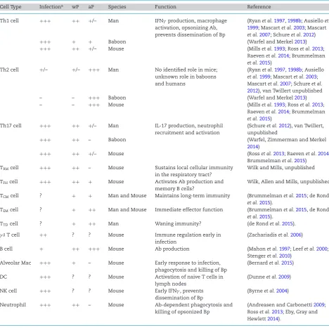
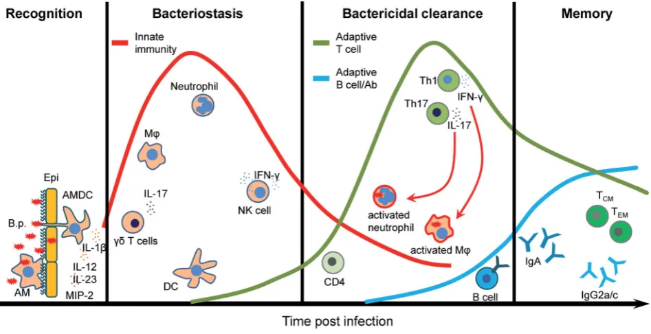
ciliated epithelial cells on the upper respiratory tract (Coutte et al.2003; Carbonettiet al.2007). These cells may also provide a first barrier, by preventing pathogen penetration and through production of mucine by secretory goblet cells which can pro-tect the lungs from pathogens that are inhaled into the respira-tory track. Two resident innate immune cell types, airway mu-cosal dendritic cells (AMDCs) and alveolar macrophages (AMs), which are in close contact with the lung epithelium, mediate the first cellular response toB. pertussis. AMDCs are strategically positioned between epithelial cells to take up antigen directly from the airway lumen and prime T cells after migration to the lymph node (Stumbleset al.1998; Jahnsenet al.2006), whereas AMs reside within the mucous layer and appear to be impor-tant for phagocytosis and killing ofB. pertussis(Lambrecht2006). These lung-resident innate immune cells provide a first line of immediate immune defense againstB. pertussisinfection. Addi-tionally, they initiate and orchestrate complex and tightly regu-lated processes that involve activation and recruitment of other immune cells and generation of long-lasting adaptive immunity (Fig.1and Table1).
[image:3.595.62.536.385.628.2]B. pertussiswas classically considered to be an extracellular pathogen that infects the upper respiratory tract, but it can also penetrate the lungs and has been found inside ciliated respira-tory epithelial cells and in lung macrophages (Paddocket al.2008; Lambertiet al.2010,2013). The recognition ofB. pertussisPAMPs and virulence factors including lipooligosacharide (LOS), adeny-late cyclase toxin (ACT), filamentous hemagglutinin (FHA) and TLR2 lipoproteins by PPRs expressed by macrophages and DCs (and other cells of the innate immune system) leads to their maturation and production of cytokines and chemokines that
mediate and regulate immune responses to the bacteria. It was also shown that PT can act as PAMP, triggering the TLR4 signal-ing pathway (Wanget al.2006; Nishidaet al.2010) and enhanc-ing IL-12 production, thereby actenhanc-ing as an adjuvant to promote Th1 responses (Ryanet al.1998a; Ausielloet al.2002; Tononet al.
2002). Binding of LOS to TLR4 results in DC maturation and pro-duction of IL-12 and IFNγ, cytokines required for development of Th1 responses (Higginset al.2003). TLR4 activation of DCs with LOS can also induce IL-10 production, consequently gen-erating IL-10-secreting type-1 regulatory T cells (Tr1) that may dampen Th1 responses and limit inflammatory pathology inB. pertussis-infected lungs (Higginset al.2003). DCs can also be acti-vated by intrinsic recognition of otherB. pertussisvirulence fac-tors and toxins. It was shown that ACT activates the NLRP3 in-flammasome and caspase-1, leading to production of mature bioactive IL-1β. Active IL-1β together with IL-23 promotes ex-pansion of murine Th17 cells, which help to recruit neutrophils that promote killing ofB. pertussis(Dunneet al.2010). Further-more, studies with human DCs have demonstrated that ACT en-hances Th17 responses but suppresses Th1 responses by inhibit-ing IL-12p70 production (Spensieriet al.2006). ACT also modu-lates TLR-induced signaling to upregulate IL-10 production and promote the development of Treg cells (Hickey, Brereton and Mills2008).
It was shown that TLR4-deficient C3H/HeJ mice had a more severe course ofB. pertussisinfection when compared with TLR4-sufficient C3H/HeN mice (Mannet al.2005; Higginset al.2006; Banuset al. 2008). TLR4 expressed by DCs and macrophages plays a central role in protective immunity to this pathogen. Furthermore, an early burst of proinflammatory cytokines and chemokines produced by AMs soon after infection depends on TLR4 signaling pathway. Mice that lack MyD88 adaptor-like pro-tein (Mal; also known as TIRAP) were unable to control infection (Bernardet al.2015). The bacteria disseminated from the lungs causing lethality in nearly 50% of MAL−/− mice. Additionally, AMs were completely depleted from the lungs of Mal−/−mice early after infection, suggesting that these cells are critical for mediating protection toB. pertussis(Bernardet al.2015).
The initial recruitment of DCs and macrophages into the lungs ofB. pertussis-infected mice is followed by recruitment or expansion ofγ δT cells and infiltration of neutrophils and NK cells. It is thought thatγ δT cells orchestrate the early cell traf-ficking into the lungs by producing IL-17 (Zachariadiset al.2006). This cytokine together with CXCL2 (MIP-2) secreted by activated macrophages and epithelial cells is required for neutrophil re-cruitment as well as induction of Th1 and Th17 responses to B. pertussis.
A primary role of neutrophils during the microbial infection is antibody-mediated phagocytosis and intracellular killing of pathogenic bacteria and formation of neutrophil extracellular traps (Andreasen and Carbonetti2009; Eby, Gray and Hewlett
2014). It was shown that ACT can inhibit neutrophil function but does not impact on protective immunity toB. pertussis. It was suggested that neutrophils are important, but not essential, in the controlling of bacteriostasis ofB. pertussis,but are critical for clearance ofB. bronchiseptica;infection of neutrophil-deficient mice withB. bronchsepticahad a lethal outcome (Harvill, Cotter and Miller1999). It was proposed thatB. bronchisepticacan kill resident and recruited phagocytes in the lungs or AMs are suf-ficient to controlB. pertussisinfection (Harvill, Cotter and Miller
1999).
NK cells play a protective role in immunity to B. pertussis through secretion of IFNγ, which enhances the antimicrobial ac-tivity of macrophages. Production of IFNγby NK cells is
depen-dent on IL-12 fromB. pertussis-activated DCs. Depletion of NK cells fromB. pertussis-infected mice resulted in the dissemina-tion of the bacteria from the respiratory tract to the liver and this was associated with reduced IFNγproduction and Th1 re-sponses (Byrneet al.2004). Thus, NK cells play a critical role in early innate immune control of infection and in shaping the adaptive immune response toB. pertussis(Fig.1).
The lungs of na¨ıve mice contain very low numbers of T cells. However, during the course of infection withB. pertus-sisthe adaptive immune response slowly develops and T cells are recruited to or expanded in the respiratory tract and me-diate subsequent bacterial clearance ofB. pertussis from the respiratory tract. Human infants without fully developed adap-tive immunity are particularly susceptible to the severe symp-toms of whooping cough, which can be lethal. Studies in mice have demonstrated that CD4+ T cells play a key role in the clearance ofB. pertussisinfection. Primary infection of athymic nu/numice, which lack all T cells, results in a persistent or even lethal infection withB. pertussis(Millset al.1993). Adoptive transfer of immune splenic T cells into athymic mice reversed chronic infection and resulted in bacterial clearance within 14 to 21 days post-challenge. Similarly, transfer of B. pertussis-specific CD4+T cells into immunosuppressed sublethally irra-diated na¨ıve BALB/c mice before infection resulted in clearance of the pathogen. In contrast, sublethally irradiated mice were not able to clear primary infection after transfer ofB. pertussis-specific CD8+cells (Millset al.1993). In addition, antigen-specific CD4+T cells from infected mice produce IFNγand/or IL-17, but not Th2-type cytokines, suggesting that Th1 and Th17 cells are involved in protective immunity toB. pertussisinfection. Indeed, IFNγ−/−mice exhibit impaired ability to clear the pathogen from the lungs (Barbicet al.1997), whereas IFNγR−/−mice develop a lethal disseminated infection. Interestingly, IFNγR−/−mice that survive the initial infection develop neither Th1 nor Th2 re-sponse, suggesting that Th17 cells might be involved in devel-oping protective immunity toB. pertussis(Mahonet al.1997). In-deed, it has been demonstrated that IL-17−/−mice infected with B. pertussishave a significantly greater bacterial load in compar-ison to wild-type (WT) mice (Rosset al.2013). This impaired abil-ity of IL-17−/−mice to clear the bacteria correlated with a signifi-cant decreased frequency of neutrophils in the lungs during the course on infection (Rosset al.2013). Taken together, the pub-lished studies demonstrate that Th1 and Th17 cells both con-tribute to the protective immunity induced by primaryB. pertus-sisinfection in mice (Fig.1).
Humoral immunity may play an auxiliary role in the clear-ance of infection from the lungs and contribute to a develop-ment of the long-lasting, cell-mediated immunity toB. pertussis. The number of B cells significantly increases in the lungs dur-ing the course of infection reachdur-ing the highest number when the infection is almost cleared. Similarly,B. pertussis-specific IgG antibodies are only detectable in the serum at significant levels when the pathogen is almost cleared from the lungs (Millset al.
possible that B cells may contribute to the induction of memory CD4+T cells by acting as the antigen-presenting cells (Leefet al.
2000; Linton, Harbertson and Bradley2000).
Convalescent mice are able to clear the pathogen from the lungs as soon as 3–7 days after reinfection (Millset al.1993). Baboons recovered from aB. pertussis infection are protected from disease and do not transmit the pathogen upon sub-sequent challenge and this was associated with a long-lived mixed Th1/Th17-cell response (Warfel and Merkel2013; Warfel, Zimmerman and Merkel 2014). Hence, natural infection in-duces strong and persistent immune protection against rein-fection withB. pertussis, although it does not result in life-long immunity.
MECHANISMS OF VACCINE-INDUCED
IMMUNITY AND LIMITATIONS OF CURRENT
AP VACCINES
Due to their high reactogenicity, wP vaccines were replaced in the 1990s in most developed countries by aP vaccines contain-ing a high dose of three to five immunogenicB. pertussis anti-gens, namely PT, Prn, FHA, Fim2 and Fim3. Both vaccine types provide protection against severe disease, however, with impor-tant underlying differences. aP vaccine-induced protective im-mune responses are less durable, and in contrast to wP vaccine-induced immunity only protect against clinical symptoms of pertussis but not against colonization and transmission ofB. per-tussis(Warfel, Zimmerman and Merkel2014).
Besides its narrow specificity, various other features of aP vaccine-induced immunity may account for these differences. First, aP-induced T-cell responses have a distinct functional pro-gramming, compared to those induced by wP vaccines, as evi-denced by both humoral and cell-mediated responses. While the two vaccine types readily induce significant levels ofB. pertussis antigen-specific IgG antibodies, as reported in humans (Edwards et al.1995; Hendrikx et al. 2011) and in mice (Redheadet al.
1993; Stengeret al.2010; Raevenet al.2015), there is a differ-ence in class switching patterns. In mice, aP vaccines induce pre-dominantly antibodies of the IgG1 subclass (Stengeret al.2010; Brummelmanet al. 2015), reflecting the induction of a Th2-type response, whereas wP vaccines induce predominantly IgG2a/b/c, as well as IgG1, and IgG3 (Raevenet al.2015), which is consistent with the strong induction of Th1 cells. A compari-son of the antibody isotype in cohorts of children that had been primed with either wP or aP vaccines, and followed between 3 and 9 years of age, showed that IgG1 was the predominant IgG subclass induced by both vaccines (Hendrikxet al.2011). In humans, however, this complement activating and opsoniz-ing IgG subclass is thought to be driven by Th1 cells (Hjelholt et al.2013). Only low levels of the IgG2 subclass were observed in both wP- and aP-primed children (Hendrikxet al.2011). The Th2-associatedB. pertussis-specific IgG4 subclass was detected only after aP vaccination, with higher concentration after a greater number of doses (Hendrikxet al.2011). Moreover, these IgG4 antibodies strongly correlate with the total IgE production in these aP-vaccinated children (Hendrikxet al.2011), indicating that Th2-skewed immune responses are induced by aP vaccina-tion in humans as well as in mice.
Further evidence for different programming of immune responses induced with aP and wP vaccines comes from studies focused on CD4+ T-cell responses and assessing the importance of their secreted cytokine profiles in the context of protective immunity in different species (Table1). In mice, aP
vaccination is associated with induction of CD4+T cells that pro-duce IL-4, IL-5 and IL-17, but relatively lower concentrations of IFNγ (Rosset al.2013; Brummelmanet al.2015), representing a mixed Th2/Th17 response, while wP vaccines induce a mixed IFNγ/IL17A (Th1/Th17) response (Rosset al.2013). Studies using cytokine-defective mice have shown that protection againstB. pertussisinduced by immunization with an aP vaccine is as ef-fective in IL-4−/−or IFNγ−/−mice as in WT mice, whereas pro-tection is significantly diminished in IL-17A−/−mice (Rosset al. 2013). This indicates that Th17 cells play an essential role in the protective immune response induced by the aP vaccine, while the Th2 cells do not contribute to protection. In contrast, Th1 are required for protection induced with wP vaccines (Rosset al.
2013).
In the baboon model, immunization with an aP vaccine, which conferred protection against disease but not infection or transmission, induces a mixed Th1/Th2 type of CD4+T-cell re-sponse. In contrast, wP vaccination, providing protection against colonization and host-to-host transmission, induced a mixed Th1/Th17 memory response (Warfel, Zimmerman and Merkel
2014), suggesting that both Th1 and Th17 cells may be impor-tant in prevention ofB. pertussiscolonization in this model.
In humans, aP vaccination has been shown to induce a Th2-dominated, yet mixed Th2/Th1/Th17 type of CD4+ T-cell re-sponse in children (Ryanet al.1998b; Ausielloet al.1999; Mascart et al.2007; Schureet al.2012). In contrast, wP vaccines induce a mostly mixed Th1/Th17-type CD4+T-cell response, similar to natural infection (Ryanet al.1998b,2000; Mascartet al.2003,
2007; Roweet al.2005; Vermeulenet al.2010; Rosset al.2013). No-tably, some of the Th1 responses in certain aP-vaccinated chil-dren may be attributed to exposure to or subclinical infections withB. pertussis(Ryanet al.1998b; Ausielloet al.1999; Mascart et al.2003; Schureet al.2012). In general, the studies in humans are consistent with the data from animal models, i.e. indicat-ing Th2 dominance associated with aP vaccination and a mixed Th1/Th17 profile after wP vaccination, implying that aP vaccines may have a poorly protective functional T-cell profile in humans. A second feature of the immune response that might con-tribute to the difference in long-term effectiveness of aP and wP vaccines has recently emerged from the series of head-to-head comparisons of the cellular immunity in aP- and wP-primed cohorts of children by Buisman and coworkers. It was found that PBMC from aP-primed 4-year-old children, three years af-ter their primary pertussis vaccine series, produced higher spe-cific levels of CD4+T-cell cytokines than those from wP-primed children; however, this response was not boosted after a fifth dose of aP vaccine, in contrast to the response in wP-primed children (Schureet al.2012). Yet, at the age of 6, two years af-ter this boosaf-ter dose, PBMC from aP-primed children produced lower levelsB. pertussis-specific IL-17 when compared with PBMC from wP-primed children (Schureet al.2013). Furthermore, as-sessment of CCR7 and CD45RA expression, which allowed CD4+ T cells to be categorized as naive T cells (TN), effector memory
T cells (TEM), central memory T cells (TCM) or terminally
differ-entiated T cells (TTD), showed that the proportion ofB. pertussis
antigen-specific TTDcells was higher, in the aP-primed cohort
(de Rondet al.2015). Together, these data suggest that aP-primed CD4+T-cell responses might be more end-stage differentiated than those induced by wP vaccines. The high antigen dose in aP vaccines compared to wP vaccines may contribute to this phe-nomenon. High antigen and adjuvant dose may induce vigorous short-term immunity, but can also exhaust the immune system and impede long-term immunity (Darrahet al.2007; Joshiet al.
2014). For example, in the Leishmania model, high dose vacci-nation with a Leishmania protein-expressing adenoviral vector in mice elicited higher numbers of total IFNγ-producing CD4+ T cells compared to low dose vaccination; however, high dose vaccination resulted in poor protective immunity (Darrahet al.
2007).
Thus, aP vaccine-induced immunity has various shortcom-ings, including immune responses of narrow specificity and with the wrong type of Th-cell subtypes and end-stage differentiated memory T cells which may collectively contribute to its subop-timal efficacy.
APPROACHES TO IMPROVE EFFICACY OF
CURRENT AP VACCINATION BY TLR LIGATION
The lesson from studies on T cells in mouse models and hu-mans to date have suggested that next-generation aP vaccines should induce CD4+T cells with a TCMcell phenotype a mixed
Th1/Th17-cell profile, like those induced by infection or immu-nization with wP vaccine. An important factor in natural infec-tion withB. pertussisand immunization with wP vaccines that steers protective adaptive immune responses is the presence of PAMPs, including LOS that binds to TLR4 and lipoproteins that bind to TLR2 (Dunneet al.2015). These PAMPs activate innate cells via PRRs and thereby promote DC maturation and produc-tion of proinflammatory cytokines that direct the inducproduc-tion of Th1 and Th17 cells (Fedeleet al.2008). Current aP vaccines do not contain classic PAMPs. Although active PT has adjuvant proper-ties and might be considered to be a PAMP, the PT preparations in aP vaccines are aldehyde treated which destroys immunos-timulatory activity (Ryanet al.1998a). Overall, PT-containing aP vaccine induces a Th2-dominated response, likely due to the Th2-promoting properties of the adjuvant alum. One approach to enhance the efficacy of aP vaccines could be to address this shortfall by replacing or complementing alum with PAMPs, which are known to induce Th1 responses. Indeed, there is evi-dence from mouse studies that several TLR agonists may be ef-fective Th1 promoting adjuvants for experimental aP vaccines.
A natural TLR2 ligand ofB. pertussis, BP1569, was recently identified (Dunneet al.2015). Immunization of mice with an experimental aP vaccine containing a corresponding synthetic lipopeptide LP1569 induced a higher level of protection against aerosolB. pertussischallenge when compared with the same vac-cine formulated with alum. Furthermore, when compared with an alum-adjuvanted vaccine, the same vaccine formulated with the TLR2 lipoprotein was found to induce significantly higher IgG2a antibodies and promote higher IL-17, IFNγand lower IL-5 production by spleen cells after antigen stimulationex vivo.
The TLR4 agonist LOS is an important PAMP in wP vaccines enhancing Th1 responsiveness, but was also the main media-tor of their high reactogenicity (Donnellyet al.2001). Although mice are less sensitive to endotoxin than humans (Munford
2010), LOS from B. pertussisis less effective in activating hu-man macrophages and DCs than LPS from other bacteria (Fedele et al.2008). Therefore, well-defined detoxified LPS derivatives from other Gram-negative bacteria with Th1-promoting capac-ity have been tested as adjuvant for an experimental aP vac-cine. Immunization of mice with an aP vaccine adjuvanted with monophosphoryl lipid A (MPL), the hydrophobic biologically ac-tive Lipid A part ofSalmonella minnesotaLPS, was more effec-tive in protecting againstB. pertussischallenge than the aP vac-cine adjuvanted with alum (Geurtsenet al.2008). Furthermore, B. pertussis-specific IL-5 production by spleen cells was lower
following immunization with MPL-adjuvanted vaccine, indicat-ing that this adjuvant can skew the immune response away from a Th2 response. Substantiating these data, the authors re-cently found, at the single T-cell level, that addition of the water-soluble genetically engineered derivative fromNeisseria meningi-tidisLPS, LpxL1 (Zariri and van der Ley2015), to the aP vaccine increased the percentage of specific IFNγand IL-17-producing CD4+T cells and diminished the percentage of IL-5-producing CD4+T cells (Brummelmanet al.2015). Combining direct MHC class II tetramer staining of Prn-specific CD4+T cells with the memory markers CD44 and CD62L in this model indicated that the ratio of TCMand TEMwas not altered by addition of the
ad-juvant; however, there was a net benefit in the number of TCM,
since higher numbers ofB. pertussis-specific memory CD4+ T cells were detected after vaccination with the LpxL1-containing aP vaccine. Hence, this analysis confirmed that there was no trade-off for the altered cytokine profile after TLR4 engaging aP vaccination by a reduction of induced TCMcells. Since there
are species differences in specificity of ligands for TLR4 (Marr et al. 2010; Bryant and Monie2012), caution should be exer-cised in translating studies on LPS-based adjuvants from mice to humans.
TLR9 agonists, including CpG oligonucleotides from bacterial DNA, can also induce Th1 and Th17 responses and have been used as adjuvants for experimental aP vaccines tested in mice. The complementation or replacement of alum in an aP vaccine by CpG increased the anti-PT IgG2a titers, and the IgG2a/IgG1 ra-tio, providing indirect evidence for induction of Th1 responses (Sugaiet al.2005). In addition,B. pertussis-specific IFNγ produc-tion by spleen cells was found to increase when PT, FHA and Prn were administered intranasally with both CpG and alum, but not with CpG alone (Asokanathan, Corbel and Xing2013). Finally, in another report substituting alum with CpG in an ex-perimental parenterally delivered aP vaccine enhanced its effi-cacy against aerosol challenge withB. pertussisand was associ-ated with strongerB. pertussis-specific IFNγand IL-17, but lower IL-4 and IL-13 production by spleen cells, and higher IgG2a con-centrations in sera (Rosset al.2013). The failure of the study by Asokanathanet al.to detect IFNγ following immunization with pertussis antigens and CpG (without alum) may be due to the route of immunization (i.n.) and the lower dose of CpG (30μg) employed. Together, these studies demonstrate that TLR agonists such as TLR2, TLR4 and TLR9 ligands can steer the aP vaccine-induced CD4+ T-cell response towards a more fa-vorable protective Th1/Th17 response, and that pertussis vac-cine formulations containing experimentally selected adjuvants could be considered as more effective alternatives for current aP vaccines.
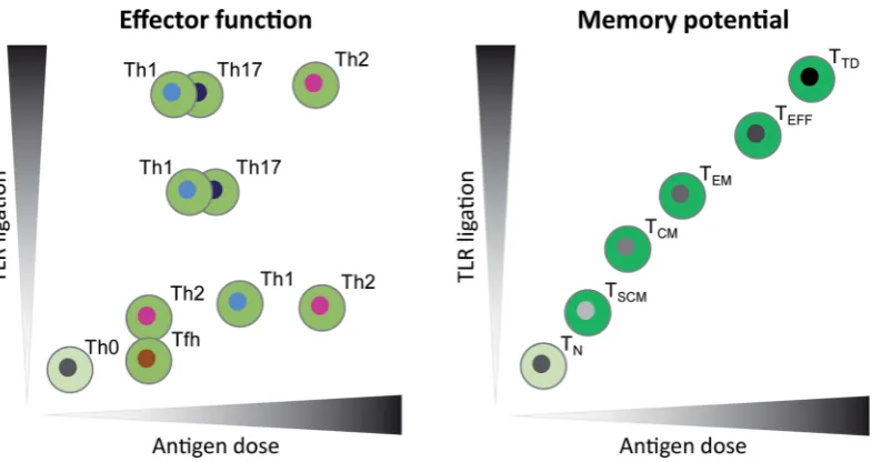
In addition to the type of adjuvant, the antigen composition and dose of both adjuvant and antigen may influence the induc-tion of T-cell response with aP vaccines. Therefore, careful eval-uation of choice of adjuvant and antigen and their dose will be required to ensure that a vaccine induces the appropriate func-tional T-cell subtype and profile of memory CD4+T cells required for complete and sustained protective immunity against infec-tion (Fig.2).
Figure 2.Models for the influence of antigen dose and the strength of TLR ligation on CD4+T-cell differentiation. Vaccine-induced CD4+T-cell responses can be
evaluated based on effector function i.e. Th type (left panel) and memory potential i.e. memory differentiation stage (right panel). Antigen dose can affect TCR signal strength, thereby influencing Th type and memory T-cell differentiation (Gattinoni, Klebanoff and Restifo2012; Tubo and Jenkins2014). Inflammatory signals, such as induced by TLR ligation, during T-cell priming can also influence the Th type and memory potential of antigen-specific T cells via innate mechanisms (Joshiet al. 2007; Tubo and Jenkins2014). Thexaxes depict the range of antigen dose and theyaxes show the amount of TLR ligation present during vaccination or infection. In vivo, antigen and TLR ligand dose may form a gradient of local concentrations, possibly inducing an array of differentiation states. In the presence of low dose TLR ligands, low antigen dose will steer towards Th2 or Tfh cells, while an intermediate dose promotes Th1 cells and a high dose Th2 cells. Increasing TLR ligation induces Th17 and Th1 cells, yet high exposure to both TLR ligation as antigen dose will promote Th2 cells (Tubo and Jenkins2014). In parallel, both low and high antigen dose and TLR ligation strength may also induce regulatory functions in cell populations (not shown). The memory differentiation stage is correlated with both the antigen dose and TLR ligation, meaning that the memory potential of the cells decreases when the cells are exposed to increasing TLR ligation and antigen dose (Joshiet al.2007; Gattinoni, Klebanoff and Restifo2012). The color gradient of memory phenotypes from light to dark indicates loss of long-term memory potential. TLR: Toll-like-receptor; TN: na¨ıve T cell; TSCM: stem-cell memory T cell; TCM: central memory T cell; TEM: effector memory T cell; TEFF: effector T cell; TTD: terminally differentiated T cell.
of IFNγ- and IL-17- producing T cells, indicating that BPZE1 should be capable of inducing the favorable Th1/Th17 response in humans (Schiavoniet al.2014). This vaccine is currently be-ing tested in the baboon model and is in clinical development (Thorstenssonet al.2014). Another vaccine concept based onB. pertussisouter membrane vesicles also has been shown to in-duce a mixed Th1/Th17-type response in mice and to inin-duce protection against B. pertussis challenge (Gaillardet al. 2014) (Raeven and Brummelman et al. in preparation). These vac-cine candidates also address other limitations of aP vacvac-cines, by expressing ‘natural’ (low) doses antigen (preventing an ex-hausted immune response) and a broader panel of antigens, im-portant to ensure maintenance of long-lasting protective immu-nity against circulatingB. pertussisstrains with altered antigenic make up.
IMMUNOLOGICAL EVALUATION OF NEW
PERTUSSIS VACCINES IN (PRE-) CLINICAL
DEVELOPMENT
The development of new pertussis vaccines requires immuno-logical tools to determine the functional differentiation, mem-ory potential, breadth and protective capacity of the induced im-mune responses in reliable preclinical animal models, impor-tant to predict efficacy in humans. Mouse and more recently baboon models have been used to evaluate candidate pertus-sis vaccines. One of the problems with these models is that the level of protection is usually assessed shortly after vaccination (van der Arket al.2012). Furthermore, mice are not the natu-ral host ofB. pertussisand relatively high vaccine doses are of-ten used in these challenge models to induce measureable
im-mune responses; thus, the results may be difficult to translate to the humans. Future studies in these models should address the issue of waning immunity by assessing the long-term pro-tection of experimental aP vaccines, and the propro-tection induced by lower doses. While it is possible to do medium-term (6–12 month) studies in the mouse model, studies of greater than 1 year will require experiments in the recently developed baboon model. Warfel and Merkel (2013) have used species-specific tools to assess waning of the vaccine induced CD4+T-cell response up to 15–24 month afterB. pertussisinfection in baboons. However, cost and animal availability will be significant limitations to ex-tensive use of this model. In both mice and baboons, identify-ing early biomarkers that predict long-term immunity would be valuable.
One such biomarker of long-term immunity in pre-clinical and clinical vaccination settings is the memory phenotype of the induced specific CD4+ T cells, since TCMare important for
long-term immunity (Sallustoet al.1999; Kaech, Wherry and Ahmed 2002; Seder and Ahmed 2003; Sallusto, Geginat and Lanzavecchia2004) (Fig.2). Furthermore, there is emerging ev-idence from other infectious diseases that follicular helper T (TFH) cells (Yoo, Fish and Braciale2012; Zimaraet al.2014) and
tis-sue resident memory (TRM) (Teijaroet al.2011; Iijima and Iwasaki
studies using prediction methods, and advanced immunopro-teomics analysis (Hanet al.2013; Stengeret al.2014), however, is still very limited (De Magistriset al.1989; Peppoloniet al.1991; Vaughanet al.2014). However, routine assessment of memory T cells induced by pertussis vaccine will require optimization and standardization of techniques. The availability of MHC class II tetramer with peptides from B. pertussisantigens together with single cell analysis using intracellular cytokine staining is one approach that may facilitate accurate quantification ofB. pertussis-specific memory T cells induced with pertussis vaccines.
Furthermore, systems biology approaches could aid in iden-tifying novel early biomarkers that predict the outcome of the adaptive immune response. Using this approach, a recent study revealed molecular and cellular immunological processes in re-sponse to liveB. pertussisinfection and may provide guidance in evaluating new candidate vaccines (Raevenet al.2014). State-of-the art tools, such as MHC class II tetramers and systems biol-ogy, are needed for mouse, baboon and humans to evaluate the quality of the immune response induced by current and poten-tial new pertussis vaccines.
CONCLUSIONS AND FUTURE PERSPECTIVE
The potential shortcomings of current aP vaccines are still a sub-ject of debate. While the switch from wP to less effective aP vac-cines is one explanation for the resurgence of pertussis, it has also been argued that recent epidemics may be part of natu-ral cycles of disease that have always been with us, even in the wP vaccine era (Riolo, King and Rohani2013). Nevertheless, the fact remains that the incidence of pertussis is higher than of any other vaccine-preventable infectious disease. There is now growing acceptance that we do have a problem and must come up with short- and long-term solutions. While a rationally de-signed third-generation pertussis vaccine would appear to be the ideal solution to improve the aP vaccine in use today, the development and especially the clinical evaluation, regulation, licensing and implementation of new aP vaccines into pediatric immunization programs will face many logistic as well as scien-tific hurdles.
In the interim, there are a number of short-term measures that are being introduced or considered. These include (1) mater-nal immunization to protect disease in the newborn by passive transfer of antibody, which has proved very effective in prevent infant death in certain countries (e.g. UK); (2) additional booster vaccination with current aP vaccines in adolescents, to tackle the problem of waning immunity; (3) immunization of adults with a vaccine that prevented infection as well as diseases to reduce the carriage and spread of the bacteria; and (4) cocoon-ing strategies to contain local outbreaks of disease.
Before considering a new pertussis vaccine for infants, the first step may be to develop an alternative aP formulation that includes a more powerful new generation adjuvant, such as a TLR agonists, instead of or in addition to alum, and to evaluate it as part of a pertussis only vaccine for boosting adolescents and maybe adults. While this will still require clear evidence of im-proved immunogenicity over the existing vaccine and stringent safety data before being licensed, it would be logistically less difficult than introducing a new pertussis vaccine for pediatric use. However, one scientific issue that may mitigate against suc-cess of this approach is that there is evidence that the immune responses is ‘set’ after the initial immunization and attempts to induce B. pertussis-specific Th1 responses in individuals
that have received a full course of aP vaccination may be diffi-cult even through the use of Th1-inducing adjuvants. So switch-ing from Th2 to Th1 by changswitch-ing the adjuvants may not be so straightforward in a primed population, but should be less of an issue in naive individuals. Although it has been argued that the immune responses is Th2 skewed in neonates (Zeppet al.
1996), it has been demonstrated that infection withB. pertussisor immunization with wP vaccines can induce potent Th1-skewed CD4 T-cells responses in human infants (Ryanet al.1998b,2000; Roweet al.2005; Mascartet al.2007), suggesting that it should be possible to induce Th1 responses in infants with an aP vaccine formulated with the appropriate Th1-promoting adjuvant.
In the longer term, a new pertussis vaccine will be required for primary and booster vaccination of infants and children. There is now convincing evidence from clinical studies that the current aP vaccine does not induce sustained protective immu-nity (Kleinet al.2012), and thatB. pertussisis evolving to escape immune responses that the vaccines generate (Mooi, Van Der Maas and De Melker2014). Furthermore, studies in mice com-plemented by those in humans have indicated that the aP vac-cine fails to induce protective Th1 responses (Ryanet al.1998b; Rosset al.2013). The development of a new aP vaccine should address these limitations and should be capable of inducing Th1 responses and sustained immunity mediated by effective T- and B-cell memory. Preliminary studies have suggested that current aP vaccines are less effective than wP vaccines in inducing TFH
and TRM cells (Wilk and Mills, unpublished). TFHplay a crucial
role in T-cell help for antibody production and in memory B-cell formation, whereas TRMappear to be critical for sustained
cellular immunity at mucosal surfaces. Therefore, new genera-tion vaccines also need to consider the inducgenera-tion of these T-cell types. The inclusion of additional antigen(s), such as ACT (Sebo, Osicka and Masin2014) or the autotransporter BrkA (Marret al.
2008), could help to broaden the immune responses and reduce the problem associated with immune escape but will add further complexity, especially for manufacturer and regulatory agency approval.
regulation of new pertussis vaccines must be overcome using lessons from immunology in order to tackle the growing threat from this vaccine-preventable infectious disease.
ACKNOWLEDGEMENTS
Kingston Mills is funded by Science Foundation Ireland (PI grant #11/PI/1036). Jolanda Brummelman is funded by a strategic re-search grant of the Ministry of Health (S/000193).
Conflict of interest.None declared.
REFERENCES
Andreasen C, Carbonetti NH. Role of neutrophils in re-sponse toBordetella pertussisinfection in mice.Infect Immun 2009;77:1182–8.
Asokanathan C, Corbel M, Xing D. A CpG-containing oligodeoxynucleotide adjuvant for acellular pertussis vaccine improves the protective response againstBordetella pertussis.Human Vaccines Immunother2013;9:325–31. Ausiello CM, Fedele G, Urbani F,et al.Native and genetically
in-activated pertussis toxins induce human dendritic cell mat-uration and synergize with lipopolysaccharide in promoting T helper type 1 responses.J Infect Dis2002;186:351–60. Ausiello CM, Lande R, Urbani F,et al.Cell-mediated immune
responses in four-year-old children after primary immu-nization with acellular pertussis vaccines. Infect Immun 1999;67:4064–71.
Banus S, Stenger RM, Gremmer ER,et al.The role of Toll-like receptor-4 in pertussis vaccine-induced immunity.BMC Im-munol2008;9:21.
Barbic J, Leef MF, Burns DL,et al.Role of gamma interferon in natural clearance ofBordetella pertussisinfection.Infect Immun 1997;65:4904–8.
Barkoff AM, Mertsola J, Guillot S,et al.Appearance of Borde-tella pertussis strains not expressing the vaccine antigen pertactin in Finland.Clin Vaccine Immunol2012;19:1703–4. Bart MJ, Harris SR, Advani A,et al.Global population structure
and evolution of Bordetella pertussis and their relationship with vaccination.mBio2014;5:e01074.
Bernard NJ, Finlay CM, Tannahill GM,et al.A critical role for the TLR signaling adapter Mal in alveolar macrophage-mediated protection againstBordetella pertussis.Mucosal Im-munol2015;8:982–92.
Black RE, Cousens S, Johnson HL,et al.Global, regional, and na-tional causes of child mortality in 2008: a systematic analy-sis.Lancet2010;375:1969–87.
Bouchez V, Brun D, Cantinelli T,et al.First report and detailed characterization ofB. pertussisisolates not expressing Pertus-sis Toxin or Pertactin.Vaccine2009;27:6034–41.
Brummelman J, Helm K, Hamstra HJ,et al.Modulation of the CD4(+) T cell response after acellular pertussis vaccination in the presence of TLR4 ligation.Vaccine2015;33:1483–91. Bryant CE, Monie TP. Mice, men and the relatives: cross-species
studies underpin innate immunity.Open Biol2012;2:120015. Byrne P, McGuirk P, Todryk S,et al.Depletion of NK cells results in
disseminating lethal infection withBordetella pertussis asso-ciated with a reduction of antigen-specific Th1 and enhance-ment of Th2, but not Tr1 cells.Eur J Immunol2004;34:2579–88. Carbonetti NH, Artamonova GV, Van Rooijen N,et al. Pertus-sis toxin targets airway macrophages to promote Borde-tella pertussisinfection of the respiratory tract.Infect Immun 2007;75:1713–20.
Coutte L, Alonso S, Reveneau N,et al.Role of adhesin release for mucosal colonization by a bacterial pathogen.J Exp Med 2003;197:735–42.
Darrah PA, Patel DT, De Luca PM,et al.Multifunctional TH1 cells define a correlate of vaccine-mediated protection against Leishmania major.Nat Med2007;13:843–50.
De Magistris MT, Romano M, Bartoloni A,et al. Human T cell clones define S1 subunit as the most immunogenic moiety of pertussis toxin and determine its epitope map.J Exp Med 1989;169:1519–32.
de Rond L, Schure RM, Ozturk K,et al.Identification of pertussis-specific effector memory T cells in preschool children.Clin Vaccine Immunol2015;22:561–9.
Donnelly S, Loscher CE, Lynch MA,et al.Whole-cell but not acel-lular pertussis vaccines induce convulsive activity in mice: evidence of a role for toxin-induced interleukin-1beta in a new murine model for analysis of neuronal side effects of vaccination.Infect Immun2001;69:4217–23.
Dunne A, Mielke LA, Allen AC,et al.A novel TLR2 agonist from Bordetella pertussisis a potent adjuvant that promotes protec-tive immunity with an acellular pertussis vaccine.Mucosal Immunol2015;8:607–17.
Dunne PJ, Moran B, Cummins RC, et al.CD11c+CD8{alpha}+ dendritic cells promote protective immunity to respiratory infection withBordetella pertussis.J Immunol2009;183:400–10. Dunne A, Ross PJ, Pospisilova E, et al. Inflammasome acti-vation by adenylate cyclase toxin directs Th17 responses and protection against Bordetella pertussis. J Immunol 2010;185:1711–9.
Eby JC, Gray MC, Hewlett EL. Cyclic AMP-mediated suppression of neutrophil extracellular trap formation and apoptosis by theBordetella pertussisadenylate cyclase toxin.Infect Immun 2014;82:5256–69.
Edwards KM, Meade BD, Decker MD,et al.Comparison of 13 acel-lular pertussis vaccines: overview and serologic response. Pe-diatrics1995;96:548–57.
Fedele G, Nasso M, Spensieri F,et al.Lipopolysaccharides from Bordetella pertussis and Bordetella parapertussis differently modulate human dendritic cell functions resulting in di-vergent prevalence of Th17-polarized responses.J Immunol 2008;181:208–16.
Feunou PF, Kammoun H, Debrie AS,et al.Heterologous prime-boost immunization with live attenuatedB. pertussisBPZE1 followed by acellular pertussis vaccine in mice. Vaccine 2014;32:4281–8.
Gaillard ME, Bottero D, Errea A,et al.Acellular pertussis vac-cine based on outer membrane vesicles capable of conferring both long-lasting immunity and protection against different strain genotypes.Vaccine2014;32:931–7.
Gattinoni L, Klebanoff CA, Restifo NP. Paths to stemness: building the ultimate antitumour T cell. Nat Rev Cancer 2012;12:671–84.
Geurtsen J, Fransen F, Vandebriel RJ, et al. Supplementa-tion of whole-cell pertussis vaccines with lipopolysaccha-ride analogs: modification of vaccine-induced immune re-sponses.Vaccine2008;26:899–906.
Greco D, Salmaso S, Mastrantonio P,et al.A controlled trial of two acellular vaccines and one whole-cell vaccine against pertussis. Progetto Pertosse Working Group.New Engl J Med 1996;334:341–8.
Han WG, Helm K, Poelen MM, et al. Ex vivo peptide-MHC II tetramer analysis reveals distinct end-differentiation pat-terns of human pertussis-specific CD4(+) T cells following clinical infection.Clin Immunol2015;157:205–15.
Han WG, van Twillert I, Poelen MC,et al.Loss of multi-epitope specificity in memory CD4(+) T cell responses toB. pertussis with age.PloS One2013;8:e83583.
Harvill ET, Cotter PA, Miller JF. Pregenomic comparative analysis between bordetella bronchiseptica RB50 and Bordetella per-tussis tohama I in murine models of respiratory tract infec-tion.Infect Immun1999;67:6109–18.
Hegerle N, Paris AS, Brun D, et al.Evolution of French Borde-tella pertussisandBordetella parapertussisisolates: increase of Bordetellae not expressing pertactin.Clin Microbiol Infect 2012;18:E340–6.
Hendrikx LH, Schure RM, Ozturk K,et al.Different IgG-subclass distributions after whole-cell and acellular pertussis infant primary vaccinations in healthy and pertussis infected chil-dren.Vaccine2011;29:6874–80.
Hickey FB, Brereton CF, Mills KH. Adenylate cycalse toxin of Bordetella pertussisinhibits TLR-induced IRF-1 and IRF-8 ac-tivation and IL-12 production and enhances IL-10 through MAPK activation in dendritic cells.J Leukocyte Biol2008;84: 234–43.
Higgins SC, Jarnicki AG, Lavelle EC,et al.TLR4 mediates vaccine-induced protective cellular immunity to Bordetella pertus-sis: role of IL-17-producing T cells. J Immunol 2006;177: 7980–9.
Higgins SC, Lavelle EC, McCann C, et al.Toll-like receptor 4-mediated innate IL-10 activates antigen-specific regulatory T cells and confers resistance toBordetella pertussisby inhibit-ing inflammatory pathology.J Immunol2003;171:3119–27. Hjelholt A, Christiansen G, Sorensen US,et al.IgG subclass
pro-files in normal human sera of antibodies specific to five kinds of microbial antigens.Pathog Dis2013;67:206–13.
Iijima N, Iwasaki A. T cell memory. A local macrophage chemokine network sustains protective tissue-resident memory CD4 T cells.Science2014;346:93–8.
Jahnsen FL, Strickland DH, Thomas JA,et al.Accelerated anti-gen sampling and transport by airway mucosal dendritic cells following inhalation of a bacterial stimulus.J Immunol 2006;177:5861–7.
Joshi NS, Cui W, Chandele A,et al.Inflammation directs mem-ory precursor and short-lived effector CD8(+) T cell fates via the graded expression of T-bet transcription factor.Immunity 2007;27:281–95.
Kaech SM, Wherry EJ, Ahmed R. Effector and memory T-cell dif-ferentiation: implications for vaccine development.Nat Rev Immunol2002;2:251–62.
Kirimanjeswara GS, Mann PB, Harvill ET. Role of antibod-ies in immunity to Bordetella infections. Infect Immun 2003;71:1719–24.
Klein NP, Bartlett J, Fireman B,et al.Comparative effectiveness of acellular versus whole-cell pertussis vaccines in teenagers. Pediatrics2013;131:e1716–22.
Klein NP, Bartlett J, Rowhani-Rahbar A,et al.Waning protection after fifth dose of acellular pertussis vaccine in children.New Engl J Med2012;367:1012–9.
Lam C, Octavia S, Ricafort L,et al.Rapid increase in pertactin-deficientBordetella pertussisisolates, Australia.Emerg Infect Dis2014;20:626–33.
Lamberti Y, Gorgojo J, Massillo C,et al. Bordetella pertussis en-try into respiratory epithelial cells and intracellular survival. Pathog Dis2013;69:194–204.
Lamberti YA, Hayes JA, Perez Vidakovics ML,et al.Intracellular trafficking ofBordetella pertussisin human macrophages. In-fect Immun2010;78:907–13.
Lambrecht BN. Alveolar macrophage in the driver’s seat. Immu-nity2006;24:366–8.
Leef M, Elkins KL, Barbic J,et al.Protective immunity to Borde-tella pertussisrequires both B cells and CD4(+) T cells for key functions other than specific antibody production.J Exp Med 2000;191:1841–52.
Liko J, Robison SG, Cieslak PR. Priming with whole-cell ver-sus acellular pertussis vaccine. New Engl J Med 2013;368: 581–2.
Linton PJ, Harbertson J, Bradley LM. A critical role for B cells in the development of memory CD4 cells.J Immunol2000;165: 5558–65.
Mahon BP, Sheahan BJ, Griffin F,et al.Atypical disease after Bor-detella pertussis respiratory infection of mice with targeted disruptions of interferon-gamma receptor or immunoglobu-lin mu chain genes.J Exp Med1997;186:1843–51.
Mann PB, Wolfe D, Latz E,et al.Comparative toll-like receptor 4-mediated innate host defense to Bordetella infection.Infect Immun2005;73:8144–52.
Marr N, Novikov A, Hajjar AM, et al. Variability in the lipooligosaccharide structure and endotoxicity among Borde-tella pertussisstrains.J Infect Dis2010;202:1897–906.
Marr N, Oliver DC, Laurent V,et al.Protective activity of the Bor-detella pertussisBrkA autotransporter in the murine lung col-onization model.Vaccine2008;26:4306–11.
Martin SW, Pawloski L, Williams M,et al.Pertactin-negative Bor-detella pertussisstrains: evidence for a possible selective ad-vantage.Clin Infect Dis2015;60:223–7.
Mascart F, Hainaut M, Peltier A,et al.Modulation of the infant immune responses by the first pertussis vaccine administra-tions.Vaccine2007;25:391–8.
Mascart F, Verscheure V, Malfroot A,et al. Bordetella pertussis in-fection in 2-month-old infants promotes type 1 T cell re-sponses.J Immunol2003;170:1504–9.
Mills KH, Barnard A, Watkins J,et al.Cell-mediated immunity to Bordetella pertussis: role of Th1 cells in bacterial clear-ance in a murine respiratory infection model.Infect Immun 1993;61:399–410.
Mooi FR, Van Der Maas NA, De Melker HE. Pertussis resurgence: waning immunity and pathogen adaptation—two sides of the same coin.Epidemiol Infect2014;142:685–94.
Munford RS. Murine responses to endotoxin: another dirty little secret?J Infect Dis2010;201:175–7.
Nishida M, Suda R, Nagamatsu Y, et al. Pertussis toxin up-regulates angiotensin type 1 receptors through Toll-like receptor 4-mediated Rac activation. J Biol Chem 2010;285: 15268–77.
Otsuka N, Han HJ, Toyoizumi-Ajisaka H,et al.Prevalence and ge-netic characterization of pertactin-deficientBordetella pertus-sisin Japan.PloS One2012;7:e31985.
Paddock CD, Sanden GN, Cherry JD,et al.Pathology and patho-genesis of fatalBordetella pertussisinfection in infants.Clin Infect Dis2008;47:328–38.
Pawloski LC, Queenan AM, Cassiday PK, et al. Prevalence and molecular characterization of pertactin-deficient Bor-detella pertussis in the United States. Clin Vaccine Immunol 2014;21:119–25.
Queenan AM, Cassiday PK, Evangelista A. Pertactin-negative variants ofBordetella pertussisin the United States.New Engl J Med2013;368:583–4.
Raeven RH, Brummelman J, Pennings JL,et al.Molecular signa-tures of the evolving immune response in mice following a Bordetella pertussisinfection.PloS One2014;9:e104548. Raeven RH, van der Maas L, Tilstra W,et al.Immunoproteomic
profiling ofBordetella pertussisouter membrane vesicle vac-cine reveals broad and balanced humoral immunogenicity.J Proteome Res2015;14:2929–42.
Redhead K, Watkins J, Barnard A,et al.Effective immunization againstBordetella pertussisrespiratory infection in mice is de-pendent on induction of cell-mediated immunity.Infect Im-mun1993;61:3190–8.
Rennels MB, Black S, Woo EJ,et al.Safety of a fifth dose of diph-theria and tetanus toxoid and acellular pertussis vaccine in children experiencing extensive, local reactions to the fourth dose.Pediatr Infect Dis J2008;27:464–5.
Riolo MA, King AA, Rohani P. Can vaccine legacy explain the British pertussis resurgence?Vaccine2013;31:5903–8. Ross PJ, Sutton CE, Higgins S,et al.Relative contribution of Th1
and Th17 cells in adaptive immunity toBordetella pertussis: towards the rational design of an improved acellular pertus-sis vaccine.PLoS Pathog2013;9:e1003264.
Rowe J, Yerkovich ST, Richmond P, et al. Th2-associated lo-cal reactions to the acellular diphtheria-tetanus-pertussis vaccine in 4- to 6-year-old children.Infect Immun 2005;73: 8130–5.
Ryan M, McCarthy L, Rappuoli R,et al.Pertussis toxin potenti-ates Th1 and Th2 responses to co-injected antigen: adjuvant action is associated with enhanced regulatory cytokine pro-duction and expression of the co-stimulatory molecules B7– 1, B7–2 and CD28.Int Immunol1998a;10:651–662.
Ryan M, Murphy G, Gothefors L,et al.Bordetella pertussis res-piratory infection in children is associated with preferen-tial activation of type 1 T helper cells.J Infect Dis1997;175: 1246–50.
Ryan M, Murphy G, Ryan E,et al.Distinct T-cell subtypes induced with whole cell and acellular pertussis vaccines in children. Immunology1998b;93:1–10.
Ryan EJ, Nilsson L, Kjellman N,et al.Booster immunization of children with an acellular pertussis vaccine enhances Th2 cytokine production and serum IgE responses against per-tussis toxin but not against common allergens.Clin Exp Im-munol2000;121:193–200.
Sallusto F, Geginat J, Lanzavecchia A. Central memory and effec-tor memory T cell subsets: function, generation, and main-tenance.Annu Rev Immunol2004;22:745–63.
Sallusto F, Lenig D, Forster R,et al.Two subsets of memory T lym-phocytes with distinct homing potentials and effector func-tions.Nature1999;401:708–12.
Schiavoni I, Fedele G, Quattrini A,et al.Live attenuated B. per-tussis BPZE1 rescues the immune functions of Respiratory Syncytial virus infected human dendritic cells by promoting Th1/Th17 responses.PloS One2014;9:e100166.
Schure RM, Hendrikx LH, de Rond LG,et al.T-cell responses be-fore and after the fifth consecutive acellular pertussis vac-cination in 4-year-old Dutch children.Clin Vaccine Immunol 2012;19:1879–86.
Schure RM, Hendrikx LH, de Rond LG,et al.Differential T- and B-cell responses to pertussis in acellular vaccine-primed versus whole-cell vaccine-primed children 2 years after preschool acellular booster vaccination.Clin Vaccine Immunol 2013;20:1388–95.
Sebo P, Osicka R, Masin J. Adenylate cyclase toxin-hemolysin relevance for pertussis vaccines. Expert Rev Vaccines 2014;13:1215–27.
Seder RA, Ahmed R. Similarities and differences in CD4+and CD8+effector and memory T cell generation.Nat Immunol 2003;4:835–42.
Sheridan SL, Ware RS, Grimwood K,et al.Number and order of whole cell pertussis vaccines in infancy and disease protec-tion.JAMA2012;308:454–6.
Spensieri F, Fedele G, Fazio C, et al. Bordetella pertussis inhi-bition of interleukin-12 (IL-12) p70 in human monocyte-derived dendritic cells blocks IL-12 p35 through adenylate cyclase toxin-dependent cyclic AMP induction.Infect Immun 2006;74:2831–8.
Stenger RM, Meiring HD, Kuipers B,et al. Bordetella pertussis pro-teins dominating the major histocompatibility complex class II-presented epitope repertoire in human monocyte-derived dendritic cells.Clin Vaccine Immunol2014;21:641–50. Stenger RM, Smits M, Kuipers B,et al.Impaired long-term
main-tenance and function ofBordetella pertussisspecific B cell memory.Vaccine2010;28:6637–46.
Stumbles PA, Thomas JA, Pimm CL, et al.Resting respiratory tract dendritic cells preferentially stimulate T helper cell type 2 (Th2) responses and require obligatory cytokine sig-nals for induction of Th1 immunity. J Exp Med 1998;188: 2019–31.
Sugai T, Mori M, Nakazawa M, et al. A CpG-containing oligodeoxynucleotide as an efficient adjuvant counter-balancing the Th1/Th2 immune response in diphtheria-tetanus-pertussis vaccine.Vaccine2005;23:5450–6.
Teijaro JR, Turner D, Pham Q,et al.Cutting edge: tissue-retentive lung memory CD4 T cells mediate optimal protection to res-piratory virus infection.J Immunol2011;187:5510–4.
Thorstensson R, Trollfors B, Al-Tawil N,et al.A phase I clin-ical study of a live attenuatedBordetella pertussis vaccine– BPZE1; a single centre, double-blind, placebo-controlled, dose-escalating study of BPZE1 given intranasally to healthy adult male volunteers.PloS One2014;9:e83449.
Tonon S, Goriely S, Aksoy E,et al. Bordetella pertussistoxin induces the release of inflammatory cytokines and dendritic cell ac-tivation in whole blood: impaired responses in human new-borns.Eur J Immunol2002;32:3118–25.
Trollfors B, Taranger J, Lagergard T,et al.A placebo-controlled trial of a pertussis-toxoid vaccine. New Engl J Med 1995;333:1045–50.
Tsang RS, Lau AK, Sill ML,et al.Polymorphisms of the fimbria fim3 gene ofBordetella pertussisstrains isolated in Canada.J Clin Microbiol2004;42:5364–7.
Tubo NJ, Jenkins MK. TCR signal quantity and quality in CD4 T cell differentiation.Trends Immunol2014;35:591–6.
van der Ark AA, Hozbor DF, Boog CJ,et al.Resurgence of pertussis calls for re-evaluation of pertussis animal models.Exp Rev Vaccines2012;11:1121–37.
van Loo IH, Heuvelman KJ, King AJ,et al.Multilocus sequence typing ofBordetella pertussisbased on surface protein genes. J Clin Microbiol2002;40:1994–2001.
Vaughan K, Seymour E, Peters B,et al.Substantial gaps in knowl-edge of Bordetella pertussis antibody and T cell epitopes rel-evant for natural immunity and vaccine efficacy.Human Im-munol2014;75:440–51.
Wang ZY, Yang D, Chen Q,et al.Induction of dendritic cell matu-ration by pertussis toxin and its B subunit differentially initi-ate Toll-like receptor 4-dependent signal transduction path-ways.Exp Hematol2006;34:1115–24.
Warfel JM, Merkel TJ. Bordetella pertussis infection induces a mucosal IL-17 response and long-lived Th17 and Th1 immune memory cells in nonhuman primates.Mucosal Im-munol2013;6:787–96.
Warfel JM, Zimmerman LI, Merkel TJ. Acellular pertussis vac-cines protect against disease but fail to prevent infection and transmission in a nonhuman primate model.P Natl Acad Sci USA2014;111:787–92.
Witt MA, Arias L, Katz PH,et al.Reduced risk of pertussis among persons ever vaccinated with whole cell pertussis vaccine compared to recipients of acellular pertussis vaccines in a large US cohort.Clin Infect Dis2013;56:1248–54.
Yoo JK, Fish EN, Braciale TJ. LAPCs promote follicular helper T cell differentiation of Ag-primed CD4+T cells during respi-ratory virus infection.J Exp Med2012;209:1853–67.
Zachariadis O, Cassidy JP, Brady J,et al. gammadelta T cells regulate the early inflammatory response to bordetella pertussis infection in the murine respiratory tract.Infect Im-mun2006;74:1837–45.
Zariri A, van der Ley P. Biosynthetically engineered lipopolysac-charide as vaccine adjuvant. Exp Rev Vaccines 2015;14: 861–76.
Zeddeman A, van Gent M, Heuvelman CJ,et al.Investigations into the emergence of pertactin-deficient Bordetella pertus-sis isolates in six European countries, 1996 to 2012. Euro Surveill2014;19:pii:20881.
Zepp F, Knuf M, Habermehl P, et al. Pertussis-specific cell-mediated immunity in infants after vaccination with a tricomponent acellular pertussis vaccine. Infect Immun 1996;64:4078–84.