Immunohistochemical Detection of Soya Protein
– Optimisation and Verification of the Method
Matej POSPIECH
2, Bohuslava TREMLOVÁ
2, Eva RENČOVÁ
1and Zdeňka RANDULOVÁ
21
Veterinary Research Institute, Brno, Czech Republic;
2Faculty of Veterinary Medicine,
University of Veterinary and Pharmaceutical Sciences, Brno, Czech Republic
Abstract
Pospiech M., Tremlová B., Renčová E., Randulová Z. (2009): Immunohistochemical detection of
soya protein – optimisation and verification of the method. Czech J. Food Sci., 27: 11–19.
A functional immunohistochemical method for soya proteins detection was developed. The procedure is based on the avidin-biotin complex (ABC) method that attains sufficient sensitivity. The method was verified by the analysis of the model samples of different forms of soya additives containing various concentrations of soya isolate. The detection limit of soya present in the model samples was 0.5%. Different possibilities of the background staining were tested. The best results were obtained with the background staining according to Calleja. The results were confirmed by the accredited indirect ELISA method. The method allows the identification of various forms of soya proteins such as isolates, texturates, concentrates, and flour.
Keywords: plant proteins; identification; model samples
Supported by the Ministry of Agriculture of the Czech Republic (Projects No. 1B53004 and MZE 00027 16201) and Ministry of Education, Youth and Sports of the Czech Republic (Project No. MSM 6215712402).
The addition of plant ingredients to meat prod-ucts is a common practice today. The need for sensitive methods for plant protein detection is associated not only with the economic aspect (product adulteration), but also with a new sig-nificant aspect: the protection of the consumers’ health. That may be endangered owing to the fact that these additives are classified as allergens according to the Czech legislation (Amendment
No.1 to Decree No.113/2005 Coll.).The European
legislation (Directive 2003/89/EC of the Euro-pean Parliament and of the Council) demands the indication of allergenic ingredients present in packaged foodstuffs.
It is necessary to have available methods that allow the detection of these ingredients. The
de-tection of the plant protein ingredients is made more difficult by low concentrations used and by the conditions of the food production that can cause soya protein structure modification.
At present, immunochemical methods are com-monly used for the detection of soya in foodstuffs, and molecular biological methods have also been
described (Meyer et al. 1996). Among
immu-nochemical methods, ELISA is most commonly used. This is also suitable for the qualitative and quantitative assessment of soya proteins in dif-ferent food products (Strahle & Roth 1996). Recently, a liquid chromatography–tandem mass spectrometry method (LC MS) has been published
for the detection of soya proteins (Leitner et al.
be mentioned; these are the oldest methods for the detection of the ingredients present in foodstuffs. The image analysis techniques can also be used nowadays for this purpose due to their simplicity and the fact that they allow for the differentiation and identification of the basic ingredients in food-stuffs by the evaluation of their microstructure
and ultrastructure (Lücker et al. 2000). These
are usually histochemical methods that are highly specific for a number of foodstuffs.
According to the literature data, different stain-ing techniques have been used for the detection of soya concentrates in meat products such as staining according to Bauer-Calleja, staining according to Grocott, or trichrom staining according to Charvat
(Heckmann et al. 1992). However, it is difficult to
recognise the presence of plant protein additives due to various shapes of different protein forms, mush-room-shaped, crescent-shaped, or circula. None of the above mentioned targeted staining procedures for plant protein additives is highly specific because these are largely based on the detection of secondary polysacharidic structures of soya cells. Soya protein is usually stained like the protein of the muscle fibres (Tremlová & Štarha 2002).
The use of different forms of soya protein in relatively small amounts can also complicate the assessment (Pedersen 1995). The diagnostic methods that are also used in other spheres are immunohistochemical methods, which associate the benefits of classical histochemical methods and highly sensitive immunological methods. The most often used methods are the following: di-rect method, indidi-rect two-stage method, indidi-rect three-stage method, peroxidase-antiperoxidase complex (PAP) method, and avidin-biotin complex
(ABC) method (Lukaš et al. 1997). Boutten et
al. (1999) described a quantitative detection of
0.05 to 5% soya presence in a liver paste by immu- nohistochemical technique based on PAP. One of the main advantages of this method is the direct detection of soya in histological preparations. However, this technique may also be valuable for the study of other proteins, e.g. collagen, and for the assessment of their concentrations and localisa-tions in the products, especially by the association of the immunohistochemical method and the image
analysis system (Belloque et al. 2002). Besides
others, immunohistochemical techniques are the following: highly sensitive indirect immunofluores-cence test allowing the detection of soya protein in salami from 0.1% (Heitmann 1987),
immunodif-fusion test, and immunoblotting Western Blot and
Dot-blot (Belloque et al. 2002).
The purpose of the present study was to develop a method for immunohistochemical detection of soya protein and to test the method on model samples with the use of different forms of soya protein.
MAterIAl AnD MethODS
Preparation of model samples. Two groups of model samples were prepared from minced pork muscle together with soya protein. The first group (samples No. 1–5) contained increasing concentrations of soya isolate A – 0.5%, 1%, 2.5%, 5%, and 10%. The second group included model samples with the addition of different types of soya protein in the concentration of 2.5%. These were soya extract (B), isolates from several producers designated C and D, defatted soya flour (E), and textured protein (F). For the establishment of the time needed for the protein visualisation, purified soya isolate (G) without musculature was used.
Sample treatment and preparation. Samples (5 g) were fixed in 10% water solution of neutral formalin (RNDr. Jan Kulich, Ltd., Prague, Czech Republic) for 24 hours. After fixation, the samples were dewatered in ascending sequence of alcohol in the autotechnicon apparatus AT-4 and embed-ded into paraffin blocks in Paraplaste (RNDr. Jan Kulich, ltd., Prague, Czech Republic); these were cut to 4 µm sections on a rotation microtome (Mikrom HM 400, Carl Zeiss, Germany). The sections were spread on the water surface and mounted on slides SuperFrost plus (Menzel Gläser, Germany). Four paraffin blocks were prepared for each sample, from which 50 µm sections were cut. We examined eight sections at magnifications of 40× and 100× under a Nikon light microscope (Nikon-alphaphot-2 YS 2, Nikon Type 119, Japan).
The examination procedure. For the assess-ment of soya protein in the products, we chose the highly sensitive indirect three-stage avidin-biotin complex method. This is an amplification method that uses the high binding affinity between avidin and biotin for the detection of antibodies. In this method, the biotin-conjugated secondary antibody binds to the primary specific antibody. The next stage is the binding of avidin-biotin peroxidase complex to the secondary biotinated antibody that significantly amplifies the signal.
The procedure used in our laboratory was a
al. 1999) developed in collaboration with the In-stitute of Animal Physiology and Genetics of the Academy of Sciences of the Czech Republic in Brno. For each sample, one slide with four sections was prepared as the negative control where the primary antibody was replaced with the antibody diluent (DakoCytomation Antibody Diluent S0809, Glostrup, Denmark).
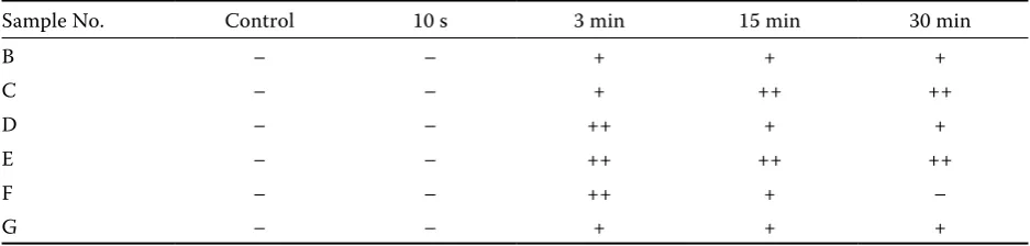
Establishment of the optimum time for the visualisation of conjugated enzyme. The es-tablishment of the optimum time needed for the visualisation is an important step in the immuno-histochemical detections. If the exposure time to the horse radish conjugated peroxidase is too short, the staining of antigen is insufficient. Vice versa, if the time is too long, the background is stained too intensively and thus it cannot be distinguished from the investigated antigen. For this purpose, we chose the model samples of minced pork contain-ing different kinds of soya additives. The negative control was prepared for each sample. We chose four values, i.e. 10 s, 3 min, 15 min, and 30 min, within the ranges declared by the manufacturer of the reagent diaminobenzidine (DAB). The result is based not only on the DAB exposure time, but also on the kind of samples. The best values were obtained after a 3-min exposure (Table 1).
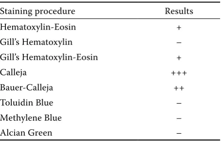
Establishment of the optimum procedure for background staining. We combined the benefits of classical histochemistry (the whole preparation or target components are well stained) with specific immunohistochemistry to stain the background. We obtained preparations in which we could assess the ingredients commonly present in foodstuffs, based on the knowledge of histology, together with the components highlighted on the basis of antigen-antibody binding. It was necessary to find a staining procedure that allows the visualisation of the background structures, however, not at the expense of the visibility of the immunohisto-chemically highlighted structures. It was difficult
to find the best staining technique because vari-ous histochemical staining procedures exist and we had to proceed empirically. We used different conventional staining procedures (Hematoxylin-Eosin, Gill’s Hematoxylin, Gill’s Hematoxylin-(Hematoxylin-Eosin, Calleja, Bauer-Calleja, Toulidin Blue, Methylene Blue, Alcian Green). The best results were ob-tained with the staining procedure according to Calleja (1897), resulting in the achievement of a good contrast between DAB chromogene and Calleja solution (Table 2).
The examination method. The following im-munohistochemical method for soya detection developed in our laboratory is based on ABC
im-munohistochemical method (Lukaš et al. 1997))
and is more sensitive than the method applied by
Boutten et al. (1999).
Sections were immersed in: (1) xylen (RNDr. Jan Kulich Ltd., Prague, Czech Republic) twice for 10 min; (2) absolute ethanol (Moravský Lihovar, Kojetín, Czech Republic) twice for 10 min, followed by 90% and subsequently by 70% aqueous ethanol (v/v) 10 min each bath; (3) tap water for 7 min; (4) distilled water for 7 min; (5) PBS – Phosphate Buffered Saline, 80 g/l NaCl (RNDr. Jan Kulich Ltd.,
Prague, Czech Republic), 2 g/l KCl, 2 g/l KH2PO4,
23.4 g/l Na2HPO4· 2 H2O, 0.16 g/l NaOH adjusted to
pH 7.4; (6) citrate buffer 21 g/l C6H8O7, 9 g/l NaOH
adjusted to pH 6 for 5 min at 650 W in a microwave;
(6) PBS for 5 min; (7) 3% (v/v) H2O2 in PBS for
[image:3.595.63.532.647.759.2]30 min and then (8) PBS twice for 5 minutes. The sections were then incubated successively: (9) for 30 min at 25°C with 5% (v/v) powdered milk diluted in TBS (Dako TBS, Glostrup, Denmark); (10) for 12 h at 8°C with an anti-soya antibody diluted 1:500 with antibody diluent (DakoCytoma-tion ref. S0809, Glostrup, Denmark) and washed in PBS twice for 5 min; (11) for 30 min at 25°C with 25 μl per section of anti-rabbit biotinylated antibody (Vector Laboratories, PK 6101, Bur-lingtone, USA) containing 10 ml TBS, 3 drops
Table 1. Establishment of optimum time for visualisation of the conjugated enzyme
Sample No. Control 10 s 3 min 15 min 30 min
B – – + + +
C – – + ++ ++
D – – ++ + +
E – – ++ ++ ++
F – – ++ + –
of normal blocking serum stock, and 1 drop of biotinylated antibody stock, and washed in PBS twice for 5 min; (12) for 30 min at 25°C with 25 μl per section of ABC reagent (Vector Laboratories, PK 6101, Burlingtone, USA) containing 5 ml TBS, 2 drops of reagent A and 2 drops of reagent B, and washed in PBS for 5 minutes.
The antibody binding was visualised by incubation in 25 μl per section of 3,3'-diaminobenzidine (DAB) (DakoCytomation, Glostrup, Denmark) for 3 min, the reaction was stopped by washing in a water bath for 5 minutes. The background was visualised in Calleja bath for 5 min and washed in water bath and then in 96% aqueous (v/v) and finally absolute ethanol twice for 5 min each, and in xylene p.a. (RNDr. Jan Kulich, Ltd., Prague, Czech Republic) twice for 5 minutes. A drop of solacryl (RNDr. Jan Kulich, Ltd., Prague, Czech Republic) and a micro coverslip were laid onto each section.
The antibodies used. For comparison, we used polyclonal antibodies from two sources as primary antibodies:
• Polyclonal self-made antibodies RASo 100/3
were obtained by immunisation of New Zealand White rabbits. Purified soya isolate was used as the immunogene. These antibodies were tested from the aspect of sensitivity and specificity by the counter-electrophoresis method. The antibodies are specific only for soya protein.
• Polyclonal antibodies purchased from
Sigma-Aldrich Company (St. Louis, USA) were des-ignated as Anti-soya protein with 2519–1ml in concentration of 9.5 mg protein supplied as solution in 0.01M phosphate buffer with pH 7.4 and preserved with 15mM sodium azide. The antibodies were also obtained from rabbits us-ing purified soya proteins as antigens. They are primarily intended for soya assessment in
foodstuffs by the indirect ELISA and the indirect dot blot immunoassay methods.
Confirmation ELISA method. The model sam-ples were simultaneously immunochemically tested for the presence of soya proteins by the accred-ited indirect competitive ELISA method for the detection of soya proteins modified in our labora-tory (Accredited Laboralabora-tory registered with the Czech Institute for Accreditation (CIA) under No. 1354). The analysis was performed using an appropriate standard operating procedure SOP 1/03–03/A. ELISA was conducted utilising 100 μl well system with the application of solid-phase soya isolate antigen followed by the addition of the sample extracts and the polyclonal New Zealand White rabbits anti soya isolate antibody of own provenance and peroxidase-labelled anti-rabbit conjugated antibody and tetramethylbenzidine (TMB) substrate. The measurement of the final absorbance was realised at 450 nm. The detection limit of the semi-quantitative ELISA method was 0.5% of the weight of the added plant protein.
reSultS AnD DIScuSSIOn testing of the method on model samples
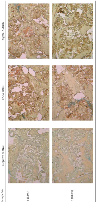
The above mentioned method was tested on two groups of model samples. In the first group, where different concentrations of one kind of soya protein were used, soya was detected in all concentrations by immunohistochemical method with comparable results (Table 3 and Figure 1). Accordingly, the immunohistochemical method confirmed the cited literature data concerning
the potential use of these methods (Boutten et
al. 1999).
[image:4.595.64.292.101.246.2]Due to the fact that different forms of soya protein can be found in the meat products (Coomaras-wamy & Flint 1973; Pipek 1998), samples were selected with the addition of different forms of soya protein used for the production of meat products as the second group of the model samples in the present study. In this group of the model samples, the targeted histochemical staining PAS-Calleja and Bauer-Calleja confirmed that it is possible to reveal soya flour and less sophisticated soya proteins by the detection of insolvable polysaccharide components, which is in accordance with a number of authors (Coomaraswamy & Flint 1973). The specificity of HE staining and staining according to Calleja showed to be low as also reported by Tremlová Table 2. Evaluation of staining procedures
Staining procedure Results
Hematoxylin-Eosin +
Gill’s Hematoxylin –
Gill’s Hematoxylin-Eosin +
Calleja +++
Bauer-Calleja ++
Toluidin Blue –
Methylene Blue –
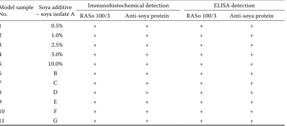
Table 3. Comparison of immunohistochemical and immunochemical detections of soy protein in samples with dif-ferent concentrations of additive
Model sample
No. – soya isolate ASoya additive
Immunohistochemical detection ELISA detection RASo 100/3 Anti-soya protein RASo 100/3 Anti-soya protein
1 0.5% + + + +
2 1.0% + + + +
3 2.5% + + + +
4 5.0% + + + +
5 10.0% + + + +
6 B + + + +
7 C + + + +
8 D + + + +
9 E + + + +
10 F + + + +
11 G + + + +
B – pork meat and soya concentrate, C – pork meat and soya isolate, D- pork meat and soya isolate, E – pork meat and defatted soya flour, F – pork meat and soya texturate, G – pure soya isolate protein
and Štarha (2002). The detection must be therefore based on the knowledge of morphological structure of the soya protein if the size of the additive is suf-ficient for light microscopy (Horn 1987). Highlight-ing by the immunohistochemical methods does not depend upon the concurrent presence of other structures (polysaccharides), and the visualisation of soya protein due to highly specific primary antibody was obtained with all the forms of soya additives (Table 3 and Figures 2–7). Based on the preliminary examinations, it seems that the obtained contrast of this staining may be sufficient even for quantitative evaluation by the image analysis system.
comparison of the results with the confirmation method
The model samples were examined by a con-firmation accredited ELISA method of our own provenance that allowed the detection of the in-vestigated ingredients in concentrations lower than 0.5%. The results obtained by the immuno-histochemical method were in consensus with the ELISA method results (Table 3).
In the first group where different concentrations of one soya protein type (soya isolate) were used soya was successfully detected by histochemical examination in all samples. In the second group, soya was detected in all model samples contain-ing different types of soya additive (texturate,
concentrate, soya flour, isolate). It was confirmed that immunochemical methods can be used for the detection of different kinds and types of soya protein (Ravestein & Driedonks 1986; Yasu-
moto et al. 1990).
cOncluSIOnS
The method developed in the present study proved to be reliable for the detection of soya present in model samples from the concentration of
0.5%. No false positivity occurred at higher
concen-trations (10%). We obtained the best results after staining the background according to Calleja, for which the image analysis system can likely be used in the future. With other staining procedures, the contrast reached was not sufficient and a higher experience and erudition of a technician would be necessary. If the image analysis system were applied for the samples stained by this method, the interaction between the technician and the image analyser would have to be higher.
Sa
m
pl
e
N
o.
N
eg
at
iv
e
co
nt
ro
l
RA
So
1
00
/3
Si
gm
a-A
ld
ri
ch
1
(0
.5
%
)
2
(1
%
)
3
(2
.5
%
Sa
m
pl
e
N
o.
N
eg
at
iv
e
co
nt
ro
l
RA
So
1
00
/3
Si
gm
a-A
ld
ri
ch
4
(5
.0
%
)
5
(1
0.
0%
)
Fi
gu
re
1
.
C
on
ne
ct
ed
w
ith
T
ab
le
3
(S
oy
a
is
ol
at
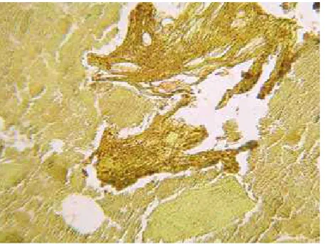
[image:7.595.65.387.81.753.2]Figure 2. Model sample No. 6-B (minced meat and soya concentrate). DAB chromogen and Calleja staining (mag-nification 25×)
[image:8.595.65.290.307.474.2]Figure 3. Model sample No. 7-C (minced meat and soya isolate). DAB chromogen and Calleja staining (magnifi-cation 25×)
[image:8.595.305.530.309.475.2]Figure 4. Model sample No. 8-D (minced meat and soya isolate). DAB chromogen and Calleja staining (magnifi-cation 25×)
Figure 5. Model sample No. 9-E (minced meat and soya flour). DAB chromogen and Calleja staining (magnifica-tion 25×)
Figure 6. Model sample No. 10-F (minced meat and soya texturate). DAB chromogen and Calleja staining (mag-nification 25×)
[image:8.595.64.293.532.704.2] [image:8.595.306.530.532.705.2]are highlighted due to the high specificity of the method. In comparison with ELISA method, immu-nohistochemical method with the counterstaining of the background also allows the detection of other components present in the product (according to the staining used and based on histology knowledge), thereby obtaining a much wider range of results. It can appear as a great advantage, above all in food-stuffs with respect to the risk of adulteration.
After the examination of a matching group of the meat products from the market, it will be possible to offer this method to the testing laboratories and to apply it to the identification of another components present in the meat products.
references
Anonymous: Amendment No. 1 to Decree No. 113/2005 Coll., on methods for labelling of foodstuffs and tobacco products. Collection of Acts: 1163–1176.
Anonymous: Directive 2003/89/EC of the European Parliament and of the Council of 10 November 2003 amending Directive 2000/13/EC as regards indica-tion of the ingredients present in foodstuffs. Ofiicial Journal L 308, 25/11/2003: 15–18.
Belloque J., Garcia M.C., Torre M., Marina M.L. (2002): Analysis of Soyabean proteins in meat prod-ucts: A review. Critical Reviews in Food Science and Nutrition, 42: 507–532.
Boutten B., Humbert C., Chelbi M., Durand P., Peyraud D. (1999): Quantification of soya proteins by association of immunohistochemistry and video image analysis. Food and Agricultural Immunology, 11: 51–59.
Calleja C. (1897): Método de tripla coloración con el car- mín litinado y el picrocarmín de índigo. Revista Tri-mestral Micrográfia, II: 100–104.
Coomaraswamy M., Flint O.F. (1973): The histo-chemical detection of soya “novel proteins” in com-minuted meat products. The Analyst, 98: 542–545. Heckman T., Neuman B., Tschirdewah B., Bent-
le W. (1992): Soya protein. Detection in raw salami and frankfurter-type sausages. Fleischwirtschaft, 72: 1423–1427.
Heitman J. (1987): Detection of soya protein in heat treated meat products by indirect immunofluores-cence. Fleischwirtschaft, 67: 62–622.
Horn D. (1987): Detection of plant protein preparations in meat products by histological examination method. Fleischwirtschaft, 67: 616–618.
Lücker E., Hildebrandt G., Horn D. (2000): Quality assurance by histological analysis of food. In: 41. Ar- beitstagung des Arbeitsgebietes „Lebensmittelhygie-ne“, Garmisch-Partenkirchen, Germany, 25.–28. 09. 2000: 588–592.
Leitner A., Castro-Rubio F., Marina M.L., Lindner W. (2006): Identification of marker proteins for the adulteration of meat products with soybean proteins multidimensional liquid chromatography-tandem mass spectrometry. Journal of Proteome Research., 5:2424–2430.
Lukáš Z., Drapelová E., Feit J., Vojtěšek B. (1997): Immunohistochemical Methods in Biology and Bioptic Diagnosis. 1st Ed. Masaryk University Brno.
Meyer R., Chardonnes F., Hűbner P., Lűthy J. (1996). Polymerase chain reaction (PCR) in the quality and safety assurance of food: detection of soya in processed meat products. Zeitschrift fűr Lebensmittel-Untersu-chung und -ForsLebensmittel-Untersu-chung, 203:339–344.
Pedersen H. E. (1995): Application of soya protein-con-centrates in processed meat-products-experience from different countries. Fleischwirtschaft, 75:1–6. Pipek P. (1998): Technologie masa II. Karmelitánske
nakladatelství v Kostelním Vydří, Praha: 158–159. Ravestein P., Driedonks R.A. (1986): Quantitative
im-munoassay for soya proteins in raw and sterilized meat products. Journal of Food Technology,21: 19–32. Strahle J., Roth M. (1996): Determination of
soya-pro-tein by enzyme-linked immunosorbent assay (ELISA). Deutsche Lebensmittel-Rundschau, 92: 247–250. Tremlová B., Štarha P. (2002): Evaluation of
histo-logical methods for detection of plant ingredients in meat products with regard to the use of image analysis systems. In: 43. Arbeitstagung des Arbeitsgebietes „Lebensmittelhygiene“, Garmisch-Partenkirchen, Ger-many, 25.–25. 09. 2001: 838–842.
Yasumoto K., Sudo M., Suzuki T. (1990): Quantita-tion of soya protein by enzyme linked immunosorbent assay of its characteristic peptide. Journal of Food Science and Agriculture, 50: 377–389.
Received for publication December 21, 2007 Accepted after corrections March 3, 2008
Corresponding author: