IDENTIFICATION AND QUANTIFICATION OF LUPEOL IN
FORMULATION CONTAINING FICUS SPECIES USING
HIGH-PERFORMANCE THIN LAYER CHROMATOGRAPHY
Valsamma Wilson*, Dr. Sugandha S. Shetye, Kawaljit Kaur, Satish N. Ambare
Department of Chemistry, KJ Somaiya College of Science and Commerce, Vidyavihar,
Mumbai-400077.
ABSTRACT
There is a growing demand for biocompatible natural products and
formulations for the treatment of various new era diseases like Cancer,
Parkinson‟s, Alzheimer‟s, and Diabetes. Plants have many
phytochemicals with different bioactivities like antioxidant,
anti-inflammatory, and anticancer activities. In the present study, we report
the identification and quantification of Lupeol, an anticancer agent,
from an Ayurvedic formulation „Nalpamaradi choorna‟ and its four
Ficus species constituents using High-Performance Thin Layer
Chromatography (HPTLC). The chromatographic separation of the
methanol extract of the samples was done on TLC alumina plates
coated with silica gel 60 F254 as the stationary phase with Toluene:
Ethyl acetate: Formic acid (5:5:0.5 v/v/v) as the mobile phase. The plates were scanned at
reflectance absorbance mode at 540 nm after derivatizing with Anisaldehyde- Sulphuric acid
reagent. The system was found to give compact spots with a Rf value of 0.72±0.02. The method was found to be linear in a range of 50-250 ng with a correlation coefficient r =
0.9982. The results showed the presence of 0.021%, 0.024%, 0.019%, 0.0.027% and 0.029%
of Lupeol in F. benghalensis, F. racemosa, F.religiosa, F. microcarpa and the formulation
respectively.
KEYWORDS: HPTLC, Nalpamaradi Choorna, Ficus species, Lupeol, Anticancer activity.
INTRODUCTION
Plant-based medicines have a broad spectrum of activities from ancient times. The plant cells
Volume 5, Issue 5, 1075-1084. Research Article ISSN 2277– 7105
*Corresponding Author
Valsamma Wilson
Department of Chemistry,
KJ Somaiya College of
Science and Commerce,
Vidyavihar,
Mumbai-400077.
Article Received on 06 March 2016,
Revised on 26 March 2016, Accepted on 15 April 2016
necessary for its survival. Secondary plant metabolites are usually synthesized by special,
differentiated cells, to function primarily in defense against predators and pathogens, and
attract pollinators and seed dispersers to help reproductive activities. Many of them are highly
toxic too.[1] The drug activity of a plant is due to these compounds. Plants have many phytochemicals with various bioactivities like antioxidant, anti-inflammatory and anticancer
activities. Extracts from natural products like fruits, vegetables, and medicinal plants have
positive effects against cancer compared with chemotherapy or hormonal treatments.[2]
Naturally occurring phytochemicals of dietary and non-dietary origin have gained interest in
the recent years due to their capability to modulate degenerative diseases like cancer,
cardiovascular diseases (CVD), diabetes, arthritis, cataract, aging,and so on. This resurgence
of interest in the bioactive phytochemicals could mainly be attributed to the large body of
scientific evidence gathered from well-designed epidemiological and experimental studies
conducted during the last two decades.[3]
Ficus species are reported as a rich source of naturally occurring antioxidants of which
phenolic compounds and flavanoids play a vital role in preventing numerous health disorders
related to oxidative stress including cardiovascular diseases, neurodegenerative diseases, and
cancer.[4] Among the many Ficus species, the most important are the four trees with milky
latex, namely Ficus racemosa L., Ficus microcarpa, Ficus religiosa L., and Ficus
benghalensis L. that constitute the group “Nalpamara” in Ayurveda. All parts- root, bark, leaf
and fruit of these trees have extensive applications in medicine. The bark of these species
forms an essential ingredient in many Ayurvedic formulations, like Nalpamaradi choorna,
Nalpamaradi tailam,Chandanasavam, Saribadyasavam,etc.[5] The bark of these four lactiferous tree species is reported to contain tannin, wax, saponin, steroid, terpenoid,
flavonoid, and alkaloid, as the major class of phytochemicals.[6] Various phytochemicals have been found to possess a broad range of activities, which may help in protection against
chronic diseases. For example, the terpenoids have been shown to decrease blood sugar level
in animal.[7] Steroids, triterpenoids, and Saponins showed the analgesic properties and central
nervous system activities.[8-9]
Lupeol, a triterpene had been reported to have anti-arthritic and anti-inflammatory
activities.[10] Investigations showed the anti-inflammatory activity of Lupeol and Lupeol
operative pain.[12] Lupeol acts as a therapeutic and chemopreventive agent for the treatment of inflammation and cancer. It is also noteworthy that Lupeol at its effective therapeutic
doses exhibits no toxicity to normal cells and tissues,[13] and protective against glutamate
toxicity.[14] In another study, Lupeol was demonstrated as an antitumor-promoting agent.[15]
Waxes of some common fruits, like apple, grape, berry, olive, and tomato,were demonstrated
as rich sources of Lupeol.[16] Another study demonstrated that lupeol/mango pulp extract are
effective in combating oxidative stress-induced cellular injury of mouse prostate; so could be
a potential chemopreventive agent against prostate cancer.[17]
Lupeol was extracted from the stem bark powder of Crataeva nurvala and Antitrypsin
(Anti-protease) activity was determined by using Folin Lowery method.[18] Even though few
research groups have developed HPTLC method for fingerprinting and quantification of
bioactive compounds in various formulations and food products,[19-23] the constituents and formulations of Nalpamara are not investigated thoroughly. So we have developed an
HPTLC densitometric method for the standardization of Nalpamaradi choorna, and the bark
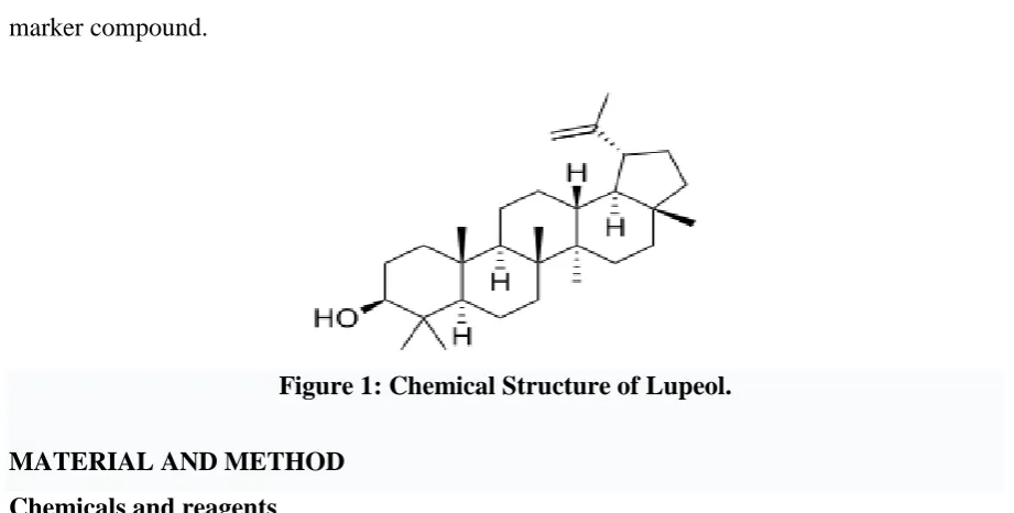
[image:3.595.68.529.391.624.2]powders of F.benghalensis, F. racemosa, F. religiosa, and F. microcarpa with Lupeol as marker compound.
Figure 1: Chemical Structure of Lupeol.
MATERIAL AND METHOD Chemicals and reagents
Lupeol was purchased from Sigma-Aldrich, Mumbai, India. Toluene, Ethyl acetate, Formic
acid, and methanol used were of HPLC grade and were procured from Merck Chemicals,
Mumbai. All the other chemicals used were of analytical grade. Silica gel 60 F254 TLC plates
(20×10cm and 10×10 cm, layer thickness 0.2 mm, Merck, Germany) were used as stationary
Sample preparation
Two samples of Nalpamaradi Choorna were purchased from a local Ayurvedic Pharmacy.
Fresh Bark of F. benghalensis, F. racemosa, F. religiosa, and F. microcarpa were collected
from Somaiya, Vidyavihar campus, Mumbai, and authenticated by Blatter Herbarium, St.
Xaviers College, Mumbai, India. They were dried in the shade, powdered and stored at 250C.
2 g each of the powdered drug and the formulations [procured (A &B) and prepared in the
laboratory by mixing equal amounts of the four constituents(C)] were extracted with
methanol (10 ml × 4) under reflux on a water bath. It was filtered through Whatman I filter
paper, filtrates were combined, concentrated under vacuum and the volume was made up to
10 ml in a volumetric flask.
Preparationof standard solution.
Lupeol (5.0mg) was weighed accurately and dissolved in 5.0 ml methanol in a standard
measuring flask. This stock solution was further diluted to a concentration of 0.05 µg/µl and
used as the standard solution for the HPTLC analysis.
Instrumentation and Chromatographic conditions
The standard and sample were spotted as bands of width 6.0 mm using a CAMAG 100.0 l
sample syringe on silica gel pre-coated Aluminium plate 60 F254 TLC plates (20×10 cm) with
0.2 mm thickness, using a CAMAG Linomat V [Switzerland] sample applicator. The plates
were activated at 110.00C for 5 minutes prior to chromatography. The distance from lower edge was 8.0 mm and from the side 15.0 mm. The distance between the tracks was 10.0 mm.
The slit dimensions were 5 mm×0.45 mm, micro and scanning speed 20 mm/s with data
resolution 100 m/step. The mobile phase consisted of Toluene: Ethyl acetate: Formic acid
5:5:0.5 (v/v/v) and 20.0 ml of it was used per chromatographic run. Linear ascending
development was done in a 20×10 cm twin trough glass chamber [CAMAG, Switzerland]
saturated with the mobile phase for 15 minutes at room temperature [250C±2]. Each
chromatogram was developed over a distance of 8.0cm followed by drying in a stream of air
with the help of hair drier. The plate was dipped in to Anisaldehyde-Sulphuric acid reagent
(1.0ml Anisaldehyde reagent added to a mixture of 10.0ml Sulphuric acid and an ice-cold
solution of 10.0 ml Acetic acid in 170.0 ml methanol) using the TLC immersion device
(CAMAG). The immersion time was 3 s. The plate was heated on a TLC Plate Heater at
CAMAG TLC scanner III and scanned at 540 nm in the absorbance mode using Tungsten
lamp. Data processing was done with the software platform winCATS (CAMAG).
Linearity and calibration curve
Linearity of the method was evaluated by constructing calibration curve at eight
concentrations. Aliquots of standard working solutions of Lupeol [0.5µl to 4.0l] were
applied to the plate to obtain concentrations in the range 50 to 200 ng/spot. The calibration
curve was developed by plotting peak area verses concentration with the help of winCATS
software.
Sample analysis
10l of each of the extracts of the three formulations and the four ingredients were spotted on
the pre-coated TLC plate in duplicate. The parameter specifications were maintained as stated
above. The samples were applied under continuous drying stream of nitrogen gas at a
constant application rate. The linearity of the detector response for the prepared standards
was assessed by means of linear regression regarding the amounts of each reference standard,
measured in ng, and area of the corresponding peak on the chromatogram. After development
and scanning of the plate densitometric estimation was carried out based on the peak areas.
The peak areas were plotted against the corresponding concentration and least square
regression analysis was performed to generate the calibration equation.
RESULT AND DISCUSSION
TLC densitometric quantification of Lupeol using HPTLC.
Terpenoids are biosynthesized from isoprene molecules (CH2=C(CH3)-CH=CH2 ), significant
in plant growth, metabolism, and growth. Terpenoids have cyclic structures with one or more
functional groups like hydroxyl, carbonyl, etc. They are found in resins, barks, and latex of
trees.[24] They can be extracted with hot methanol. The detection is slightly difficult as they
are colourless compounds and not sensitive to universal chromogenic compounds. However,
can be detected on TLC plate with H2SO4 and heating.
In the present study, a slightly modified method was developed. Initial fingerprinting studies
were performed on the pure compound and the conditions were optimized. HPTLC
chromatograms of methanol extracts of all the test samples were developed under identical
conditions. The visualization of Lupeol bands were made possible with the derivatization
resolved TLC profile of the marker compound corresponding to the individual constituents
and the formulation (violet coloured bands) authenticate the presence of the marker in all the
samples (Figure 3). The correlation coefficient was found to be 0.9982, which is indicative of
peak purity. In the linearity study, response was found to be a linear function of the amount
applied on the range 25-250 ng/spot (Table 1). The detectability, ie, LOD and LOQ were
determined to be 18.0 and 61.25 ng/spot respectively.
The amount (%) of the bio-active marker in different samples was determined on the basis of
the calibration curve as given in Table 2.
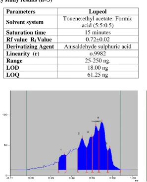
Table 1 Linearity study results (n=3)
[image:6.595.154.452.273.642.2]
Figure 2: A Representative HPTLC Chromatogram of Formulation C.
Parameters Lupeol
Solvent system Touene:ethyl acetate: Formic
acid (5:5:0.5)
Saturation time 15 minutes
Rf value Rf Value 0.72±0.02
Derivatizing Agent Anisaldehyde sulphuric acid
Linearity (r) o.9982
Range 25-250 ng.
LOD 18.00 ng
Figure 3: HPTLC Fingerprint profile of Standard and samples at 540 nm after derivatization.
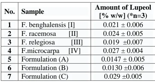
Table 2: Analysis of sample.
*n= no. of times procedure repeated.
Among the four Ficus species, F. microcarpa contain the highest amount (0.027%) and F.
religiosa the least quantity of Lupeol (0.019%). But this is comparable to that reported for F.
religiosa by another group (0.020).[25] The Lupeol content in the formulations A & B were
found to be lesser than the individual constituents. But C, the one prepared in the laboratory
using fresh bark from Somaiya, Vidyavihar Campus demonstrated to be much better (0.029
%) than the marketed samples. It contains a better percentage of Lupeol than the constituents
even indicating a strong synergistic effect.
CONCLUSION
A simple HPTLC method was developed for the quantification of Lupeol, a triterpenoid from
Nalpamaradi choorna, an Ayurvedic formulation and its Ficus constituents. Lupeol, has been
reported to have a broad range of medicinal properties that include strong antioxidant,
anti-No. Sample Amount of Lupeol
[% w/w] (*n=3)
1 F. benghalensis [I] 0.021 ± 0.006
2 F. racemosa [II] 0.024 ± 0.005
3 F. relegiosa [III] 0.019 ±0.007
4 F.microcarpa [IV] 0.027 ± 0.004
5 Formulation (A) 0.0147 ± 0.005
6 Formulation (B) 0.0130 ±0.006
[image:7.595.167.426.328.465.2]presence of Lupeol in the tested samples. The medicinal properties of these Ficus species
plants may be related to their bioactive compounds. Lupeol being one of the bioactive
compounds, could be responsible for the biological activities of these plants. This feature
makes these common plants promising candidates for further studies. Application of this
developed method on isolation and pharmacological studies of this compound could lead to
the discovery of modern medicine with less side effects for the treatment of inflammation and
cancer as it is a chemopreventive and therapeutic agent.
ACKNOWLEDGEMENT
The autours are grateful to Anchrom Laboratories Ltd, Mulund, Mumbai, India for their
training and support.
REFERENCES
1. Chinnaswamy, N. K. Identification and quantification of Lupeol in Dipteracanthus
Patulus (JACQ) Nees by High-performance Liquid Chromatography-Photo Diode Array
Method. International Journal of Pharma and Bio Sciences, 2013; 4(4): 660-6.
2. Ji-Young Lee, W.-I. H. Antioxidant and anticancer activities of organic extracts from
Platycodon grandiflorum A. De Candolle roots. Journal of Ethnopharmacology., 2004;
93: 409-15.
3. Arumughan C, Renuka Devi R. Phytochemical characterization of defatted
Phytochemical characterization of defatted rice bran and optimization of a process for
their extraction and enrichment. Bioresource Technology, 2007; 98: 3037-43.
4. Shirisha, N. Antioxidant properties of Ficus species- A review. International Journal of
Pharm Tech Research, 2010; 2(4): 2174-82.
5. Sivarajan V.V, Balachandran I. Ayurvedic drugs and their sources. New Delhi: Oxford &
IBH publishing company Pvt. Ltd., 1994.
6. S.Nesamony, Dr. Oushadha Sasyangal(medicinal plants). Kerala: The State Institute of
language, Kerala, 2008.
7. Mandal, S. C., Maity, T.K., Das j., Saba B.P. and Pal M. Anti- inflammatory evaluation
of Ficus racemosa Linn. Leaves extract. . J.Ethnopharmacol, 2009; 72: 87-92.
8. Argal, A. and Pathak. KCNS activity of Calotropis gigantean roots.
9. Shaikh, T., Rub, R., Kiran, B., Pimprikar R.B., Sufiyan A. Antibacterial activity of Ficus racemosaLinn. Leaves on Actinomyces viscosus. J. Pharm. Sci. & Research, 2010; 2(1): 41-4.
10.R.B. Agarval, V.D. Rangari. Antiinflammatory and antiarthritic activities of Lupeol and
19a-h Lupeol isolated from Strobilanthus Callosus and Strobilanthus Ixiocephala roots. Indian Journal of Pharmacology, 2003; 35: 384-7.
11.T. Geeta, P. Varalakshmi. Anti-inflammatory activity of Lupeol and Lupeol linoleate in
rats. Journal of Ethnopharmacology, 2001; 76(1): 77-80.
12.de Lima FO., et al. Antinociceptive effect of Lupeol: evidence for a role of cytokines
inhibition. Phytotherapy Research, 2013; 27(10): 1557-63.
13.Saleem, Mohammad. Lupeol, a novel anti-inflammatory and anti-cancer dietary
triterpene. Cancer Letters, 2009; 285(2): 109-15.
14.James M. Brimson, Sirikalaya J. Brimson, Christopher A. Brimson, et al. Rhinacanthus
nasutus Extracts Prevent Glutamate and Amyloid-β Neurotoxicity in HT-22 Mouse
Hippocampal Cells: Possible active compounds include Lupeol, Stigmasterol, and
β-Sitosterol. Int. J. Mol. Sci, 2012; 13(4): 5074-97.
15.Mukhtar HH., Bhat MS. Lupeol Anti-Tumor Agent and uses thereof. Patent Publication,
2014.
16.Anna Szakiel, Flora Pensec. Fruit cuticular waxes as a source of biologically active
triterpenoids. Phytochem Rev, 2012; 11: 263–84.
17.Sahdeo Prasad, Neetu Kalra, Madhulika Singh, Yogeshwer Shukla. Protective effects of
Lupeol and mango extract against androgen-induced oxidative stress in Swiss albino
mice. Asian Journal of Androl, 2008; 10(2): 313-8.
18.Swati Balapure, Varsha Wadegaonkar. Determination of Antitrypsin Activity of Lupeol
by Folin Lowery method. International Journal of Science and Technology, 2014; 2(4): 1-6.
19.SC Verma, E Vashishth, S Subhani, R Singh, P Pant and M M Padhi. Comparative
Phytochemical Study of Stem Bark Versus Small Branches of Ficus Religiosa Linn Using
High-Performance Thin Layer Chromatographic Ultra Violet Detection Method. World
Journal of Pharmaceutical Research, 2014; 3(9): 1154-62.
20.Lakshmi HimaBindu M R, Angala Parameswari S, Gopinath C. Determination of
Flavanoidal Content by Ficus religiosa Linn Leaf Extract by TLC and HPTLC.
21.Varsha Bhoir, Viraj Hibare, Mrinalini C. Damle. Development and Validation of
Stability-Indicating HPTLC Method For The Standardization Of Perindopril And
Indapamide. International Journal of Pharmacy and Pharmaceutical Science, 2014; 6(7):
621-5.
22.Gertrude E. Morlock, Stephanie Meyer, Benno F. Zimmermann, Jean-Marc Roussel.
High-performance thin-layer chromatography analysis of steviol glycosides in Stevia
formulations and sugar-free food products, and benchmarking with (ultra)
high-performance liquid chromatography. Journal of Chromatography A, 2014; 102-11.
23.Charushila C. Shinde, Shakuntala Chopade, Suneela S. Dhaneshwar. Development and
Validation of HPTLC Method for Estimation of Voglibose in Pharmaceutical Dosage
Forms. International Journal of Pharmacy and Pharmaceutical Sciences., 2015; 7(5):
288-93.
24.Kulshreshtha MJ., Kulsheshtha DK., and Rastogi RP. The Triterpenoids. Biochem, 1972;
11: 2369-81.
25.Ajay Kumar Singh Rawat, Shashi Shankar Tiwari, Amit Srivastava, and Sharad
Srivastava. Comparative Botanical And Phytochemical Evaluation of Medicinally
Important Stem Bark of Ficus Species. Asian Pacific Journal of Tropical Disease, 2012;