Acta Cryst.(2003). E59, o227±o229 DOI: 10.1107/S1600536803001569 Bilal R. Kaafaraniet al. C32H21NO2
o227
organic papers
Acta Crystallographica Section E Structure Reports
Online
ISSN 1600-5368
p
±
p
-Stacking and nitro±
p
-stacking
interactions of
1-(4-nitrophenyl)-4-phenyl-2,4-bis(phenylethynyl)butadiene
Bilal R. Kaafarani,aBrigitte
Wex,aAllen G. Oliver,b
Jeanette A. Krause Bauercand
Douglas C. Neckersa*
aCenter for Photochemical Sciences, Bowling
Green State University, Bowling Green, OH 43403, USA,bCollege of Chemistry, University of California-Berkeley, Berkeley, CA 94720-1460, USA, andcDepartment of Chemistry, University of Cincinnati, Cincinnati, OH 45221-0172, USA
Correspondence e-mail: [email protected]
Key indicators
Single-crystal synchrotron study
T= 173 K
Mean(C±C) = 0.004 AÊ
Rfactor = 0.059
wRfactor = 0.156
Data-to-parameter ratio = 10.9
For details of how these key indicators were automatically derived from the article, see http://journals.iucr.org/e.
#2003 International Union of Crystallography Printed in Great Britain ± all rights reserved
The ®rst observed side product, C32H21NO2, in the Sonoga-shira coupling reaction is reported. The molecular packing shows a high degree of -stacking interactions in the solid state.
Comment
In this paper, we report the X-ray structure and stacking of 1-(4-nitrophenyl)-4-phenyl-2,4-bis(phenylethynyl)butadiene, (4). This compound was isolated as a side product during the Sonogashira (Sonogashira et al., 1975) coupling reaction of
,-dibromo-p-nitrostyrene, (1), and phenylacetylene to afford Y-enyne (3) (Kaafarani & Neckers, 2001; Kaafaraniet al., 2001; Kaafarani, Wex, Krause Bauer & Neckers, 2002; Kaafarani, Wex, Strehmer & Neckers, 2002). A diyne, formed by reductive elimination, is a classical side product in the course of the Sonogashira coupling reaction [in this case, 1,4-diphenylbutadiyne (3)] (Nicolaou & Sorensen, 1996). It appears to us that compound (4) is formed after the addition of another phenylacetylene to Y-enyne (2). To the best of our knowledge, this is the ®rst report of such a side product in the Sonogashira coupling reaction.
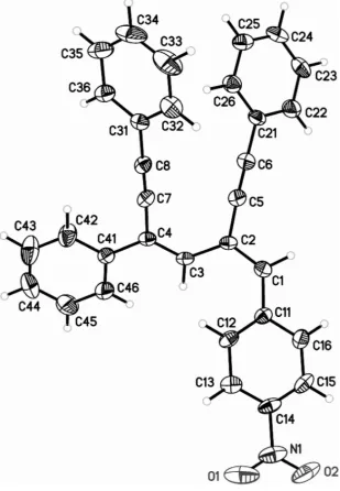
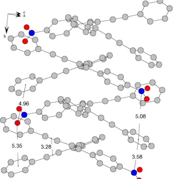
The molecular structure of (4) is shown in Fig. 1. Nitro substitution has been reported to induce±-stacking inter-actions (Garden et al., 2002). The nitro-substitution-induced
±-stacking and nitro±-stacking interactions in (4) are shown in Fig. 2. The distance between the centroids of the phenyl rings alternates between 4.96 and 5.35 AÊ. The distance between the centers of the acetylene triple bonds is 3.28 AÊ. The strong electron-withdrawing NO2group interacts with the
-system of the phenyl ring of a neighboring molecule. The distance between the N atom and the center of the phenyl rings varies between 3.58 and 5.08 AÊ for the alternating layers
Received 13 January 2003 Accepted 17 January 2003 Online 24 January 2003
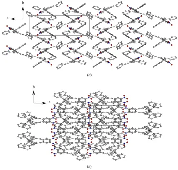
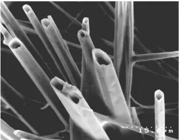
(Fig. 2). The molecular packing of (4) is shown in Fig. 3. The scanning electron micrograph (SEM) of (4) shows its crystal habit in the form of complex, cactus-like needles with a diameter of several micrometers (Fig. 4). Interestingly, these tubes seem hollow; however, a shadow effect induced by the restricted lateral placement of the detector in the SEM can not be excluded and further analysis will be required to con®rm this observation.
Experimental
Single crystals were obtained from methanol±methylene chloride
small crystal size, it was necessary to collect data using synchrotron radiation rather than a laboratory source. A suitable crystal was mounted on the tip of a glass ®ber with paratone-N and immediately transferred to the goniostat bathed in a cold stream.
Crystal data
C32H21NO2
Mr= 451.50
Monoclinic,P21=c
a= 13.583 (3) AÊ
b= 7.6515 (14) AÊ
c= 23.399 (5) AÊ
= 100.093 (11)
V= 2394.2 (9) AÊ3
Z= 4
ÿ3
Synchrotron radiation
= 0.885 AÊ
Cell parameters from 4292 re¯ections
= 6.9±29.0
= 0.08 mmÿ1
T= 173 (2) K Needle, orange 0.070.060.05 mm
Figure 2
±±Stacking and nitro±interactions of (4).
Figure 3
The molecular packing of (4), viewed (a) in thebcplane and (b) in theab plane.
Figure 4
An SEM micrograph of (4).
Figure 1
Data collection
Bruker Proteum300 diffractometer
!scans
Absorption correction: multi-scan (SADABS; Sheldrick, 2001)
Tmin= 0.995,Tmax= 0.996
19213 measured re¯ections 3438 independent re¯ections
2579 re¯ections withI> 2(I)
Rint= 0.095 max= 29.5
h=ÿ15!15
k=ÿ7!8
l=ÿ25!25
Re®nement
Re®nement onF2
R[F2> 2(F2)] = 0.059
wR(F2) = 0.156
S= 1.07 3438 re¯ections 316 parameters
H-atom parameters constrained
w= 1/[2(F
o2) + 0.5111P]
whereP= (Fo2+ 2Fc2)/3
(/)max< 0.001 max= 0.19 e AÊÿ3 min=ÿ0.24 e AÊÿ3 Table 1
Selected geometric parameters (AÊ,).
O1ÐN1 1.224 (3) O2ÐN1 1.227 (3) N1ÐC14 1.461 (3) C1ÐC2 1.354 (3) C1ÐC11 1.454 (3) C2ÐC5 1.437 (4) C2ÐC3 1.449 (4)
C3ÐC4 1.348 (3) C4ÐC7 1.436 (3) C4ÐC41 1.473 (4) C5ÐC6 1.193 (3) C6ÐC21 1.436 (4) C7ÐC8 1.192 (3) C8ÐC31 1.431 (3) O1ÐN1ÐO2 124.3 (3)
O1ÐN1ÐC14 117.7 (3) O2ÐN1ÐC14 118.0 (3) C2ÐC1ÐC11 128.3 (3) C1ÐC2ÐC5 116.8 (2) C1ÐC2ÐC3 123.3 (2) C5ÐC2ÐC3 119.6 (2)
C4ÐC3ÐC2 128.6 (2) C3ÐC4ÐC7 119.9 (2) C6ÐC5ÐC2 176.8 (3) C5ÐC6ÐC21 179.5 (3) C8ÐC7ÐC4 176.8 (3) C7ÐC8ÐC31 176.7 (2)
Intensity data were collected at 173 K, using a Proteum300 detector at Beamline 11.3.1 at the Advanced Light Source (Lawrence Berkeley National Laboratory). The detector was set at a distance of 6.6 cm from the crystal. A series of 1 s data frames measured at 0.2
increments of!were collected to calculate a unit cell and to measure a hemisphere of intensity data. Data were corrected for absorption usingSADABS(6, 1 harmonics) and a high resolution limit of 0.85 AÊ applied, based on examination of the ®nal merged listing from SAINT. A highjIÿ hIijerror was also applied (10 s.u.) to remove outliers from the data set.
All H atoms were included in calculated geometries (CÐ H = 0.95 AÊ), riding on the parent atom. The isotropic
displa-cement parameters for the H atoms were set as 1.2Ueqof the adjacent atom.
Data collection:SMART(Bruker, 2001); cell re®nement:SAINT (Bruker, 2001); data reduction: SAINT; program(s) used to solve structure: SHELXTL (Bruker, 2001); program(s) used to re®ne structure: SHELXTL; molecular graphics: SHELXTL and DIAMOND(Crystal Impact, 1997); software used to prepare mate-rial for publication:SHELXTL.
The authors thank the Center for Microscopy and Micro-analysis and Dr M. Cayer for helpful discussion and guidance while using the scanning electron microscope. BRK thanks the McMaster Endowment for a research fellowship. This work was supported by NSF (grant No. DMR-0091689). Samples for crystallographic analysis at the synchrotron were submitted through the SCrAPS-West (Service Crystallography at Advanced Photon Source) program. Crystallographic data were collected at the Small-Crystal Crystallography Beamline 11.3.1 at the Advanced Light Source (ALS), developed by Albert Thompson of the Experimental Systems Group of the ALS. The ALS is supported by the US Department of Energy, Of®ce of Basic Energy Sciences under contract DE-AC03-76SF00098.
References
Bruker (2001). SMART (Version 5.625), SAINT (Version 6.06) and
SHELXTL(Version 6.1). Bruker AXS Inc., Madison, Wisconsin, USA. Crystal Impact (1997). DIAMOND. Version 2.1e. Crystal Impact, Bonn,
Germany.
Garden, S. J., da Cunha, F. R., Wardell, J. L., Skakle, J. M. S., Low, J. N. & Glidewell, C. (2002).Acta Cryst.C58, o463±o466.
Kaafarani, B. R. & Neckers, D. C. (2001).Tetrahedron Lett.42, 4099±4102. Kaafarani, B. R., Pinkerton, A. A. & Neckers, D. C. (2001).Tetrahedron Lett.
42, 8137±8139.
Kaafarani, B. R., Wex, B., Krause Bauer, J. A. & Neckers, D. C. (2002).
Tetrahedron Lett.43, 8227±8230.
Kaafarani, B. R., Wex, B., Strehmel, B. & Neckers, D. C. (2002).Photochem. Photobiol. Sci.1, 942±950.
Nicolaou, K. C. & Sorensen, E. J. (1996).Classics in Total Synthesis: Targets, Strategies, Methods, pp. 582±586. Weinheim, New York: VCH.
Sheldrick, G. M. (2001).SADABS. Version 2.03. University of GoÈttingen, Germany.
Sonogashira, K., Tohda, Y. & Hagihara, N. (1975).Tetrahedron Lett.50, 4467± 4470.
supporting information
Acta Cryst. (2003). E59, o227–o229 [doi:10.1107/S1600536803001569]
π
–
π
-Stacking and nitro
–
π
-stacking interactions of
1-(4-nitrophenyl)-4-phenyl-2,4-bis(phenylethynyl)butadiene
Bilal R. Kaafarani, Brigitte Wex, Allen G. Oliver, Jeanette A. Krause Bauer and Douglas C.
Neckers
S1. Comment
In this paper, we report the X-ray structure and stacking of 1-(4-nitrophenyl)-4-phenyl-2,4-bis(phenylethynyl)butadiene,
(IV). This compound was isolated as a side product during the Sonogashira (Shonogashira et al., 1975) coupling reaction of β,β-dibromo-p-nitrostyrene, (I), and phenylacetylene to afford Y-enyne (II) (Kaafarani & Neckers, 2001; Kaafarani et al., 2001; Kaafarani, Wex, Krause Bauer & Neckers, 2002; Kaafarani, Wex, Strehmel & Neckers, 2002). Diyne, formed by reductive elimination, is a classical side product in the course of the Sonogahsira coupling reaction [in this case,
1,4-diphenylbutadiyne (III)] (Nicholaou & Sorensen, 1996). It appears to us that compound (IV) is formed after the addition
of another phenylacetylene to Y-enyne (II). To the best of our knowledge, this is the first report of such a side product in
the Sonogashira coupling reaction.
The molecular structure of (IV) is shown in Fig. 1. Nitro substitution was reported to induce π–π-stacking interactions (Garden et al., 2002). The nitro-substitution-induced π–π-stacking and nitro–π-stacking interactions in (IV) are shown in Fig. 2. The distance between the centroids of the phenyl rings alternates between 4.96 and 5.35 Å. The distance between
the centers of the acetylene triple bonds is 3.28 Å. The strong electron-withdrawing NO2 group interacts with the π
-system of the phenyl ring of a neighboring molecule. The distance between the N atom and the center of the phenyl rings
varies between 3.58 and 5.08 Å for the alternating layers (Fig. 2). The molecular packing of (IV) is shown in Fig. 3.
The scanning electron micrograph (SEM) of (IV) shows its crystal habit in the form of complex, cactus-like needles
with a diameter of several micrometers (Fig. 4). Interestingly, these tubes seem hollow; however, a shadow effect induced
by the restricted lateral placement of the detector in the SEM can not be excluded and further analysis will be required to
confirm this observation.
S2. Experimental
Single crystals were obtained from methanol–methylene chloride solutions. Due to the weakly diffracting nature of the
sample and the small crystal size, it was necessary to collect data using synchrotron radiation rather than a laboratory
source. A suitable crystal was mounted on the tip of a glass fiber with paratone-N and immediately transferred to the
goniostat bathed in a cold stream. Intensity data were collected at 173 K using a Proteum300 detector at Beamline 11.3.1
at the Advanced Light Source (Lawrence Berkeley National Laboratory). The detector was set at a distance of 6.6 cm
supporting information
sup-2
Acta Cryst. (2003). E59, o227–o229
S3. Refinement
All H atoms were included in calculated geometries (C—H = 0.95 Å) riding on the atoms to which they are bonded. The
[image:5.610.152.461.124.570.2]isotropic displacement parameters for the H atoms were set as 1.2Ueq of the adjacent atom.
Figure 1
Figure 2
supporting information
sup-4
[image:7.610.125.484.70.413.2]Acta Cryst. (2003). E59, o227–o229
Figure 3
Figure 4
An SEM micrograph of (IV).
2,4-bis(phenylethynyl)-4-phenyl-1-(4-nitrophenyl)butadiene
Crystal data
C32H21NO2
Mr = 451.50
Monoclinic, P21/c
a = 13.583 (3) Å
b = 7.6515 (14) Å
c = 23.399 (5) Å
β = 100.093 (11)°
V = 2394.2 (9) Å3
Z = 4
F(000) = 944
Dx = 1.253 Mg m−3
Synchrotron radiation, λ = 0.88500 Å Cell parameters from 4292 reflections
θ = 6.9–29.0°
µ = 0.08 mm−1
T = 173 K Needle, orange 0.07 × 0.06 × 0.05 mm
Data collection
Proteum 300 detector diffractometer
Radiation source: Beamline 11.3.1 ALS at LBL Channel cut SiIII monochromator
ω scans
Absorption correction: multi-scan (SADABS; Sheldrick, 2001)
Tmin = 0.995, Tmax = 0.996
19213 measured reflections 3438 independent reflections 2579 reflections with I > 2σ(I)
Rint = 0.095
θmax = 29.5°, θmin = 1.9°
h = −15→15
k = −7→8
supporting information
sup-6
Acta Cryst. (2003). E59, o227–o229 Refinement
Refinement on F2
Least-squares matrix: full
R[F2 > 2σ(F2)] = 0.059
wR(F2) = 0.156
S = 1.07 3438 reflections 316 parameters 0 restraints
Primary atom site location: structure-invariant direct methods
Secondary atom site location: difference Fourier map
Hydrogen site location: inferred from neighbouring sites
H-atom parameters constrained
w = 1/[σ2(F
o2) + 0.5111P]
where P = (Fo2 + 2Fc2)/3
(Δ/σ)max < 0.001
Δρmax = 0.19 e Å−3
Δρmin = −0.24 e Å−3
Special details
Experimental. Single crystals were obtained from methanol-methylene chloride solutions. Due to the weakly diffracting nature of the sample and the small crystal size, it was necessary to collect data using synchrotron radiation rather than a lab source. A suitable crystal was mounted on the tip of a glass fiber with paratone-N and immediately transferred to the goniostat bathed in a cold stream. Intensity data was collected at 173 K using a Proteum300 detector at Beamline 11.3.1 at the Advanced Light Source (Lawrence Berkeley National Laboratory). The detector was set at a distance of 6.6-cm from the crystal. A series of 1 − s data frames measured at 0.2° increments of ω were collected to calculate a unit cell and to measure a hemisphere of intensity data. Data was corrected for absorption using SADABS (6, 1 harmonics) and a high resolution limit of 0.85 Å applied, based on examination of the final merged listing from SAINT. A high |I-<I>| error was also applied (10 su) to remove outliers from the dataset. No formal measure of the extent of decay is printed out by this program.
The final unit cell is obtained from the refinement of the XYZ weighted centroids of reflections above 20 σ(I). Note that the absorption correction parameters Tmin and Tmax also reflect beam corrections, etc. As a result, the numerical values for Tmin and Tmax may differ from expected values based solely absorption effects and crystal size.
Geometry. All e.s.d.'s (except the e.s.d. in the dihedral angle between two l.s. planes) are estimated using the full covariance matrix. The cell e.s.d.'s are taken into account individually in the estimation of e.s.d.'s in distances, angles and torsion angles; correlations between e.s.d.'s in cell parameters are only used when they are defined by crystal symmetry. An approximate (isotropic) treatment of cell e.s.d.'s is used for estimating e.s.d.'s involving l.s. planes.
Refinement. Refinement of F2 against ALL reflections out to 0.85 Å. The weighted R-factor wR and goodness of fit S are
based on F2, conventional R-factors R are based on F, with F set to zero for negative F2. The threshold expression of F2 >
σ(F2) is used only for calculating R-factors(gt) etc. and is not relevant to the choice of reflections for refinement. R
-factors based on F2 are statistically about twice as large as those based on F, and R-factors based on ALL data will be
even larger.
Fractional atomic coordinates and isotropic or equivalent isotropic displacement parameters (Å2)
x y z Uiso*/Ueq
O1 0.02775 (16) 0.9713 (3) 0.13521 (12) 0.0680 (8) O2 −0.05727 (15) 0.8194 (3) 0.18773 (11) 0.0639 (7) N1 0.02171 (19) 0.8735 (3) 0.17582 (13) 0.0453 (7) C1 0.38061 (18) 0.6603 (3) 0.32111 (12) 0.0304 (6)
H1 0.3725 0.6362 0.3598 0.037*
C2 0.47420 (17) 0.6394 (3) 0.30964 (11) 0.0253 (6) C3 0.49742 (17) 0.6450 (3) 0.25153 (11) 0.0253 (6)
H3 0.4447 0.6119 0.2212 0.030*
C12 0.29205 (18) 0.8179 (3) 0.23300 (12) 0.0289 (6)
H12 0.3544 0.8525 0.2235 0.035*
C13 0.20470 (19) 0.8709 (3) 0.19798 (12) 0.0316 (7)
H13 0.2062 0.9412 0.1647 0.038*
C14 0.11478 (18) 0.8188 (3) 0.21268 (12) 0.0322 (7) C15 0.11026 (19) 0.7179 (3) 0.26037 (13) 0.0370 (7)
H15 0.0476 0.6833 0.2694 0.044*
C16 0.19769 (19) 0.6675 (3) 0.29493 (12) 0.0348 (7)
H16 0.1952 0.5989 0.3285 0.042*
C21 0.68427 (18) 0.4998 (3) 0.44801 (11) 0.0291 (6) C22 0.6596 (2) 0.3797 (3) 0.48760 (13) 0.0403 (7)
H22 0.5942 0.3315 0.4824 0.048*
C23 0.7305 (2) 0.3304 (3) 0.53474 (12) 0.0428 (8)
H23 0.7137 0.2473 0.5616 0.051*
C24 0.8246 (2) 0.4003 (3) 0.54289 (12) 0.0389 (7)
H24 0.8727 0.3662 0.5755 0.047*
C25 0.8495 (2) 0.5194 (3) 0.50418 (12) 0.0364 (7)
H25 0.9148 0.5683 0.5101 0.044*
C26 0.78007 (19) 0.5686 (3) 0.45645 (12) 0.0335 (7)
H26 0.7981 0.6499 0.4294 0.040*
C31 0.81010 (18) 0.9151 (3) 0.34704 (11) 0.0247 (6) C32 0.7951 (2) 1.0115 (3) 0.39437 (12) 0.0396 (7)
H32 0.7291 1.0308 0.4013 0.048*
C33 0.8757 (3) 1.0803 (4) 0.43174 (14) 0.0557 (9)
H33 0.8652 1.1466 0.4645 0.067*
C34 0.9715 (3) 1.0527 (4) 0.42150 (15) 0.0556 (10)
H34 1.0268 1.1006 0.4472 0.067*
C35 0.9872 (2) 0.9574 (4) 0.37489 (14) 0.0456 (8)
H35 1.0533 0.9382 0.3683 0.055*
C36 0.90747 (19) 0.8890 (3) 0.33745 (12) 0.0328 (7)
H36 0.9187 0.8234 0.3047 0.039*
C41 0.60290 (18) 0.6674 (3) 0.17517 (11) 0.0262 (6) C42 0.6912 (2) 0.7229 (3) 0.15912 (14) 0.0442 (8)
H42 0.7403 0.7774 0.1874 0.053*
C43 0.7102 (3) 0.7023 (4) 0.10406 (16) 0.0577 (9)
H43 0.7713 0.7435 0.0946 0.069*
C44 0.6417 (3) 0.6226 (4) 0.06259 (14) 0.0528 (9)
H44 0.6538 0.6104 0.0240 0.063*
C45 0.5545 (2) 0.5600 (4) 0.07773 (14) 0.0584 (9)
H45 0.5070 0.5019 0.0494 0.070*
C46 0.5354 (2) 0.5805 (4) 0.13287 (13) 0.0456 (8)
H46 0.4753 0.5348 0.1425 0.055*
Atomic displacement parameters (Å2)
U11 U22 U33 U12 U13 U23
supporting information
sup-8
Acta Cryst. (2003). E59, o227–o229
O2 0.0229 (12) 0.0813 (15) 0.0836 (19) 0.0082 (11) −0.0016 (11) −0.0222 (13) N1 0.0337 (16) 0.0364 (13) 0.0584 (19) 0.0103 (12) −0.0120 (13) −0.0161 (13) C1 0.0300 (16) 0.0351 (13) 0.0251 (16) −0.0014 (12) 0.0019 (12) −0.0020 (12) C2 0.0222 (14) 0.0266 (12) 0.0248 (16) −0.0013 (10) −0.0026 (11) −0.0020 (11) C3 0.0211 (14) 0.0261 (12) 0.0260 (15) 0.0003 (10) −0.0036 (11) 0.0015 (11) C4 0.0232 (15) 0.0169 (11) 0.0275 (16) 0.0009 (10) −0.0011 (11) 0.0025 (10) C5 0.0277 (15) 0.0374 (14) 0.0311 (17) −0.0013 (12) 0.0036 (13) −0.0008 (13) C6 0.0313 (16) 0.0412 (15) 0.0293 (17) 0.0024 (12) 0.0028 (14) 0.0022 (13) C7 0.0223 (14) 0.0178 (11) 0.0364 (17) 0.0010 (11) 0.0059 (12) 0.0030 (11) C8 0.0252 (15) 0.0210 (11) 0.0324 (17) −0.0025 (11) 0.0055 (12) 0.0023 (11) C11 0.0227 (14) 0.0252 (12) 0.0304 (16) −0.0007 (10) 0.0035 (11) −0.0055 (12) C12 0.0202 (14) 0.0238 (12) 0.0418 (18) −0.0043 (10) 0.0034 (12) −0.0023 (12) C13 0.0324 (16) 0.0222 (12) 0.0376 (18) −0.0001 (11) −0.0010 (13) −0.0011 (11) C14 0.0213 (15) 0.0257 (12) 0.0460 (19) 0.0049 (11) −0.0041 (12) −0.0125 (13) C15 0.0209 (15) 0.0399 (15) 0.052 (2) 0.0010 (12) 0.0104 (13) −0.0086 (14) C16 0.0311 (16) 0.0399 (14) 0.0354 (17) 0.0003 (12) 0.0115 (13) −0.0008 (13) C21 0.0309 (15) 0.0329 (13) 0.0217 (15) 0.0017 (12) −0.0003 (11) −0.0025 (12) C22 0.0381 (17) 0.0481 (16) 0.0335 (18) −0.0099 (13) 0.0030 (13) 0.0020 (14) C23 0.060 (2) 0.0428 (15) 0.0252 (17) −0.0025 (15) 0.0053 (14) 0.0072 (13) C24 0.0455 (19) 0.0398 (15) 0.0260 (17) 0.0042 (14) −0.0084 (13) −0.0035 (13) C25 0.0315 (16) 0.0377 (14) 0.0368 (18) −0.0009 (12) −0.0024 (13) −0.0059 (13) C26 0.0363 (16) 0.0320 (13) 0.0311 (17) −0.0027 (12) 0.0026 (12) 0.0050 (12) C31 0.0301 (15) 0.0159 (10) 0.0260 (16) −0.0014 (10) −0.0006 (11) 0.0040 (11) C32 0.0485 (18) 0.0324 (13) 0.0373 (19) −0.0004 (13) 0.0058 (14) −0.0035 (13) C33 0.087 (3) 0.0383 (15) 0.037 (2) −0.0080 (17) −0.0037 (18) −0.0118 (14) C34 0.064 (2) 0.0416 (17) 0.048 (2) −0.0186 (16) −0.0258 (18) 0.0075 (16) C35 0.0334 (17) 0.0461 (16) 0.051 (2) −0.0066 (13) −0.0113 (15) 0.0056 (16) C36 0.0294 (16) 0.0311 (13) 0.0359 (17) −0.0020 (11) 0.0001 (12) 0.0003 (12) C41 0.0262 (14) 0.0207 (11) 0.0312 (17) 0.0010 (11) 0.0036 (11) 0.0047 (11) C42 0.0440 (18) 0.0455 (16) 0.046 (2) −0.0193 (14) 0.0157 (15) −0.0096 (14) C43 0.069 (2) 0.0522 (18) 0.061 (3) −0.0232 (17) 0.0359 (19) −0.0114 (17) C44 0.076 (2) 0.0515 (18) 0.036 (2) 0.0057 (17) 0.0239 (18) 0.0065 (15) C45 0.055 (2) 0.087 (2) 0.032 (2) −0.0006 (18) 0.0016 (16) −0.0109 (17) C46 0.0319 (17) 0.0696 (19) 0.0344 (19) −0.0056 (14) 0.0031 (14) −0.0047 (15)
Geometric parameters (Å, º)
O1—N1 1.224 (3) C23—C24 1.368 (4)
O2—N1 1.227 (3) C23—H23 0.9500
N1—C14 1.461 (3) C24—C25 1.368 (4)
C1—C2 1.354 (3) C24—H24 0.9500
C1—C11 1.454 (3) C25—C26 1.382 (4)
C1—H1 0.9500 C25—H25 0.9500
C2—C5 1.437 (4) C26—H26 0.9500
C2—C3 1.449 (4) C31—C32 1.376 (4)
C3—C4 1.348 (3) C31—C36 1.394 (4)
C3—H3 0.9500 C32—C33 1.380 (4)
C5—C6 1.193 (3) C33—H33 0.9500
C6—C21 1.436 (4) C34—C35 1.360 (5)
C7—C8 1.192 (3) C34—H34 0.9500
C8—C31 1.431 (3) C35—C36 1.371 (4)
C11—C12 1.388 (4) C35—H35 0.9500
C11—C16 1.403 (4) C36—H36 0.9500
C12—C13 1.379 (3) C41—C42 1.385 (4)
C12—H12 0.9500 C41—C46 1.394 (4)
C13—C14 1.384 (4) C42—C43 1.367 (4)
C13—H13 0.9500 C42—H42 0.9500
C14—C15 1.367 (4) C43—C44 1.364 (4)
C15—C16 1.370 (4) C43—H43 0.9500
C15—H15 0.9500 C44—C45 1.380 (4)
C16—H16 0.9500 C44—H44 0.9500
C21—C26 1.386 (4) C45—C46 1.369 (4)
C21—C22 1.387 (4) C45—H45 0.9500
C22—C23 1.384 (4) C46—H46 0.9500
C22—H22 0.9500
O1—N1—O2 124.3 (3) C23—C24—C25 120.1 (3)
O1—N1—C14 117.7 (3) C23—C24—H24 119.9
O2—N1—C14 118.0 (3) C25—C24—H24 119.9
C2—C1—C11 128.3 (3) C24—C25—C26 120.2 (3)
C2—C1—H1 115.8 C24—C25—H25 119.9
C11—C1—H1 115.8 C26—C25—H25 119.9
C1—C2—C5 116.8 (2) C25—C26—C21 120.2 (2)
C1—C2—C3 123.3 (2) C25—C26—H26 119.9
C5—C2—C3 119.6 (2) C21—C26—H26 119.9
C4—C3—C2 128.6 (2) C32—C31—C36 119.0 (2)
C4—C3—H3 115.7 C32—C31—C8 121.8 (2)
C2—C3—H3 115.7 C36—C31—C8 119.2 (2)
C3—C4—C7 119.9 (2) C31—C32—C33 120.1 (3)
C3—C4—C41 123.3 (2) C31—C32—H32 119.9
C7—C4—C41 116.7 (2) C33—C32—H32 119.9
C6—C5—C2 176.8 (3) C34—C33—C32 119.9 (3)
C5—C6—C21 179.5 (3) C34—C33—H33 120.0
C8—C7—C4 176.8 (3) C32—C33—H33 120.0
C7—C8—C31 176.7 (2) C35—C34—C33 120.4 (3)
C12—C11—C16 117.9 (2) C35—C34—H34 119.8
C12—C11—C1 123.7 (2) C33—C34—H34 119.8
C16—C11—C1 118.3 (2) C34—C35—C36 120.0 (3)
C13—C12—C11 121.5 (2) C34—C35—H35 120.0
C13—C12—H12 119.3 C36—C35—H35 120.0
C11—C12—H12 119.3 C35—C36—C31 120.5 (3)
C12—C13—C14 118.2 (3) C35—C36—H36 119.7
C12—C13—H13 120.9 C31—C36—H36 119.7
supporting information
sup-10
Acta Cryst. (2003). E59, o227–o229
C15—C14—C13 122.2 (2) C42—C41—C4 121.5 (2)
C15—C14—N1 119.0 (3) C46—C41—C4 121.8 (2)
C13—C14—N1 118.7 (3) C43—C42—C41 122.4 (3)
C14—C15—C16 118.8 (3) C43—C42—H42 118.8
C14—C15—H15 120.6 C41—C42—H42 118.8
C16—C15—H15 120.6 C44—C43—C42 120.3 (3)
C15—C16—C11 121.3 (3) C44—C43—H43 119.9
C15—C16—H16 119.4 C42—C43—H43 119.9
C11—C16—H16 119.4 C43—C44—C45 118.8 (3)
C26—C21—C22 119.2 (2) C43—C44—H44 120.6
C26—C21—C6 120.6 (2) C45—C44—H44 120.6
C22—C21—C6 120.3 (2) C46—C45—C44 121.0 (3)
C23—C22—C21 119.8 (3) C46—C45—H45 119.5
C23—C22—H22 120.1 C44—C45—H45 119.5
C21—C22—H22 120.1 C45—C46—C41 120.9 (3)
C24—C23—C22 120.5 (3) C45—C46—H46 119.5
C24—C23—H23 119.8 C41—C46—H46 119.5