organic papers
o1116
Erika Kaiser-Morriset al. C6H8N2 DOI: 10.1107/S1600536801017846 Acta Cryst.(2001). E57, o1116±o1117 Acta Crystallographica Section EStructure Reports Online
ISSN 1600-5368
2,6-Dimethylpyrazine at 5 K: a neutron-diffraction
study
Erika Kaiser-Morris,a* Alain Cousson,aWerner Paulusband Francois Fillauxc
aLaboratoire LeÂon Brillouin, CEA Saclay, 91191
Gif-sur-Yvette CEDEX, France,bUniversite de
Rennes 1, LCSIM/UMR 6511, Campus de Beaulieu, Avenue du GeÂneÂral Leclerc, 35042 Rennes Cedex, France, andcLADIR, 2 rue Henry
Dunant, 94320 Thiais, France
Key indicators
Single-crystal neutron study
T= 5 K
Mean(C±C) = 0.002 AÊ
Rfactor = 0.032
wRfactor = 0.016 Data-to-parameter ratio = 7.9
For details of how these key indicators were automatically derived from the article, see http://journals.iucr.org/e.
#2001 International Union of Crystallography Printed in Great Britain ± all rights reserved
Single-crystal neutron-diffraction techniques are used to determine the crystal structure of 2,6-dimethylpyrazine (DMP), C6H8N2, at 5 K. The space group is P21/awith Z = 4, as at room temperature. The methyl groups are ordered. There are two crystallographically inequivalent methyl groups in the unit cell. Different rotational dynamics may account for the two rotational tunnelling transitions observed with inelastic neutron-scattering techniques.
Comment
As found for the structure of this material, (I), at 20 K (Kaiser-Morriset al., 2001), the space group isP21/a(monoclinic) with four molecules per unit cell. There is no evidence for any phase transition between 20 and 5 K, and no signi®cant changes of the lattice parameters below 20 K.
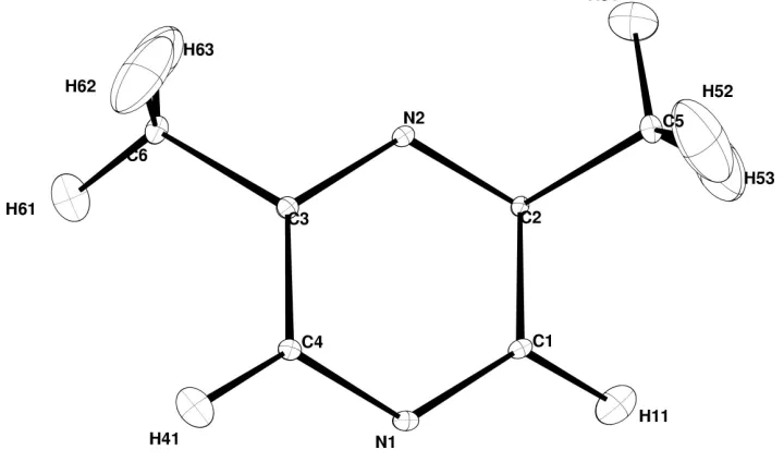
The structure consists of parallel layers of planar molecules perpendicular to the (201) plane (Kaiser-Morriset al., 2001). The protons of the methyl groups are quite localized at all temperatures. For each methyl group, one of the protons is almost in the molecular plane. The displacement ellipsoids for both methyl groups correspond quite well to those anticipated for hindered rotors with a rather high potential barrier and threefold symmetry (Fig. 1). There are two different crystal-lographic environments for the methyl groups linked to the same pyrazine ring. The different local potentials may account for the different tunnelling frequencies. This is con®rmed by further inelastic neutron-scattering measurements performed on single crystals (NicolaõÈet al., 1998).
Experimental
2,6-Dimethylpyrazine (DMP) is hygroscopic and melts at 311 K. We performed neutron-diffraction experiments with a single crystal at 5 K on the four-circle neutron diffractometer 5-C2 at the LLB (Saclay, France). A large single crystal (115 cm) was obtained at low temperature. A small single crystal (555 mm) was cut, glued on a goniometer head and oriented on 5-C2. The measurements were performed with the!scan mode and an incident wavelength close to 0.83 AÊ selected with the Cu (220) monochromator.
Crystal data
C6H8N2 Mr= 108.14
Monoclinic,P21=a a= 7.287 (7) AÊ b= 10.725 (9) AÊ c= 7.452 (8) AÊ = 90.37 (9) V= 582.4 AÊ3 Z= 4
Dx= 1.23 Mg mÿ3
Neutron radiation = 0.8308 AÊ
Cell parameters from 16 re¯ections
= 9.8±21.5 = 0.08 mmÿ1 T= 5 K Prism, white 5.05.05.0 mm
Data collection
OrpheÂe reactor (Saclay, France): 5-C2 four-circle
!scans
Absorption correction: none 2026 measured re¯ections 1605 independent re¯ections 1153 re¯ections withI> 3(I) Rint= 0.022
max= 35 h=ÿ10!10 k=ÿ14!4 l=ÿ10!4 2 standard re¯ections
frequency: 450 min intensity decay: none
Re®nement
Re®nement onF R= 0.032 wR= 0.016 S= 1.04 1153 re¯ections 146 parameters
All H-atom parameters re®ned Weighting scheme: Chebychev
polynomial with 5 parameters: 0.951,ÿ3.12, 0.00654,ÿ0.885, ÿ0.649 (Carruthers & Watkin, 1979)
(/)max< 0.001
max= 0.78 e AÊÿ3
min=ÿ0.73 e AÊÿ3
Extinction correction: Larson (1970)
Extinction coef®cient: 2.39 (18) Atomic scattering factors from
Sears (1992)
Table 1
Selected geometric parameters (AÊ,).
N1ÐC1 1.3341 (11)
N1ÐC4 1.3397 (11)
N2ÐC2 1.340 (1)
N2ÐC3 1.3394 (11)
C1ÐC2 1.4023 (12)
C2ÐC5 1.4991 (13)
C3ÐC4 1.3982 (13)
C3ÐC6 1.4980 (12)
C1ÐN1ÐC4 116.27 (7) C2ÐN2ÐC3 117.44 (7) N1ÐC1ÐC2 122.04 (8) N2ÐC2ÐC1 121.07 (8) N2ÐC2ÐC5 118.56 (8)
C1ÐC2ÐC5 120.37 (8) N2ÐC3ÐC4 120.71 (8) N2ÐC3ÐC6 117.74 (8) C4ÐC3ÐC6 121.55 (8) N1ÐC4ÐC3 122.47 (8)
Data collection:DIF4N(modi®ed Linux version ofDIF4; Stoe & Cie; 2000); cell re®nement:DIF4N; data reduction:PRON(modi®ed version of REDU4; Stoe & Cie, 2000); program(s) used to re®ne structure: CRYSTALS (Watkin et al., 1996); molecular graphics:
CAMERON(Watkinet al., 1996); software used to prepare material for publication:CRYSTALS.
We thank J. Godard from the Parc d'Orsay, France for providing the single crystals.
References
Carruthers, J. R. & Watkin, D. J. (1979).Acta Cryst.A35, 698±699.
Kaiser-Morris, E., NicolaõÈ, B., Cousson, A., Paulus, W. & Fillaux, F. (2001). Acta Cryst.E57, o1113±o1114.
Larson A. (1970).Crystallographic Computing, edited by F. R. Ahmed, pp. 291±294. Copenhagen: Munksgaard.
NicolaõÈ, B., Kaiser, E., Fillaux, F., Kearley, G. J., Cousson, A. & Paulus, W. (1998).Chem. Phys.26, 1±13.
Sears, V. F. (1992).Neutron News,3, 26±37.
Stoe & Cie (2000). DIF4 andPRON Software. Stoe & Cie, Darmstadt, Germany.
Watkin, D. J., Prout, C. K. & Pearce, L. J. (1996).CAMERON. Chemical Crystallography Laboratory, Oxford, England.
Watkin, D. J., Prout, C. K., Carruthers, J. R. & Betteridge, P. W. (1996). CRYSTALS. Issue 10. Chemical Crystallography Laboratory, Oxford, England.
Figure 1
supporting information
sup-1
Acta Cryst. (2001). E57, o1116–o1117
supporting information
Acta Cryst. (2001). E57, o1116–o1117 [doi:10.1107/S1600536801017846]
2,6-Dimethylpyrazine at 5
K: a neutron-diffraction study
Erika Kaiser-Morris, Beatrice Nicola
ï
, Alain Cousson, Werner Paulus and Francois Fillaux
S1. Comment
As found for the structure of this material, (I), at 20 K (Kaiser-Morris et al., 2001), the space group is P21/a (monoclinic)
with four molecules per unit cell. There is no evidence for any phase transition between 20 and 5 K, and no significant
changes of the lattice parameters below 20 K.
The structure consists of parallel layers of planar molecules perpendicular to the (201) plane (Kaiser-Morris et al., 2001). The protons of the methyl groups are quite localized at all temperatures. For each methyl group, one of the protons
is almost in the molecular plane. The displacement ellipsoids for both methyl groups correspond quite well to those
anticipated for hindered rotors with a rather high potential barrier and threefold symmetry (Fig. 1). There are two
different crystallographic environments for the methyl groups linked to the same pyrazine ring. The different local
potentials may account for the different tunnelling frequencies. This is confirmed by further inelastic neutron scattering
measurements performed on single crystals (Nicolaï et al., 1998).
S2. Experimental
2,6-Dimethylpyrazine (DMP) is hygroscopic and melts at 311 K. We performed neutron-diffraction experiments with a
single-crystal at 5 K on the four-circle neutron diffractometer 5 C2 at the LLB (Saclay, France). A large single-crystal (1
× 1 × 5 cm) was obtained at low temperature. A small single-crystal (5 × 5 × 5 mm) was cut, glued on a goniometer head
Figure 1
The molecular structure of (I) at 5 K, with 50% probability displacement ellipsoids. For a packing diagram, see the
preceding paper (Kaiser-Morris et al., 2001).
2,6 dimethylpyrazine
Crystal data C6H8N2
Mr = 108.14
Monoclinic, P21/a
a = 7.287 (7) Å b = 10.725 (9) Å c = 7.452 (8) Å β = 90.37 (9)° V = 582.4 Å3
Z = 4
F(000) = 114.73
Dx = 1.23 Mg m−3
Melting point: not measured K Neutron radiation, λ = 0.8308 Å Cell parameters from 16 reflections θ = 9.8–21.5°
µ = 0.08 mm−1
T = 5 K Prism, white 5.0 × 5.0 × 5.0 mm
Data collection
Orphée reactor (Saclay, France): 5C2 four-circle diffractometer
Radiation source: Orphée reactor Saclay France Cu (220) monochromator
ω scans
2026 measured reflections 1605 independent reflections 1153 reflections with I > 3σ(I)
Rint = 0.022
θmax = 35°, θmin = 1°
h = −10→10 k = −14→4 l = −10→4
2 standard reflections every 450 min intensity decay: 0.0%
Refinement Refinement on F
Least-squares matrix: full R[F2 > 2σ(F2)] = 0.032
wR(F2) = 0.016
S = 1.04 1153 reflections
146 parameters
All H-atom parameters refined
Chebychev polynomial with 5 parameters: 0.951, -3.12, 0.00654, -0.885, -0.649 (Carruthers & Watkin, 1979)
supporting information
sup-3
Acta Cryst. (2001). E57, o1116–o1117
Δρmax = 0.78 e Å−3
Δρmin = −0.73 e Å−3
Extinction correction: Larson (1970) Extinction coefficient: 2.39 (18)
Fractional atomic coordinates and isotropic or equivalent isotropic displacement parameters (Å2)
x y z Uiso*/Ueq
N1 0.12244 (9) 0.98280 (6) 0.23650 (9) 0.0077
N2 0.19686 (9) 0.74311 (6) 0.36758 (8) 0.0067
C1 0.19508 (13) 0.96467 (8) 0.39921 (12) 0.0070
C2 0.23301 (13) 0.84503 (8) 0.46568 (12) 0.0058
C3 0.12436 (13) 0.76015 (8) 0.20379 (12) 0.0057
C4 0.08834 (13) 0.88017 (8) 0.13934 (13) 0.0070
C5 0.31588 (14) 0.82873 (9) 0.64883 (12) 0.0094
C6 0.08275 (14) 0.64663 (9) 0.09368 (12) 0.0089
H11 0.2236 (4) 1.0470 (2) 0.4804 (3) 0.0234
H41 0.0295 (3) 0.8937 (2) 0.0054 (3) 0.0217
H51 0.3166 (6) 0.7316 (3) 0.6874 (4) 0.0439
H52 0.4577 (4) 0.8614 (4) 0.6497 (4) 0.0484
H53 0.2416 (5) 0.8827 (4) 0.7475 (4) 0.0447
H61 0.0098 (6) 0.6710 (3) −0.0284 (4) 0.0420
H62 0.2071 (4) 0.5992 (3) 0.0547 (5) 0.0449
H63 −0.0003 (5) 0.5821 (3) 0.1693 (4) 0.0367
Atomic displacement parameters (Å2)
U11 U22 U33 U12 U13 U23
N1 0.0115 (3) 0.0048 (3) 0.0068 (3) 0.0008 (2) −0.0009 (2) 0.0009 (2)
N2 0.0095 (3) 0.0043 (3) 0.0062 (3) −0.0002 (2) −0.0008 (2) −0.0003 (2)
C1 0.0105 (4) 0.0037 (4) 0.0068 (4) 0.0002 (3) −0.0008 (3) −0.0002 (3)
C2 0.0098 (4) 0.0039 (3) 0.0036 (3) 0.0000 (3) −0.0006 (3) −0.0004 (3)
C3 0.0071 (4) 0.0051 (3) 0.0051 (4) 0.0000 (3) −0.0000 (3) −0.0000 (3)
C4 0.0104 (4) 0.0052 (4) 0.0055 (4) −0.0001 (3) −0.0010 (3) 0.0006 (3)
C5 0.0137 (4) 0.0084 (4) 0.0059 (4) 0.0001 (3) −0.0022 (3) −0.0006 (3)
C6 0.0128 (4) 0.0068 (4) 0.0071 (4) −0.0007 (3) −0.0011 (3) −0.0017 (3)
H11 0.0364 (12) 0.0124 (8) 0.021 (1) 0.0004 (8) −0.0038 (9) −0.0034 (7)
H41 0.0304 (11) 0.0195 (9) 0.0152 (9) 0.0004 (8) −0.0078 (8) 0.0022 (8)
H51 0.079 (2) 0.018 (1) 0.0341 (14) −0.0036 (13) −0.0250 (16) 0.007 (1)
H52 0.0269 (12) 0.084 (3) 0.0342 (13) −0.0246 (15) −0.0110 (11) 0.0167 (15)
H53 0.0570 (17) 0.0588 (19) 0.018 (1) 0.0343 (17) −0.0023 (11) −0.0116 (12)
H61 0.075 (2) 0.0251 (13) 0.0257 (12) 0.0038 (13) −0.0272 (14) −0.002 (1)
H62 0.0262 (12) 0.0372 (14) 0.071 (2) 0.0044 (11) 0.0037 (12) −0.0332 (15)
H63 0.0532 (17) 0.0244 (12) 0.0327 (13) −0.0212 (11) 0.0139 (12) −0.0052 (9)
Geometric parameters (Å, º)
N1—C1 1.3341 (11) C3—C6 1.4980 (12)
N1—C4 1.3397 (11) C4—H41 1.093 (2)
N2—C3 1.3394 (11) C5—H52 1.092 (3)
C1—C2 1.4023 (12) C5—H53 1.083 (3)
C1—H11 1.090 (2) C6—H61 1.083 (3)
C2—C5 1.4991 (13) C6—H62 1.081 (3)
C3—C4 1.3982 (13) C6—H63 1.080 (3)
C1—N1—C4 116.27 (7) C3—C4—H41 120.48 (15)
C2—N2—C3 117.44 (7) C2—C5—H51 110.82 (18)
N1—C1—C2 122.04 (8) C2—C5—H52 110.11 (17)
N1—C1—H11 117.35 (15) H51—C5—H52 107.8 (4)
C2—C1—H11 120.61 (15) C2—C5—H53 110.83 (17)
N2—C2—C1 121.07 (8) H51—C5—H53 109.6 (3)
N2—C2—C5 118.56 (8) H52—C5—H53 107.6 (3)
C1—C2—C5 120.37 (8) C3—C6—H61 111.12 (17)
N2—C3—C4 120.71 (8) C3—C6—H62 111.25 (17)
N2—C3—C6 117.74 (8) H61—C6—H62 107.2 (3)
C4—C3—C6 121.55 (8) C3—C6—H63 110.30 (17)
N1—C4—C3 122.47 (8) H61—C6—H63 108.6 (3)