Original Article
An elevated serum level of nesfatin-1 may predict poor
outcomes in patients with aneurysmal
subarachnoid hemorrhages
Weijian Luo, Dong Chen, Hao Wang, Jiliang Hu
Department of Neurosurgery, Shenzhen People’s Hospital, Second Clinical Medical College of Ji’nan University, Shenzhen, China
Received August 27, 2018; Accepted January 12, 2019; Epub May 15, 2019; Published May 30, 2019
Abstract: Objective: To investigate the role of nesfatin-1 in patients with aneurysmal subarachnoid hemorrhage (aSAH). Method: A retrospective study was performed. There were 97 aSAH patients treated in Shenzhen People’s Hospital from October 1, 2014 to December 30, 2016 in this study. Patient demographics, neurological presenta-tions, in hospital complicapresenta-tions, and clinical outcomes were recorded in detail. Serum samples on admission were collected and quantitative assay techniques were used to detect the serum level of nesfatin-1. The association between nesfatin-1 and the clinical outcome was analyzed using a multivariate regression analysis. In addition, we used a receiver operating characteristic (ROC) curve investigate the value of nesfatin-1 in predicting poor outcomes. Results: Among the 97 included patients, 72 patients achieved good outcomes and 25 patients achieved poor out-comes. The serum concentration of nesfatin-1 in patients with poor outcomes was higher than patients with good outcomes. After analyzing with multivariate regression analysis, we found that GCS, Hunt-Hess III-V, DCI, and an elevated level of nesfatin-1 were significantly associated with poor outcome. The area under the ROC curve of Fisher grade, GCS, Hunt-Hess and nesfatin-1 were 0.721, 0.748, 0.808, and 0.852, respectively. Conclusion: Elevated se-rum levels of nesfatin-1 significantly correlated with the clinical severity of aSAH patients. Sese-rum nesfatin-1 in aSAH patients might be a potential biomarker for predicting the outcome of patients with aSAH.
Keywords: Subarachnoid hemorrhage, Nesfatin-1, biomarker, outcome
Introduction
Aneurysmal subarachnoid hemorrhage (aSAH) is a fateful neurological disorder which is caused by the rupture of an intracranial aneu-rysm. aSAH accounted for 5%-10% of all strokes and its estimated annual incidence is about 9/100,000 person/year [1]. It was reported that 90 day case-fatality rates of patients with aSAH was still as high as 29% in 2010 despite of an obvious decline in case-fatality compared
with 1988 [2]. The World Federation of Neu-rological Surgeons grade (WFNS), the
Hunt-Hess grade, and postoperative complications (such as cerebral vasospasm (CVS), delayed cerebral ischemia (DCI)) are reported to be independent risk factors associated with the poor outcomes of patients with aSAH. But their values in predicting poor outcomes weren’t very high and were sometimes inaccurate.
Many molecules are involved in the pathologi-cal process after aSAH and some of them played important roles in causing neurological complications. In recent years, more and more
studies found that inflammatory biomarkers
[3-5] (C-reactive protein (CRP), interleukin 6 (IL-6), Neutrophil), Thromboelastography
parame-ters [6] and other cytokines [7] were significant -ly associated with the poor outcomes of aSAH patients. Using biomarkers to predict the out-come of patients with aSAH might be more reli-able, and previous studies have demonstrated their incomparable roles in predicting progn- osis.
Nesfatin-1 is an anorexigenic peptide derived from the calcium and DNA-binding protein, nu- cleobindin2 (NUCB2) [8]. Nesfatin-1 can be
me studies revealed that serum nesfatin-1 was
significantly associated with clinical severity
and could be used as a new biochemical mark-er of various diseases, including polycystic ov- ary syndrome [10], chronic obstructive pulmo-nary disease [11], preeclampsia [12], and trau-matic brain injury [13]. That nesfatin-1 acts as a biochemical marker for many diseases might
be partly due to its role in the inflammatory cas
-cade of diseases. The inflammatory response
in brain tissue after subarachnoid hemorrhage has been observed and a previous study indi-cated that the level of nesfatin-1 in peripheral blood was elevated in aSAH patients. Until now, little was known about the prognostic role of nesfatin-1 in patients with aSAH. So we per-formed this retrospective study to explore the prognostic role of nesfatin-1 in patients with aSAH.
Method
Patients
This retrospective study was performed in the Department of Neurosurgery of Shenzhen People’s Hospital from October 1, 2014 to December 30, 2016. This study was approved by the Ethics Committee of Shenzhen People’s Hospital and informed consents from all pa- tients included were signed by their relatives. The inclusion criteria were: (1) Diagnosed with a subarachnoid hemorrhage using head com-puted tomography (CT), and (2) A responsible intracranial aneurysm was present on the head and found using CT angiography (CTA) or digital subtraction angiography (DSA). The exclusion criteria were: (1) Time from symptom onset to hospitalization > 72 hours; (2) Peri-mesencep- halic non-aneurysmal SAH or SAH caused by head trauma, vascular anomaly, vascular mal-formation and moyamoya disease. All patients were treated with a therapeutic regimen includ-ing hemostatic, prophylactic, anti-epilepsy, and nimodipine drugs. All patients received coiling or clipping within 72 hours of admission. Data collection
The demographic information of the patients included was collected. The details of the acc- ompanying diseases were acquired by ques-tioning the patients or their relatives in detail. The Glasgow Coma Scale (GCS) and the Hunt-Hess grade were used to assess the
neurologi-cal status of the patients included. The Fisher
Grade according to the presentations on the
head CT on admission was used. Postoperative complications including neurological (DCI, re-bleeding, epilepsy) and systematic complica-tions (pneumonia, urinary tract infection (UTI)) were all recorded in detail. A head CT was
rou-tinely performed for all patients on the first day
and at any time during the hospital stay if the patients suffered a clinical deterioration. Definition
Delayed cerebral ischemia: DCI is defined as a
unexplained neurological deterioration or a new infarct on head CT after clipping or coiling but does not refer to surgery-related cerebral
ischemia. A neurological infection is defined as:
if patients had complications, such as enceph-alitis or meningitis during a hospital stay. A poor
outcome is defined as a Glasgow Outcome
Sc-ale (GOS) score of 1-3, and a good outcome
was defined as a GOS score of 4-5 at 6 months.
Blood samples
Blood samples were collected on admission. All samples were centrifuged at 3000 rpm for 5
min, and the supernatant fluid was stored at
-80°C for future assays. ELISA kits (ELISA Kit for Nesfatin-1 (CEA242Hu, Wuhan USCN busi-ness Co., Ltd.)) were used to detect the serum level of nesfatin-1. All operations were perfor- med according to the manufacturer’s instruc- tions.
Statistical analysis
The mean, the standard deviation, and the me- dian were used for the continuous variables and percentage was used for the categorical variables. A t test was used for comparisons between two groups and a one-way ANOVA analysis was used for three or more group com-parisons. A box-plot was used to present the levels of Nesfastin-1 between different groups. Univariate and multivariate regression analy-ses were used to analyze the independent risk factors of poor outcomes. Receiver operating characteristic (ROC) curves were used to acce- ss the variables’ prediction abilities. The statis-tical analyses were conducted using SPSS 21. Results
Baseline information
aver-age aver-age was 58.226 ± 10.453 years old. There were 28 patients with hypertension, 11 patients with diabetes, and 23 patients who smoked. Among these patients, 31 patients were
Hunt-Hess grade III-IV and 53 patients were Fisher
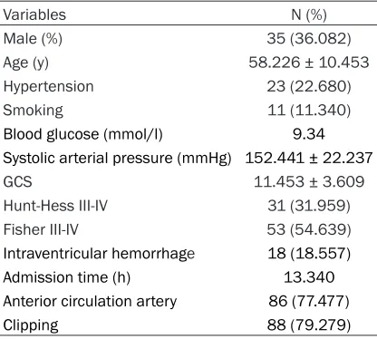
grade III-IV. In all, there were 111 intracranial aneurysms, 88 of which received microsurgical clipping. The basic information of the included patients is presented in Table 1.
Serum levels of nesfatin-1 in aSAH patients
The patients with poor outcomes had higher levels of nesfatin-1 when compared with the patients with good outcomes (11.342 ± 1.826 vs 9.118 ± 0.278, P < 0.01, Figure 1A). Also, the patients with DCI had higher levels of nes-fatin-1 than the patients with non-DCI (11.736 ± 1.844 vs 8.842 ± 2.629, P < 0.01, Figure 1B). Because nesfatin-1 plays an important
role in the process of inflammation, we
perfor-med an additional analysis. But the results revealed that there was no difference bet- ween patients with infectious diseases and patients with non-infectious during their hospi-tal stays (Figure 1C).
The correlation between nesfatin-1 and clini-cal severity
The serum concentration of nesfatin-1 was positively correlated with the Hunt-Hess grade (r = 0.417, P < 0.01 95% CI [0.237-0.569]) and a higher Hunt-Hess grade (Figure 2B, 2C). But serum nesfatin-1 failed to show any
correla-tions with the GCS or Fisher grade. CRP, an
important acute phase stress protein, has a
significant correlation with the clinical progno
-sis of patients with aSAH [14]. Our preliminary
analysis showed a significantly positive correla -tion between nesfatin-1 and CRP (Figure 2A). Risk factors associated with poor outcome
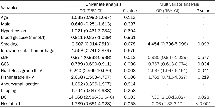
Univariate and multivariate analyses were used to analyze the risk factors associated with the poor outcomes of patients with aSAH. The re- sults showed that GCS, Hunt-Hess III-V, DCI,
and an elevated level of nesfatin-1 were signifi -cantly associated with poor outcomes. The re- sults of the univariate and multivariate analy-ses are shown in Table 2.
ROC of nesfatin-1 in predicting poor outcome
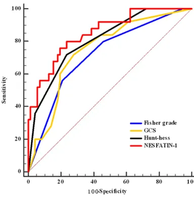
ROC was used to accessed the predictive po- wer of nesfatin-1. The area under the curve (AUC) was 0.721, 0.748, 0.808, and 0.852 of
the Fisher grade, GCS, Hunt-Hess and
nesfa-tin-1, respectively. The sensitivity and specifici -ty of nesfatin-1 in predicting a poor outcome were 0.760 and 0.806, respectively. The ROC cures of the variants are presented in Figure 3. Discussion
In this retrospective study, we found that patients with poor outcomes had higher serum levels of nesfatin-1 when compared with pa- tients with good outcomes. What’s more,
nes-fatin-1 was significantly elevated in patients
who suffered DCI. The serum level of nesfatin-1 was positively correlated with Hunt-Hess grade
and serum CRP level. Further multivariate
regression analysis revealed Hunt-Hess III-IV, GCS, DCI, and sBP on admission, and elevated
levels of nesfatin-1 were significantly associat -ed with the poor outcomes of patients with aSAH. The predictive values of the variates were evaluated using ROC, and the results sh- owed that nesfatin-1 was better than
Hunt-Hess, GCS, and Fisher grade in predicting the
poor outcomes of patients with aSAH.
Inflammatory response was one of the most
[image:3.612.91.299.84.271.2]important causes of secondary brain injury, cerebral vasospasm and DCI [15, 16]. Serum biomarkers, such as CRP and D-dimer had been reported to be independent risk factors associated with the poor outcomes of patients with aSAH in several studies [17, 18]. The role of nesfatin-1 was mainly investigated in pa- tients with traumatic brain injury [13] and few studies were performed to demonstrate its role in hemorrhagic stroke. Previous studies found
Table 1. Baseline characteristics
Variables N (%)
Male (%) 35 (36.082)
Age (y) 58.226 ± 10.453
Hypertension 23 (22.680)
Smoking 11 (11.340)
Blood glucose (mmol/l) 9.34
Systolic arterial pressure (mmHg) 152.441 ± 22.237
GCS 11.453 ± 3.609
Hunt-Hess III-IV 31 (31.959)
Fisher III-IV 53 (54.639)
Intraventricular hemorrhage 18 (18.557)
Admission time (h) 13.340
Anterior circulation artery 86 (77.477)
that nesfatin-1 plays an important role in the
process of inflammation, especially in central
nervous system (CNS) diseases. Wistar albino rats were randomly divided into a saline-treat-ed SAH group and a nesfatin-1-treatsaline-treat-ed SAH group, and each group had 18 rats. 48 hours after SAH induction, the rats in the nesfatin-1 treated SAH group had better conditions of neurological impairment and oxidative brain
injury [19]. What’s more, pro-inflammatory cyto -kines in the peripheral blood were also depre-
ssed by treatment with nesfatin-1 [20]. Another study conducted by Chon-hui Tang et al. showed
that 20 g/kg nesfatin-1 treatment could signifi
-cantly suppress inflammation after traumatic
brain injury in rats [19]. The above studies indi-cated that nesfatin-1 might play an important
role in the inflammatory processes of central
[image:4.612.91.518.73.177.2]nervous system diseases, including aSAH. Recently, Cakir et al. reported that aSAH pa- tients in non-survival group had higher level of Table 2. Risk factors associated with poor outcome
Variables Univariate analysis Multivariate analysis
OR (95% CI) P value OR (95% CI) P value
Age 1.035 (0.990-1.097) 0.113 -
-Male 0.640 (0.251-1.613) 0.337 -
-Hypertension 1.221 (0.461-3.284) 0.694 -
-Blood glucose (mmol/l) 0.911 (0.827-1.039) 0.961 -
-Smoking 2.607 (0.914-7.510) 0.078 4.454 (0.796-5.098) 0.093
Intraventricular hemorrhage 1.563 (0.741-2.879) 0.675
sBP 0.977 (0.938-0.988) 0.012 0.980 (0.947-1.029) 0.577
GCS 0.789 (0.690-0.911) 0.008 0.767 (0.613-0.974) 0.034
Hunt-Hess grade III-IV 5.240 (2.569-10.584) 0.008 2.537 (1.047-6.191) 0.041 Fisher grade III-IV 2.668 (1.503-4.757) 0.006 1.761 (0.713-4.327) 0.219
Aneurysmal location 1.062 (0.396-1.907) 0.914 -
-Clipping 1.794 (0.647-4.933) 0.258 -
-DCI 14.668 (2.586-32.640) 0.003 7.35 (2.18-16.82) 0.028
[image:4.612.92.531.216.318.2]Nesfatin-1 1.789 (0.651-4.928) 0.058 2.06 (1.33-3.17) < 0.001
Figure 2. Correlation between nesfatin-1 and clinical severities.
[image:4.612.91.523.375.589.2]nesfatin-1 than patients in survival group, wh- ich indicated that nesfatin-1 was an useful bio-marker in the management of aSAH [21]. Our study found that levels of nesfatin-1 in the blood of the poor outcome patients was higher than the good outcome patients and an obvi-ous difference was observed between patients with DCI and non-DCI. Although nesfatin-1 was
a vital protein in the inflammation-mediated
process, it failed to show any advantages in the
identification of systemic infections and
non-infected aSAH patients. We usually use the Hu-
nt-Hess grade, the Fisher grade, and the GCS
score to assess the severity of aSAH. These indicators have been used clinically for a long time. In recent years, the assessment of the severity of aSAH by serum biochemical mark-ers has gradually gained attention. A higher
level of NSE was significantly associated with a high Hunt-Hess grade, the WFNS score, and
GCS, which indicated that NSE was a promising tool in screening with a high risk of having a poor outcome after spontaneous SAH [22].
CRP was a highly sensitive inflammatory mark -er, and its serum level related to severity of aSAH. In this study, we found that serum nesfa-tin-1 positively correlated with CRP and the Hunt-Hess grade, but no correlation was
observed between nesfatin-1 and the Fisher
grade. Our results were not in line with previous studies. In a clinical study conducted by Cakir, the serum levels of nesfatin-1 were found to be
significantly correlated with WFNS and the Fisher grade (r2 = 0.521, r2 = 0.602, respec
-tively). But the relationship between the serum nesfatin-1 levels and CRP was consistent between these two studies.
There were some limitations in this retrospec-tive study. This was a retrospecretrospec-tive study and the number of patients was relatively small. We just evaluated the value of pre-operative nesfa-tin-1 in predicting poor outcomes, but the dynamic change of nesfatin-1 after aSAH is still unknown. Moreover, patients with aSAH might have received either clipping or coiling. Diffe- rent surgical approaches might have different effects on nesfatin-1 elevation, because the inevitable brain tissue damage could be induc- ed by microsurgery. So post-operative markers may be more reliable in predicting neurological
complications and functional outcome. Finally,
little is known about relationship between its peak level and poor outcome, so in our opinion, more clinical studies should be conducted to explore the role of serum nesfatin-1 in aSAH. Conclusion
Nesfatin-1 is significantly elevated in aSAH
patients with poor outcomes. Nesfatin-1 is
sig-nificantly associated with clinical severity, and
it is an independent prognostic factor for poor outcome in patients with aSAH.
Disclosure of conflict of interest
None.
Address correspondence to: Jiliang Hu, Depart- ment of Neurosurgery, Shenzhen People’s Hospital, Second Clinical Medical College of Ji’nan Univer- sity, No. 1017, Dongmen North Road, Luohu Distri- ct, Shenzhen 518020, China. Tel: 0755-25533018; E-mail: [email protected]
References
[1] Rincon F, Rossenwasser RH and Dumont A. The epidemiology of admissions of nontrau-matic subarachnoid hemorrhage in the United States. Neurosurgery 2013; 73: 217-222; dis-cussion 212-213.
[image:5.612.91.283.70.265.2][2] Mackey J, Khoury JC, Alwell K, Moomaw CJ, Kissela BM, Flaherty ML, Adeoye O, Woo D, Ferioli S, De Los Rios La Rosa F, Martini S, Khatri P, Broderick JP, Zuccarello M and Kle- indorfer D. Stable incidence but declining ca- se-fatality rates of subarachnoid hemorrhage in a population. Neurology 2016; 87: 2192-2197.
[3] Srinivasan A, Aggarwal A, Gaudihalli S, Moh- anty M, Dhandapani M, Singh H, Mukherjee KK and Dhandapani S. Impact of early leukocy-tosis and elevated high-sensitivity C-reactive protein on delayed cerebral ischemia and neu-rologic outcome after subarachnoid hemorrh- age. World Neurosurg 2016; 90: 91-95. [4] Csajbok LZ, Nylen K, Ost M, Sonander H and
Nellgard B. In-hospital C-reactive protein pre-dicts outcome after aneurysmal subarachnoid haemorrhage treated by endovascular coiling. Acta Anaesthesiol Scand 2015; 59: 255-264. [5] Badjatia N, Monahan A, Carpenter A,
Zimmer-man J, Schmidt JM, Claassen J, Connolly ES, Mayer SA, Karmally W and Seres D. Inflamma-tion, negative nitrogen balance, and outcome after aneurysmal subarachnoid hemorrhage. Neurology 2015; 84: 680-687.
[6] Ramchand P, Nyirjesy S, Frangos S, Doerfler S, Nawalinski K, Quattrone F, Ju C, Patel H, Dri- scoll N, Maloney-Wilensky E, Stein SC, Levine JM, Kasner SE and Kumar MA. Thromboelas-tography parameter predicts outcome after subarachnoid hemorrhage: an exploratory an- alysis. World Neurosurg 2016; 96: 215-221. [7] Kubo Y, Koji T, Yoshida J, Ogawa A and
Ogas-awara K. Predicting neurological deficit severi-ty due to subarachnoid haemorrhage: soluble CD40 ligand and platelet-derived growth fac-tor-BB. Crit Care Resusc 2016; 18: 242-246. [8] Wei Y, Li J, Wang H and Wang G.
NUCB2/nesfa-tin-1: expression and functions in the regula-tion of emoregula-tion and stress. Prog Neuropsycho-pharmacol Biol Psychiatry 2018; 81: 221-227. [9] Goebel-Stengel M and Wang L. Central and pe-ripheral expression and distribution of NUCB2/ nesfatin-1. Curr Pharm Des 2013; 19: 6935-6940.
[10] Alp E, Gormus U, Guducu N and Bozkurt S. Nesfatin-1 levels and metabolic markers in po- lycystic ovary syndrome. Gynecol Endocrinol 2015; 31: 543-547.
[11] Leivo-Korpela S, Lehtimaki L, Hamalainen M, Vuolteenaho K, Koobi L, Jarvenpaa R, Kankaanranta H, Saarelainen S and Moilanen E. Adipokines NUCB2/nesfatin-1 and visfatin as novel inflammatory factors in chronic ob-structive pulmonary disease. Mediators In-flamm 2014; 2014: 232167.
[12] Zhang C, Wang Y, Wang Y, Li J, Liu R and Liu H. Decreased levels of serum nesfatin-1 in pa-tients with preeclampsia. Biomarkers 2014; 19: 402-406.
[13] Wu GQ, Chou XM, Ji WJ, Yang XG, Lan LX, Sheng YJ, Shen YF, Li JR, Huang GZ, Yu WH, Dong XQ, Du Q, Yang DB, Zhang ZY, Wang H, Shen YF and Jiang L. The prognostic value of plasma nesfatin-1 concentrations in patients with traumatic brain injury. Clin Chim Acta 2016; 458: 124-128.
[14] Hollig A, Stoffel-Wagner B, Clusmann H, Velde-man M, Schubert GA and Coburn M. Time courses of inflammatory markers after aneu-rysmal subarachnoid hemorrhage and their possible relevance for future studies. Front Neurol 2017; 8: 694.
[15] Zheng VZ and Wong GKC. Neuroinflammation responses after subarachnoid hemorrhage: a review. J Clin Neurosci 2017; 42: 7-11. [16] Lucke-Wold BP, Logsdon AF, Manoranjan B,
Turner RC, McConnell E, Vates GE, Huber JD, Rosen CL and Simard JM. Aneurysmal sub-arachnoid hemorrhage and neuroinflamma-tion: a comprehensive review. Int J Mol Sci 2016; 17: 497.
[17] Fukuda H, Lo B, Yamamoto Y, Handa A, Yama-moto Y, Kurosaki Y and Yamagata S. Plasma D-dimer may predict poor functional outcomes through systemic complications after aneurys-mal subarachnoid hemorrhage. J Neurosurg 2017; 127: 284-290.
[18] Liu JH, Li XK, Chen ZB, Cai Q, Wang L, Ye YH and Chen QX. D-dimer may predict poor out-comes in patients with aneurysmal subarach-noid hemorrhage: a retrospective study. Neural Regen Res 2017; 12: 2014-2020.
[19] Tang CH, Fu XJ, Xu XL, Wei XJ and Pan HS. The anti-inflammatory and anti-apoptotic effects of nesfatin-1 in the traumatic rat brain. Peptides 2012; 36: 39-45.
[20] Ozsavci D, Ersahin M, Sener A, Ozakpinar OB, Toklu HZ, Akakin D, Sener G and Yegen BC. The novel function of nesfatin-1 as an anti-inflam-matory and antiapoptotic peptide in subarach-noid hemorrhage-induced oxidative brain dam-age in rats. Neurosurgery 2011; 68: 1699-1708; discussion 1708.
[21] Cakir M, Calikoglu C, Yilmaz A, Akpinar E, Bayraktutan Z and Topcu A. Serum nesfatin-1 levels: a potential new biomarker in patients with subarachnoid hemorrhage. Int J Neurosci 2017; 127: 154-160.