R E S E A R C H
Open Access
Deciphering composition and function of
the root microbiome of a legume plant
Kyle Hartman
1,2, Marcel GA van der Heijden
1,2,3, Valexia Roussely-Provent
4, Jean-Claude Walser
5and Klaus Schlaeppi
1*Abstract
Background:Diverse assemblages of microbes colonize plant roots and collectively function as a microbiome. Earlier work has characterized the root microbiomes of numerous plant species, but little information is available for legumes despite their key role in numerous ecosystems including agricultural systems. Legumes form a root nodule symbiosis with nitrogen-fixingRhizobiabacteria and thereby account for large, natural nitrogen inputs into soils. Here, we describe the root bacteria microbiome of the legumeTrifolium pratensecombining
culture-dependent and inculture-dependent methods. For a functional understanding of individual microbiome members and their impact on plant growth, we began to inoculate root microbiome members alone or in combination to
Trifoliumroots.
Results:At a whole-root scale,Rhizobiabacteria accounted for ~70% of the root microbiome. Other enriched members included bacteria from the generaPantoea,Sphingomonas,Novosphingobium, andPelomonas. We built a reference stock of 200 bacteria isolates, and we found that they corresponded to ~20% of the abundant root microbiome members. We developed a microcosm system to conduct simplified microbiota inoculation experiments with plants. We observed that while an abundant root microbiome member reduced plant growth when inoculated alone, this negative effect was alleviated if thisFlavobacteriumwas co-inoculated with other root microbiome members.
Conclusions:TheTrifoliumroot microbiome was dominated by nutrient-providingRhizobiabacteria and enriched for bacteria from genera that may provide disease protection. First microbiota inoculation experiments indicated that individual community members can have plant growth compromising activities without being apparently pathogenic, and a more diverse root community can alleviate plant growth compromising activities of
its individual members. A trait-based characterization of the reference stock bacteria will permit future microbiota manipulation experiments to decipher overall microbiome functioning and elucidate the biological mechanisms and interactions driving the observed effects. The presented reductionist experimental approach offers countless opportunities for future systematic and functional examinations of the plant root microbiome.
Keywords:Clover, Root, Microbiome, 16S rRNA sequencing, Microcosm
Background
Plant roots in soil are in contact with the most microbially diverse biome on the planet, with estimates of bacteria diversity as high as 38,000 taxa per gram of soil [1]. The
root bacteria microbiome typically consists of
Proteobac-teria,Actinobacteria, andBacteroidetes[2]. Recent studies have highlighted the root bacteria microbiome of several
plant species, includingArabidopsis [3, 4] and a number
of crop species, like barley [5], maize [6], sugarcane [7], and rice [8]. However, the microbiome of nitrogen-fixing plants, in particular legumes such as red clover, has received little attention in microbiome studies.
Trifolium pratense (red clover, hereafter: Trifolium) is an important forage legume and grown on approxi-mately four million hectares worldwide [9]. Because of its beneficial symbiosis with N-fixing rhizobia, Trifolium is cultivated in grass/clover mixtures or as a cover crop
in crop rotations [10]. While the species’genetic
diver-sity has been characterized using morphological traits [11], DNA marker polymorphism [12], and genome * Correspondence:[email protected]
1Plant-Soil Interactions, Agroscope, Institute for Sustainability Sciences,
Reckenholzstrasse 191, CH-8046 Zürich, Switzerland
Full list of author information is available at the end of the article
analyses [13], its root microbiome has not been investi-gated using high-throughput sequencing tools.
Further-more, Trifolium’s association with rhizobia suggests its
microbiome may differ from non-legumes in that rhizo-bia are expected to be highly abundant [14].
The N-provision by rhizobia represents a well-established service to their host. Similarly, other microbiome members were found to assist their host plant in nutrient uptake, protection from pathogens, or modulating immunity re-sponses [15, 16]. However, how microbial functions affect plants if a service-providing member is in a diverse com-munity, and how entire microbial communities affect their host, remains poorly understood [16]. One limitation of ribosomal RNA-based root microbiota characterizations is that such approaches only provide indirect information, based upon taxonomic classification, about the function(s) of its members. One suggested approach for the functional examination of the root microbiome relies on isolating root microbes to build microbe collections [17]. The availability of bacterial isolates offers the opportunity for genome sequencing to obtain insights into their potential functions, but more importantly, the activity of these strains can be empirically tested in host-microbiota interaction experiments.
Microbe collections have been assembled [18–22]
des-pite that the recalcitrance to cultivation of many bacteria
taxa—with estimates that more than 99% of soil bacteria
cannot be cultured [23]—was often seen as a limitation.
This recalcitrance does not necessarily apply to bacteria of the root microbiome as evidenced by an earlier study of Chelius and Triplett [24], who reported a phylogen-etic overlap of 48% between their bacteria isolate collec-tion and a 16S ribosomal RNA (rRNA) clone library from maize roots. More recently, Bai et al. [21] reported a collection of nearly 6000 root-derived bacteria isolates
and a remarkable 54–65% isolation rate compared to the
abundant (>0.1% relative abundance) operational
taxo-nomic units (OTUs) in Arabidopsis thaliana roots.
However, it required considerable effort including large-scale isolation using serial dilutions (seven different bac-teria isolation media were used!) and subsequent high-throughput taxonomy identification.
Experimental manipulation of the microbiome and assays with plants require contained systems in which host-microbiota interaction experiments can be conducted with-out with-outside microbial contamination. Recently, microcosm systems have been used in combination with bacteria refer-ence stocks to examine the dynamic process of root micro-biome assembly from a defined input community under microcosm conditions [21, 22]. In these experiments, stable and reproducible community assembly was observed. How-ever, these experiments were not designed to clarify how root communities compare to plants grown in artificial sub-strate in microcosms or in natural soil conditions.
Here, we addressed some of the aforementioned re-search gaps and report a detailed characterization of the Trifolium root bacteria microbiome. We sampled the whole-root system including nodules, removed the rhizosphere and investigated the entire root bacterial communities consisting of rhizoplane and endosphere habitats. We utilized a multi-step approach to investi-gate the composition and culturable fraction of its root microbiome (Fig. 1). We also move towards a func-tional understanding of specific members of the Trifo-lium root microbiome and developed a microcosm
system (Additional file 1: Figure S1a–d) in which we
conducted multi-strain inoculation experiments with Trifolium germinated from surface-sterilized seeds and investigated the inoculation-induced effects on plant growth.
Results
Composition of the Trifolium root microbiome
The 16S amplicon sequencing of 24 Trifolium root sam-ples and 15 soil samsam-ples from climate chamber and nat-ural site growth experiments (Fig. 1, Table 1, Additional file 1: Figure S2,) yielded 9,923,925 high-quality, non-chimeric sequences across all samples, with a median of
153,072 (range 21,731–981,922) sequences per sample
(Additional file 2). We rarefied the dataset to an even se-quencing depth of 20,000 sequences and identified 3495 bacteria OTUs and one archaea OTU.
We confirmed in the Trifolium root microbiome the typical patterns that are often observed in microbial ecol-ogy. The soil microbiome is richer and phylogenetically more diverse than the root microbiome (Additional file 1: Figure S3; Table S1). We quantified the major components driving differences between samples (ß-diversity) using unconstrained principal coordinates analysis (PCoA) on weighted UniFrac distances and found a clear separation along axis 1 (explaining 69.7% of the overall variation) and confirmed the general pattern that soil and roots harbor distinct microbiomes (Fig. 2). Axis 2 explained 15.5% of the variation overall and separated mainly the root but not the soil samples, and we did not notice an obvious cluster-ing whether the plants were grown in the same soil in a climate chamber or in the field, suggesting negligible
ef-fects of thegrowth conditiononβ-diversity.We detected a
significant effect of growth condition on OTU richness
only (Additional file 1: Figure S3; Table S1). However, experiment-to-experiment variation (especially climate chamber experiment 2) largely explained the variability between root samples (Additional file 1: Figure S4). Pos-sible effects due to differences in climatic conditions were generally not detected and would have an effect size smaller than replicate experimental variation.
evident in the taxonomic profiles of the samples. Soil
samples contained abundant Proteobacteria,
Actinobac-teria, andAcidobacteria accounting for a mean of 54.7, 24.7, and 6.9%, respectively (Additional file 1: Figure S5). The Trifolium root microbiome was dominated by
Proteobacteria that accounted for a mean abundance of 90.7% across both experimental conditions (Additional file 1: Figure S5).
For the detailed characterization of the Trifolium root microbiome (Fig. 1, step I), we first identified the OTUs
Fig. 1Characterization of the root microbiome. We collected a natural field soil and used it in a series of Trifolium growth experiments. (I) We investigated the composition of the root bacteria microbiome using 16S rRNA sequencing of root samples. (II) We utilized the same root material for an isolation effort to explore the culturable fraction of root bacteria microbiome and assembled a reference stock of bacteria isolates. (III) We subsequently developed a microcosm system to explore plant-microbiota interactions and (IV) investigated the composition of the Trifolium root microbiome in the system by inoculating microbiota extracted from the field soil. (V) We conducted microbiota manipulation experiments in which we inoculated culturable, abundant members of the root microbiome and scored their effects on plant growth
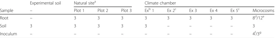
Table 1Overview of the number of replicate samples by sample type, experiment, and experimental replicate, or plot
Experimental soil Natural sitea Climate chamber
Sample – Plot 1 Plot 2 Plot 3 Exb1 Ex 2c Ex 3 Ex 4 Ex 5c Microcosms
Root – 3 3 3 3 3 3 3 3 8d/12e
Soil 3 3 3 3 3 – – – – 3
Inoculum – – – – – – – – – 4f/3g
a
Bacteria isolates from natural site plants were cultured from plants collected from within and outside the experimental plots
b
Experiment
c
Bacteria isolates from climate chamber plants were cultured from these experiments, plus one non-sequenced growth experiment
d
Total number of samples collected from the soil extract experiment. One root sample was collected from each replicate microcosm
e
Total number of samples from the simplified community experiments. Four root samples were collected from each of the three experiments
f
Independently prepared soil extract samples used as the experimental start inoculum. See Additional file1for details
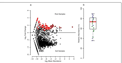
g
that were significantly higher in relative abundance in root compared to soil samples and discovered a total of 61 OTUs significantly enriched in root samples (Fig. 3), 15 of which were abundant with a mean relative abundance of at least 0.1% across all root samples. These 15 OTUs accounted for 74.5% of rarefied sequences, and we termed
them “RootOTUs”—referring to the abundant and
root-specific members of the Trifolium root microbiome. The
RootOTUs consisted mostly ofProteobacteria(14 OTUs,
Additional file 1: Table S2) and represented six different
orders: Rhizobiales(6),Sphingomonadales(3),
Enterobac-teriales (2), Burkholderiales (1),Caulobacterales (1), and Rhodospirillales (1). The remaining non-Proteobacteria
RootOTU belonged to theFirmicutesand was classified in
the genusSyntrophomonas.We noted that one RootOTU
(OTU1, matchingRhizobium leguminosarum) dominated
the Trifolium root microbiome and explained the high
prevalence ofProteobacteria(Additional file 1: Figure S5).
OTU1 ranged from 35.4 to 89.7% in samples from both
growth conditions and accounted for a median of 73.5% of the root community (Fig. 3b). We confirmed that
the high abundance of OTU1 in the overall root
com-munity was due to the rhizobia bacteria present in
root nodules (Additional file 1: Supplementary
methods), and we noted a few non-OTU1 sequences
inside the nodules, suggesting additional
within-nodule bacteria diversity (Additional file 1: Supple-mentary results, Figure S6).
Fig. 2Sample type, growth conditions, and experiment explain much of the variation in soil and root bacteria communities. Unconstrained principal coordinates analysis (PCoA) of weighted UniFrac distances of root and soil samples from climate chamber (CC Root,CC Soil) and natural site growth experiments (NS Root,NS Soil), as well as the unplanted experimental field soil (Exp. Soil). See Additional file 1: Figure S4 for points colored by the replicate experiment
In summary, root bacterial communities did not differ substantially whether the plants were grown under con-trolled or field conditions, thereby validating our approach using climate chamber experiments. The abundant and root-specific members of the Trifolium root microbiome
consisted mainly ofProteobacteriaand nodule-inhabiting
rhizobia bacteria accounted for ~70% of the root microbiome.
Isolated members of the Trifolium root microbiome We isolated bacteria from Trifolium roots of two climate chamber experiments and from plants grown at the nat-ural site (Table 1) and characterized a total of 200 cultured
bacteria (Fig. 1, step II).Proteobacteriadominated the
cul-ture collection, being represented by 78.5% isolates while Actinobacteria, Firmicutes, and Bacteroidetes accounted for 8, 8, and 5.5% of isolates, respectively (Fig. 4a). The isolates were assigned to 34 different genera (Fig. 4b). The
19 genera of the Proteobacteria (157 isolates) included
abundant Pseudomonas (83 isolates), Janthinobacterium
(19), and Stenotrophomonas (9). We found seven genera
in the phylumActinobacteria(16 isolates) with
Microbac-terium(7),Micrococcus(3), andMicromonospora(2)
hav-ing more than one representative isolate. In theFirmicutes
(16 isolates), we noted five different genera, withBacillus
(9), Staphylococcus (3), and Paenibacillus (2) being the
most abundant. Finally, we found three genera in the
Bac-teroidetes(11 isolates):Flavobacterium(8),
Mucilaginibac-ter(2), andPedobacter(1).
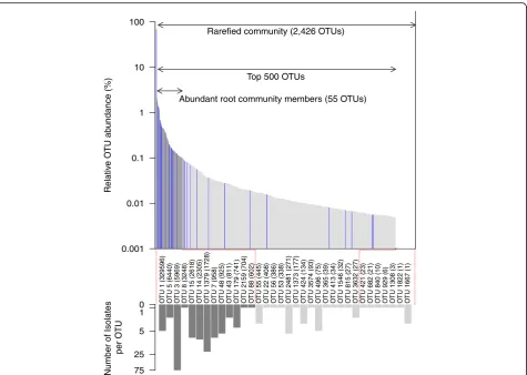
We clustered the bacteria isolate sequences to the rep-resentative sequences of the OTUs of the Trifolium root
community profiles at ≥97% sequence similarity (see
Additional file 1: Supplementary methods) and deter-mined whether a bacteria isolate constituted an abundant and root-enriched member of the Trifolium microbiome. Overall, out of the 200 bacteria isolates, 181 (90.5%) iso-lates clustered to 34 OTUs of the root community profile while for 19 (8.5%) isolates, we did not find a matching community member. All of the 34 isolated OTUs were present in the rarefied root community (2426 OTUs), cor-responding to an isolation rate of 1.4% (Fig. 5). The isola-tion rate increased to 23.6% when comparing to the abundant community members: 55 abundant OTUs had a
mean relative abundance of≥0.1% across all root samples,
and for 13 of these, we were able to culture bacteria strains. We identified 11 bacteria isolates for 2 of the 15 RootOTUs (Fig. 3; Additional file 1: Table S2). The
cul-tured RootOTUs included the dominant OTU1 (R.
Pantoea sp. (3) Serratia sp. (3) Rahnella sp. (3) Serratia sp. (2)
Enterobacter sp. (5)
Leclercia sp. (1) Erwinia sp. (4)
Bradyrhizobium sp. (9)
Mesorhizobium sp. (1)
Rhizobium sp. (6)
Flavobacterium sp. (8)
Pedobacter sp. ( 1)
Mucilaginibacter sp. (2
)
Micrococcus sp.
(3)
)
1(
.
p
s
ai
v
o
k
sr
e
O
Curtobacte
riu
m
sp.
(1) Herbiconiux sp.
(1) Micr
obacterium
sp. (7) Mycobacterium sp. (1) Micromonospora sp. (2)
Paenibacillus sp. (2)
Bacillus sp. (1) Staphyl
ococcus sp.
(3) Bacillus sp. (8) Sporosarcina sp. (1) Strept
ococcus sp.
(1)
Delftia sp.
(1) Variovorax sp. (1)
Cupriavidus sp. (1) Collimonas sp.
(2) Rugamonas
sp. (2)
Janthinobacterium sp. (19)
Dyella sp. (1)
Rudaea sp. (1)
Stenotrophomonas sp. (9) Pseudomonas sp. (83) 0.05
Proteobacteria
Actinobacteria Firmicutes
200 isolates
Bacteroidetes 78.5%
8% 8%
5%
a
b
leguminosarum; 5 isolates), as well as OTU48 (Pantoea agglomerans; 6 isolates).
We concluded that almost a quarter of the abundant root community members can be obtained in culture, and we achieved this with a manageable effort (200 strains) and straightforward microbiological techniques. By linking to the information of the root community profiles, we have characterized the bacteria strains of the reference stock with rank and relative abundance in the Trifolium root microbiome, and thereby the reference stock represents a toolbox for future microbiota ma-nipulation experiments.
Towards functional investigations of the Trifolium root microbiota
Finally, we developed microcosms (Fig. 1, step III) and evaluated their potential to conduct plant-microbiota interaction experiments. Recent microbiota inoculation experiments [21, 22] revealed that approximately half of
the inoculated bacteria strains previously isolated from
roots of soil-grownArabidopsiseither completely failed or
failed to robustly colonize the roots of their host plant under microcosm conditions. We speculate that this could partly be due to the different physical and chemical condi-tions in the microcosms compared to soil and that these conditions are unfavorable for certain isolates. Therefore, we performed a soil extract experiment to pre-screen for possible microcosm-adapted bacteria strains. For this, we characterized the root microbiome of Trifolium that as-sembled after inoculation of a diverse soil microbiota ex-tracted from the experimental field soil (Additional file 1: Figure S7a, b, Figure S8; Supplementary methods and re-sults). We defined the root bacteria community (Fig. 1, step IV) and determined which bacteria isolates (from the reference stock, Fig. 5) corresponded to abundant OTUs on the roots under microcosm conditions (Additional file 1: Figure S8; Supplementary methods). See the Additional file 1: Supplementary results for a
0.001 0.01 0.1 1 10 100
Relativ
e O
TU ab
undance (%)
Rarefied community (2,426 OTUs)
Top 500 OTUs
Abundant root community members (55 OTUs)
75 25 5 1
0 O
TU 1 (329596)
O
TU 5 (6440)
O
TU 3 (5969)
O
TU 8 (3248)
O
TU 15 (2618)
O
TU 14 (2305)
O
TU 1379 (1728)
O
TU 7 (958)
O
TU 48 (925)
O
TU 43 (811)
O
TU
179
(741)
O
TU 2159 (704)
O
TU 88 (602)
O
TU 55 (445)
O
TU 22 (408)
O
TU 56 (386)
O
TU 53 (338)
O
TU 2481 (271)
O
TU 1373 (177)
O
TU 424 (134)
O
TU 3574 (93)
O
TU 496 (75)
O
TU 365 (39)
O
TU 413 (34)
O
TU 1546 (32)
O
TU 815 (27)
O
TU 3632 (27)
O
TU 421 (23)
O
TU 682 (21)
O
TU 840 (10)
O
TU 929 (6)
O
TU 1308 (3)
O
TU 1822 (1)
O
TU 1667 (1)
Number of Isolates
per O
TU
comparison between microcosm and soil-grown root communities (Additional file 1: Figure S9a, b; Supple-mentary methods and results).
We then conducted microcosm experiments in which we inoculated Trifolium in the microcosms with bacteria strains isolated from its root microbiome. The goal was not to screen strains or to test specific functions but instead to combine all our tools (reference stock, microcosms, com-munity sequencing, and soil extract information) and valid-ate the overall experimental approach for future microbiota inoculation experiments. We assembled a simplified com-munity, choosing strains from the reference stock that corresponded to abundant OTUs on the roots under
microcosm conditions and belonged to well-represented
bacterial genera in the collection (Additional file 1: Figure
S9; strains per OTU were randomly chosen): a
Flavobacter-ium(F;Bacteroidetes, #8 isolates for this genus in the
refer-ence stock; KHB002), a Pseudomonas (P; Proteobacteria,
#83; KHB004), and a Janthinobacterium(J;Proteobacteria,
#19, KHB023; Table 2). We also included aMicrobacterium
(M;Actinobacteria, #7; strain KHB073) because this genus was well-represented in the reference stock (numerous iso-lates could indicate that these bacteria were abundant on roots; Fig. 4b) and because we wanted the inoculated com-munity to broadly reflect the abundant bacterial phyla of
plant root microbiomes (Actinobacteria,Bacteroidetes, and
Proteobacteria; [2, 25]). We inoculated these bacteria alone or in combination to the autoclaved microcosms (Fig. 1,
step V) at densities of 106cells mL−1and planted
surface-sterilized Trifolium seeds. We then monitored the commu-nity dynamics of the inoculated simplified commucommu-nity and scored effects of the bacteria inoculation on plant growth in three replicate experiments.
After 25 days, we harvested the experiments and
counted≥106bacterial colony forming units of the
inoc-ulated strains on the roots (Table 2). This confirmed that the chosen strains are also able to successfully colonize roots under the artificial growth conditions in the micro-cosms. We noted a lower biomass in one experiment compared to the two others, and this experiment-to-experiment variation indicated to us that numerous rep-licates are also needed when highly controlled conditions
are used. With regard to the effects of individual bacteria
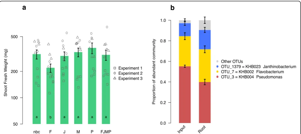
inoculation on the plants, we found that the
Flavobacter-iumnegatively affected the growth of Trifolium, while the
other bacteria did not have an effect on shoot biomass production (Fig. 6a). The combined application of the
bac-teria (FJMP) also did not have an apparent effect on
bio-mass production but alleviated the negative impact of the Flavobacterium when grown alone. We measured the composition of the simplified community upon inocula-tion and after 25 days on the roots (Addiinocula-tional file 1:
Sup-plementary methods for details). The Microbacterium
could not be captured with the community quantification method, and we noted a small proportion of additional OTU sequences possibly representing sequencing errors or contamination, or in root samples, being derived from seed endophytes. Despite these limitations, the analysis re-vealed that the three other inoculated members retained similar proportions on the roots during 25 days of incuba-tion as compared to when they were inoculated (Fig. 6b). This observation indicated that the alleviation of the
nega-tive impact of the Flavobacterium was not due to
out-competition of this community member, but rather that
its negative activities may have been “buffered” by the
other bacteria in the simplified community.
Discussion
Root microbiome composition
Here, we have characterized the bacterial communities
on roots ofT. pratensewith respect to their composition
and reported first steps towards experimentally testing their functions. Trifolium harbors a diverse root micro-biome that differs qualitatively and quantitatively from that of the surrounding bulk soil (Fig. 2), confirming
studies with other plant species [3–5, 26]. We found that
OTU1, matchingR. leguminosarum, accounted for a
me-dian 73.5% of the root microbiome (Fig. 3b). We separ-ately inspected root nodules and confirmed that
Trifolium nodules were primarily inhabited byR.
legumi-nosarum(Additional file 1: Figure S6) but also contained other bacteria taxa. This is in agreement with earlier
work revealing within-nodule diversity in Trifolium
repensand Trifolium fragiferum, which consisted of the
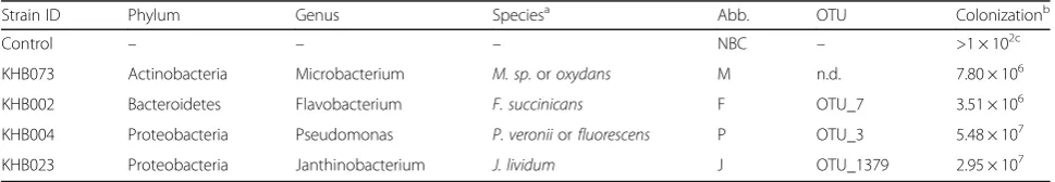
Table 2Bacterial strains used in the microcosm experiments
Strain ID Phylum Genus Speciesa Abb. OTU Colonizationb
Control – – – NBC – >1 × 10²c
KHB073 Actinobacteria Microbacterium M. sp.oroxydans M n.d. 7.80 × 10⁶
KHB002 Bacteroidetes Flavobacterium F. succinicans F OTU_7 3.51 × 10⁶
KHB004 Proteobacteria Pseudomonas P. veroniiorfluorescens P OTU_3 5.48 × 10⁷
KHB023 Proteobacteria Janthinobacterium J. lividum J OTU_1379 2.95 × 10⁷
Abb.Abbreviation,n.d.not detected in the Trifolium root microbiome using high-throughput sequencing
a
Taxonomy based on Greengenes 16S database [51]
b
Mean bacterial cell number on roots after 25 days in the microcosms in experiment 3
c
dominantR. leguminosarumand the less-frequent
rhizo-bia species Bradyrhizobium japonicum, Sinorhizobium
sp., and Mesorhizobium[27, 28]. For the purpose of the
microcosm experiments, we described the root micro-biome of Trifolium at a whole-root scale, sampling the entire root system including nodules. For a broader de-scription of legume microbiomes, future work investigat-ing the variation in multiple soil types and comparisons with non-legume plants is needed. Additionally, an in-depth spatial assessment of legume root microbiomes would be insightful, e.g., by profiling the bacteria com-munities of root tissues with the nodules removed as well as inside the root nodules.
The large number of DNA sequences allowed us to thoroughly characterize the Trifolium root microbiome beyond the dominant rhizobia members. In addition to Rhizobium, Trifolium supports enriched OTUs from
the genera Pantoea, Sphingomonas, Novosphingobium,
and Pelomonas, among others, in its root microbiome (Additional file 1: Table S2). A review of relevant litera-ture reveals that bacteria isolates of some of these gen-era have been found to be antagonistic to pathogens (Additional file 1: Table S2). This could possibly suggest a partitioning of complementary host services in the
Trifolium root microbiome with “disease protection”
and “nutrient provision” provided by the mentioned
root-enriched genera and the nodule-inhabiting
Rhizobia, respectively. However, because it is notori-ously problematic to infer bacteria function from a tax-onomy assignment [29], approaches other than 16S community sequencing are required for the functional un-derstanding of the root microbiome. As a next step, such an indicative observation from cultivation independent microbiome analysis could be examined by testing refer-ence stock bacteria belonging to these OTUs for their abil-ity to suppress pathogens.
Reference stocks and microcosms to study functions of the root microbiome
With the isolation of root microbiome members (Fig. 5),
setting up an experimental microcosm system
(Additional file 1: Figure S1a–d) and testing for
micro-biota effects on plant growth (Fig. 6a), we delineate a possible approach to advance the functional understand-ing of the root microbiome. We built our reference stock (Fig. 4b) using one bacteria isolation medium, and at a sampling depth of 200 bacteria strains, we captured close to a quarter of the abundant members of the Trifo-lium root microbiome. Therefore, we believe that our work presents an encouraging example especially for smaller laboratories with limited resources. For future work, additional isolation media and growth conditions would likely permit us to broaden the reference stock
and contribute to a targeted cultivation of “missing” Trifolium root microbiome members.
Experimentation with inoculated plants
We conducted multi-strain inoculation experiments with members of the Trifolium root microbiome to evaluate the suitability of microcosm growth system for plant-microbiota inoculation experiments. However, we first conducted the soil extract experiment (Additional file 1: Figure S7a, b, Figure S8; Supplementary methods and re-sults) as a proof-of-concept to pre-screen microcosm-adapted bacteria strains. We subsequently tested four bacteria strains, three of which were culturable members of the abundant root community (Fig. 5) and were also abundant members of the root microbiome in the soil extract experiment (Additional file 1: Figure S8). We
chose to include aMicrobacteriumisolate because of its
abundance in our reference stock (seven isolates, Fig. 4b)
and its classification in the Actinobacteria, a phylum
shown to be abundant in plant root microbiomes [25]. We confirmed that these strains successfully colonized plant roots as suggested by the higher abundances on roots compared to their initial inoculated density to the microcosms (Table 2).
We could not capture the Microbacterium strain with
the community quantification method (Fig. 6b), and simi-larly, none of the seven isolates from the reference stock clustered to any OTU in the entire dataset. A first possible
explanation is that theMicrobacteriumis a rare but easily
culturable microbiome member. Alternatively, the
Micro-bacteriumcould be an abundant microbiome member, as indicated by the numerous isolates in the reference stock, but absent in the community profiles because of an ob-served mismatch in the priming site of the PCR primer 799F. A third possible explanation for the microcosms is that although the titer quantification revealed that the Microbacterium strain successfully colonized the plant roots in mono-associations, this strain was outcompeted in the simplified community by the other tested strains. Future experiments need to clarify these possibilities, but nevertheless, this is an example where cultivation and DNA-based approaches do not overlap, and a reminder that both methods have inherent limitations. While it is often discussed that PCR primers are biased towards cer-tain bacterial taxa [30], the same is also true for isolation media, which have a specificity by favoring growth of cer-tain bacterial groups [31].
We quantified the fresh weight of the shoot biomass in response to the bacteria in mono-associations or when the four bacteria were combined to a simplified community. We found that plants grew smaller when
in-oculated with the Flavobacterium strain in a
mono-association, but that this negative plant growth response
was alleviated when the Flavobacterium was inoculated
in a community with the other strains (Fig. 6a). Since we
measured that the Flavobacterium comprised roughly a
third of the community (Fig. 6b), we excluded the possi-bility that the loss of the negative growth effect was due to the bacterium being outcompeted by the other inocu-lated strains. Instead, the growth compromising activities
of the Flavobacterium were possibly counteracted by
one or more of the co-inoculated isolates, or alterna-tively, it did not reach a sufficient cell density in the sim-plified community treatment.
The reference stock bacteria and microcosms present valuable resources for future microbiota manipulation experiments in which the contribution of the plant root microbiome to plant growth can be investigated. One next step would be to identify the functional traits, e.g., related to bio-control or plant growth promotion, of the reference stock bacteria using bioassays and/or genome sequencing. We expect that different strains that map-ping to the same OTU would interact differently with the host plant, and thus the testing of the functional range among bacteria within an OTU presents another next step. In summary, there are countless opportunities for microcosm experiments. For example, the microbiota of Trifolium can be manipulated with regard to its taxo-nomic or trait composition or with regard to its diversity and tested for effects on plant growth. Furthermore, the interplay among community members or the dynamics of community assembly can be examined in more detail. Finally, microbiota induced effects on plant growth under stress conditions such as high salinity, reduced nutrient availability, or pathogens can be investigated.
Conclusions
We have reported a multi-step approach (Fig. 1) com-bining cultivation-dependent and independent methods to describe and functionally examine the root micro-biome of Trifolium. The need to experimentally manipu-late a microbiota requires reference stocks of isomanipu-lates, and we believe that reductionist plant-microbiota sys-tems will permit a systematic examination of the root microbiome functions. Further studies employing tar-geted manipulations of the root microbiome can help in the development of new tools to increase the sustainabil-ity of other agricultural plant species [17] and investigate the relationship between microbiome diversity and plant performance [16].
Methods
Preparation of experimental soil, plant cultivation, and harvest
Experimental soil
Systems and Tillage (FAST) experiment (47° 26′ 20″ N
8° 31′ 40″ E). The experimental soil is a loamy sand
with the following physicochemical characteristics: pH 6.11; 16/31/51% clay/silt/sand; 19.37/1.25/4.88 mg/kg N/ P/K (measured in 1:10 water extract by Eric Schweizer AG, Thun, Switzerland). In March 2013, we manually
ex-cavated three 1 m2 plots to a depth of 30 cm. The top
layer of vegetation (5 cm) was removed, and the remaining bulk soil was collected, passed through a 2-mm sieve, homogenized and stored at 4 °C until use.
Plants
Seeds of T. pratense var. Milvus were surface-sterilized
(10 min. in 70% ethanol, then 10 min. in 5% bleach and
two washes with sterile H2O) and cultivated under
con-trolled conditions (16 h/25 °C days, 8 h/16 °C nights; Additional file 1: Table S3) in climate chambers (Sanyo MLR-352H; Panasonic, Osaka, Japan) and natural condi-tions in a field experiment. For the climate chamber
ex-periments, pots (8 × 8 × 8.5 cm) were filled with
experimental soil, 15–20 sterilized seeds were sown in
the center of each pot, and after 1 week of growth, the germinated seedlings were thinned until one plant per pot remained. The plants were watered two to three
times per week with distilled H2O. We conducted five
independent replicate climate chamber growth experi-ments (Additional file 1: Figure S2). We also conducted a field experiment in April 2013 using the three exca-vated plots from the soil collection effort (see above). A
polycarbonate plastic ring (∅ 30 cm, height 20 cm) was
placed in the center of each plot and filled with the ex-perimental soil (homogenized, sieved to 2 mm). The remaining area outside the plastic ring was filled with regular field soil. A few sterilized seeds were sown in each plot and covered with a thin layer of experimental soil (Additional file 1: Figure S2). During the growth period, the plots were weeded twice but otherwise ex-posed to natural conditions and not managed.
Harvest
The climate chamber plants were harvested after 9 weeks, and the field experiment was harvested once the plants reached the same growth stage as the plants in the climate chamber (14 weeks, Additional file 1: Figure S2). The en-tire soil volume inside the plastic ring with the above-ground plants was harvested and brought to the laboratory where the plants were processed. The roots were shaken to remove bulk soil and rinsed with distilled
H2O to remove the rhizosphere (adhering soil particles),
and we then sampled the 5-cm fragment of the root
sys-tem corresponding to the soil depth between −1 and
−6 cm using a scalpel in a Petri dish. The 5-cm root
frag-ment presented the same sampling unit used for DNA ex-traction and for isolation of bacteria. Because our
sampling method does not discriminate between microbes inhabiting the inner root tissue, root nodules, or the root
surface, we refer to the profiled community as“root”
-asso-ciated or simply“root”microbiome and do not
differenti-ate between the different compartments. We also collected soil aliquots of the climate chamber and plots of the field experiment by sampling plant root-free bulk soil into 2-mL plastic tubes. The soil samples were
flash-frozen in liquid nitrogen and stored at−20 °C until further
processing.
16S rRNA community profiling
Detailed information regarding the sequencing approach is available in Additional file 1: Supplementary methods.
DNA extraction
Three 5-cm root fragments were combined into a 15-mL plastic tube making up one DNA sample, and we prepared three replicate DNA samples per experiment (nine root samples total). Similarly, for the field experi-ment, nine plants per plot were sampled and divided equally to make three replicate samples per plot. DNA was extracted using the FastDNA® SPIN Kit for Soil (MP Biomedicals, Solon, OH, USA) according to the
manu-facturer’s instructions (Additional file 1: Supplementary
methods for further details).
PCR, library preparation, and sequencing
We used the primers 799F [24] and 1193R [32] flanking
the variable regions V5–V7 of the 16S rRNA gene [33].
The 5′ end of the forward primer was amended with a
unique 6-mer barcode selected from Faircloth and Glenn [34] (Additional file 2). See Additional file 1: Supplemen-tary methods for details related to PCR and purification. Library preparation and sequencing were conducted at the Functional Genomics Centre Zurich (http://www. fgcz.ch) on the Illumina MiSeq Personal Sequencer (Illumina, San Diego, CA, USA).
Sequence processing
The raw reads were processed using an in-house-developed bioinformatics pipeline, which is available in Additional file 3. Briefly, the raw paired-end reads were
quality filtered and trimmed at the 3′-end to 280 bp
using PRINSEQ v0.20.4 [35] to improve the merging success and reduce error rate [36]. The trimmed paired-end reads were merged with FLASH v.1.2.9 [37]. Sequences from individual samples were de-multiplexed according to the forward barcode using Cutadapt v1.4.2 [38]. The merged 16S sequences were quality filtered with PRINSEQ and for OTU delineation truncated at a fixed length of 360 bp, sorted by abundance, de-replicated, and
clustered to operational taxonomic units (OTU, ≥97%
v8.0.1623 [39]. Amplicons were chimera-screened against the GOLD database v.5 [40] and removed. Taxonomy assignment of the OTU representative sequences was per-formed using the SILVA 16S v119 database [41] with the RDP classifier as implemented in QIIME v1.8 [42].
Statistical analysis of community profiles
All analyses were performed using R v3.1.2 [43] and different R packages. The R code and input files re-quired to replicate all analyses and figures is available in Additional file 4, and the approach is outlined in Additional file 1: Supplementary methods. Briefly, the OTU and taxonomy tables were filtered to exclude OTUs classified as eukaryotes, chloroplasts, and mito-chondria. The OTU table was rarefied to 20,000 se-quences per sample (Additional file 1: Supplementary methods, Figure S10), and the abundance of each OTU was expressed as percentages of the total num-ber of counts in a sample. All statistical analyses were
performed on log2+ 1 transformed data. All P values
were adjusted for multiple comparisons with the false discovery rate (FDR) correction using the Benjamini-Hochberg method [44]. We made use of the R pack-ages vegan v2.3-5 [45], picante v1.6-2 [46], and the
Bioconductor package phyloseq v1.14 [47].
Bacteria reference stock
Detailed information regarding isolation, sequencing, and taxonomic assignment of bacteria isolates is avail-able in Additional file 1: Supplementary methods.
We isolated root-associated bacteria from two climate chamber experiments and from Trifolium individuals col-lected from the field site by plating serial dilutions of a root slurry onto flour medium agar [48] plates amended
with 10μg mL−1cycloheximide (to inhibit fungal growth;
Sigma Aldrich, St. Louis, MO, USA). DNA extracted from single colony isolates was subjected to PCR using the primers 27F [49] and 1401R [50] and Sanger sequenced with 1401R as the sequencing primer by Microsynth AG (Balgach, Switzerland). These sequences were used for taxonomy assignment using the RDP classifier against the SILVA (v119) [41] database as implemented in QIIME [42]. Twenty-three isolates could not be assigned using SILVA and were further classified against the 16S riboso-mal RNA database using NCBI BLAST. Additional file 5 gives the unique ID, source of isolation, taxonomy infor-mation, and 16S rRNA sequence and for each isolate.
Microcosm experiments
Detailed information regarding the design of the micro-cosms and bacteria community experiments is available in Additional file 1: Supplementary methods.
We constructed experimental microcosms from
Magenta GA-7 boxes (Sigma Aldrich, St. Louis, MO,
USA) and filled them with 70 g of a calcined clay
mar-keted as OilDri (Damolin GmbH, Oberhausen,
Germany) (Additional file 1: Supplementary methods, Figure S1a, b). Microcosms containing the artificial soil substitute were covered with aluminum foil and steril-ized by autoclaving (2 × 99 min at 121 °C). We pre-germinated surface-sterilized Trifolium seeds (see above) for 4 days under controlled conditions in a climate chamber (Additional file 1: Table S3) on square Petri dishes containing 0.5× Murashige and Skoog basal medium (Sigma Aldrich, St. Louis, MO, USA) supple-mented with 1% sucrose. Seedlings with roots of ~1 cm length that were free of visible contaminations, but po-tentially containing endophytes, were used to conduct a microcosm experiment to assess the effect of four bac-teria strains, inoculated individually and in combination, on plant growth (Additional file 1: Figure S1c, d). We determined the community profiles of the start inocu-lum of the combination treatment samples (three inde-pendent preparations) and the root samples using the 16S rRNA sequencing approach described above. The sequences of samples from all microcosm experiments were co-clustered with the sequences of the field- and climate chamber-grown Trifolium for community com-parisons across experiments. We subsequently assessed the effect of the bacteria treatments on plant shoot bio-mass in the three replicate experiments using two-way analysis of variance (ANOVA). Significant differences between the different treatments were assessed with
Tukey’s honest significant differences (HSD) test and
were considered significant atP< 0.05.
Additional files
Additional file 1:Supplementary methods. Expanded description of all experimental methods. Supplementary results. Results of the clone library analysis and soil extract microcosm experiment. Supplementary discussion. Discussion of root microbiome assembly in microcosms.Figure S1.Photos documenting the setup and planting of the microcosm experiments. Figure S2.Time-course photos of climate chamber and natural site Trifolium growth experiments.Figure S3.Rarefaction curves andα-diversity of root and soil samples in both growth conditions.Figure S4.PCoA plot colored individually by replicate Trifolium growth experiment. See Fig. 2 in the main text.Figure S5.Weighted UniFrac clustering of Trifolium root and soil samples linked to differences in phyla abundances.Figure S6.Clone library sequences clustering to OTUs from the root community profiles. Figure S7.Quantitative and qualitative comparisons of soil extract inoculum
Additional file 2:Sample name, experiment, barcode sequences, and sequence counts of the Trifolium root and soil samples and the simplified community and soil extract microcosm experiments. (XLSX 47 kb)
Additional file 3:Command line code and necessary input files needed to replicate bioinformatic analysis. (RAR 145 kb)
Additional file 4:R code and necessary input files needed to replicate all statistical analyses and reproduce R-generated figures. (RAR 2550 kb)
Additional file 5:Unique ID, taxonomy, isolation source, and FASTA sequence of the isolates in the bacteria reference stock. (XLSX 88 kb)
Abbreviations
ANOVA:Analysis of variance; FAST: Farming Systems and Tillage; FDR: False discovery rate; HSD: Honest significant differences; OTU: Operational taxonomic unit; PCoA: Principal coordinates analysis
Acknowledgements
We thank Dr. Beat Boller from the Agroscope Institute for Sustainability Sciences for Trifolium seeds, Michael Gétaz for harvesting assistance, and Dr. Lucy Poveda from the Functional Genomics Centre Zurich for technical support in MiSeq sequencing.
Funding
This work was supported by a grant from the Swiss National Science Foundation (grant PDFMP3_137136) awarded to MvdH and Bernhard Schmid.
Availability of data and materials
The MiSeq 16S sequencing data is stored at the European Nucleotide Archive database (accession no. PRJEB15152). All other files needed to replicate the analysis are available in Additional files 2, 3, 4, and 5. Raw sequencing files of the bacteria isolates and clone library are available from the authors upon request.
Authors’contributions
KH, MVDH, and KS conceived of the study, participated in its design, and wrote the manuscript. KH, VRP, and KS conducted the experiments and analyzed the data. JCW developed the bioinformatics analysis. All authors read and approved the final manuscript.
Competing interests
The authors declare that they have no competing interests.
Consent for publication Not applicable
Ethics approval and consent to participate Not applicable
Author details
1Plant-Soil Interactions, Agroscope, Institute for Sustainability Sciences,
Reckenholzstrasse 191, CH-8046 Zürich, Switzerland.2Department for Evolutionary Biology and Environmental Studies, University of Zürich, Zürich, Switzerland.3Plant-Microbe Interactions, Institute of Environmental Biology, Faculty of Science, Utrecht University, Utrecht, The Netherlands.4ISARA-Lyon, Lyon, France.5Genetic Diversity Centre, ETH Zürich, Zürich, Switzerland.
Received: 6 September 2016 Accepted: 8 December 2016
References
1. Curtis TP, Sloan WT, Scannell JW. Estimating prokaryotic diversity and its limits. Proc Natl Acad Sci U S A. 2002;99:10494–9.
2. Hacquard S, Garrido-Oter R, González A, Spaepen S, Ackermann G, Lebeis S, et al. Microbiota and host nutrition across plant and animal kingdoms. Cell Host Microbe. 2015;17:603–16.
3. Bulgarelli D, Rott M, Schlaeppi K, Ver Loren van Themaat E, Ahmadinejad N, Assenza F, et al. Revealing structure and assembly cues for Arabidopsis root-inhabiting bacterial microbiota. Nature. 2012;488:91–5.
4. Lundberg DS, Lebeis SL, Paredes SH, Yourstone S, Gehring J, Malfatti S, et al. Defining the core Arabidopsis thaliana root microbiome. Nature. 2012;488:86–90. 5. Bulgarelli D, Garrido-Oter R, Münch PC, Weiman A, Dröge J, Pan Y, et al.
Structure and function of the bacterial root microbiota in wild and domesticated barley. Cell Host Microbe. 2015;17:392–403.
6. Peiffer JA, Spor A, Koren O, Jin Z, Tringe SG, Dangl JL, et al. Diversity and heritability of the maize rhizosphere microbiome under field conditions. Proc Natl Acad Sci U S A. 2013;110:6548–53.
7. Yeoh YK, Paungfoo-Lonhienne C, Dennis PG, Robinson N, Ragan MA, Schmidt S, et al. The core root microbiome of sugarcanes cultivated under varying nitrogen fertiliser application. Environ Microbiol. 2015;18: 1338–51.
8. Edwards J, Johnson C, Santos-Medellín C, Lurie E, Podishetty NK, Bhatnagar S, et al. Structure, variation, and assembly of the root-associated microbiomes of rice. Proc Natl Acad Sci. 2015;112:E911–20.
9. Isobe S, Kölliker R, Boller B, Riday H. Red clover. In: Cai H, Yamada T, Kole C, editors. Genetics,Genomics and Breeding of Forage Crops. Boca Raton: CRC Press; 2014. p. 220–49.
10. Taylor NL, Quesenberry KH. Red Clover Science. Dordrecht: Kluwer Academic Publishers; 1996.
11. Dias PMB, Julier B, Sampoux J-P, Barre P, Dall’Agnol M. Genetic diversity in red clover (Trifolium pratense L.) revealed by morphological and microsatellite (SSR) markers. Euphytica. 2008;160:189–205.
12. Kölliker R, Herrmann D, Boller B, Widmer F. Swiss Mattenklee landraces, a distinct and diverse genetic resource of red clover (Trifolium pratense L.). Theor Appl Genet. 2003;107:306–15.
13. De Vega JJ, Ayling S, Hegarty M, Kudrna D, Goicoechea JL, Ergon Å, et al. Red clover (Trifolium pratense L.) draft genome provides a platform for trait improvement. Sci Rep. 2015;5:17394.
14. Aleklett K, Leff JW, Fierer N, Hart M. Wild plant species growing closely connected in a subalpine meadow host distinct root-associated bacterial communities. Peer J. 2015;3:e804.
15. Berendsen RL, Pieterse CMJ, Bakker PAHM. The rhizosphere microbiome and plant health. Trends Plant Sci. 2012;17:478–86.
16. van der Heijden MGA, Hartmann M. Networking in the plant microbiome. PLoS Biol. 2016;14:e1002378.
17. Schlaeppi K, Bulgarelli D. The plant microbiome at work. Mol Plant Microbe Interact. 2015;28:212–7.
18. Berg G, Opelt K, Zachow C, Lottmann J, Götz M, Costa R, et al. The rhizosphere effect on bacteria antagonistic towards the pathogenic fungus Verticillium differs depending on plant species and site. FEMS Microbiol Ecol. 2006;56:250–61.
19. Berg G, Roskot N, Steidle A, Eberl L, Zock A, Smalla K. Plant-dependent genotypic and phenotypic diversity of antagonistic rhizobacteria isolated from different Verticillium host plants. Appl Environ Microbiol. 2002;68:3328–38.
20. Zachow C, Tilcher R, Berg G. Sugar beet-associated bacterial and fungal communities show a high indigenous antagonistic potential against plant pathogens. Microb Ecol. 2008;55:119–29.
21. Bai Y, Müller DB, Srinivas G, Garrido-Oter R, Potthoff E, Rott M, et al. Functional overlap of the Arabidopsis leaf and root microbiota. Nature. 2015;528:364–9.
22. Lebeis SL, Paredes SH, Lundberg DS, Glavina T, Jones CD. Salicylic acid modulates colonization of the root microbiome by specific bacterial taxa. Science. 2015;349:1678–81.
23. Hugenholtz P, Pace NR. Identifying microbial diversity in the natural environment: a molecular phylogenetic approach. Trends Biotechnol. 1996;14:190–7. 24. Chelius MK, Triplett EW. The diversity of archaea and bacteria in association
with the roots of Zea mays L. Microb Ecol. 2001;41:252–63.
25. Bulgarelli D, Schlaeppi K, Spaepen S, Ver Loren van Themaat E, Schulze-Lefert P. Structure and functions of the bacterial microbiota of plants. Annu RevPlant Biol. 2013;64:807–38.
26. Zarraonaindia I, Owens SM, Weisenhorn P, West K, Hampton-Marcell J, Lax S, et al. The soil microbiome influences grapevine-associated microbiota. MBio. 2015;6:e02527–14.
27. Liu XY, Wang ET, Li Y, Chen WX. Diverse bacteria isolated from root nodules of Trifolium, Crotalaria and Mimosa grown in the subtropical regions of China. Arch Microbiol. 2007;188:1–14.
29. Langille MGI, Zaneveld J, Caporaso JG, McDonald D, Knights D, Reyes JA, et al. Predictive functional profiling of microbial communities using 16S rRNA marker gene sequences. Nat Biotechnol. 2013;31:814–21.
30. Klindworth A, Pruesse E, Schweer T, Peplies J, Quast C, Horn M, et al. Evaluation of general 16S ribosomal RNA gene PCR primers for classical and next-generation sequencing-based diversity studies. Nucleic Acids Res. 2013;41(1):e1.
31. Tabacchioni S, Chiarini L, Bevivino A, Cantale C, Dalmastri C. Bias caused by using different isolation media for assessing the genetic diversity of a natural microbial population. Microb Ecol. 2000;40:169–76.
32. Bodenhausen N, Horton MW, Bergelson J. Bacterial communities associated with the leaves and the roots of Arabidopsis thaliana. PLoS One. 2013;8:e56329. 33. Yarza P, Yilmaz P, Pruesse E, Glockner FO, Ludwig W, Schleifer K-H, et al.
Uniting the classification of cultured and uncultured bacteria and archaea using 16S rRNA gene sequences. Nat Rev Micro. 2014;12:635–45. 34. Faircloth BC, Glenn TC. Not all sequence tags are created equal: designing
and validating sequence identification tags robust to indels. PLoS One. 2012;7:e42543.
35. Schmieder R, Edwards R. Quality control and preprocessing of metagenomic datasets. Bioinformatics. 2011;27:863–4.
36. Schirmer M, Ijaz UZ, D’Amore R, Hall N, Sloan WT, Quince C. Insight into biases and sequencing errors for amplicon sequencing with the Illumina MiSeq platform. Nucleic Acids Res. 2015;43:e37.
37. MagočT, Salzberg SL. FLASH: fast length adjustment of short reads to improve genome assemblies. Bioinformatics. 2011;21:2957–63. 38. Martin M. Cutadapt removes adapter sequences from high-throughput
sequencing reads. EMBnet J. 2011;17:10–2.
39. Edgar RC. UPARSE: highly accurate OTU sequences from microbial amplicon reads. Nat Methods. 2013;10:996–8.
40. Reddy TBK, Thomas AD, Stamatis D, Bertsch J, Isbandi M, Jansson J, et al. The Genomes OnLine Database (GOLD) v. 5: a metadata management system based on a four level (meta)genome project classification. Nucleic Acids Res. 2015;43:D1099–106.
41. Quast C, Pruesse E, Yilmaz P, Gerken J, Schweer T, Yarza P, et al. The SILVA ribosomal RNA gene database project: improved data processing and web-based tools. Nucleic Acids Res. 2013;41:D590–6.
42. Caporaso JG, Kuczynski J, Stombaugh J, Bittinger K, Bushman FD, Costello EK, et al. QIIME allows analysis of high-throughput community sequencing data. Nat Methods. 2010;7:335–6.
43. R Core Team. R: a language and environment for statistical computing. Vienna: R Foundation for Statistical Computing; 2015.
44. Benjamini Y, Hochberg Y. Controlling the false discovery rate: a practical and powerful approach to multiple testing. J R Stat Soc Ser B. 1995;57:289–300. 45. Oksanen J, Blanchet FG, Kindt R, Legendre P, Minchin PR, O’Hara RB, et al.
vegan: community ecology package. 2015.
46. Kembel SW, Cowan PD, Helmus MR, Cornwell WK, Morlon H, Ackerly DD, et al. Picante: R tools for integrating phylogenies and ecology. Bioinformatics. 2010;26:1463–4.
47. McMurdie PJ, Holmes S. Phyloseq: an R package for reproducible interactive analysis and graphics of microbiome census data. PLoS One. 2013;8:e61217. 48. Coombs JT, Franco CMM. Isolation and identification of actinobacteria from
surface-sterilized wheat roots. Appl Environ Microbiol. 2003;69:5603–8. 49. Lane D. 16S/23S rRNA sequencing. In: E S, Goodfellow M, editors. Nucleic
acid Tech. Bact. Syst. New York: John Wiley and Sons; 1991. p. 115–75. 50. Nübel U, Engelen B, Felske A, Snaidr J, Wieshuber A, Amann RI, et al.
Sequence heterogeneities of genes encoding 16S rRNAs in Paenibacillus polymyxa detected by temperature gradient gel electrophoresis. J Bacteriol. 1996;178:5636–43.
51. DeSantis TZ, Hugenholtz P, Larsen N, Rojas M, Brodie EL, Keller K, et al. Greengenes, a chimera-checked 16S rRNA gene database and workbench compatible with ARB. Appl Environ Microbiol. 2006;72:5069–72.
• We accept pre-submission inquiries
• Our selector tool helps you to find the most relevant journal
• We provide round the clock customer support
• Convenient online submission
• Thorough peer review
• Inclusion in PubMed and all major indexing services
• Maximum visibility for your research
Submit your manuscript at www.biomedcentral.com/submit