Open Access
R E S E A R C H
© 2010 Jové et al; licensee BioMed Central Ltd. This is an Open Access article distributed under the terms of the Creative Commons At-tribution License (http://creativecommons.org/licenses/by/2.0), which permits unrestricted use, disAt-tribution, and reproduction in any medium, provided the original work is properly cited.
Research
When cholesterol is not cholesterol: a note on the
enzymatic determination of its concentration in
model systems containing vegetable extracts
Mariona Jové
1, José CE Serrano
1, Maria Josep Bellmunt
1, Anna Cassanyé
1, Neus Anglès
2, Jordi Reguant
2,
José R Morelló
2, Reinald Pamplona
1and Manuel Portero-Otín*
1Abstract
Background: Experimental evidences demonstrate that vegetable derived extracts inhibit cholesterol absorption in the gastrointestinal tract. To further explore the mechanisms behind, we modeled duodenal contents with several vegetable extracts.
Results: By employing a widely used cholesterol quantification method based on a cholesterol oxidase-peroxidase coupled reaction we analyzed the effects on cholesterol partition. Evidenced interferences were analyzed by studying specific and unspecific inhibitors of cholesterol oxidase-peroxidase coupled reaction. Cholesterol was also quantified by LC/MS. We found a significant interference of diverse (cocoa and tea-derived) extracts over this method. The interference was strongly dependent on model matrix: while as in phosphate buffered saline, the development of unspecific fluorescence was inhibitable by catalase (but not by heat denaturation), suggesting vegetable extract derived H2O2 production, in bile-containing model systems, this interference also comprised cholesterol-oxidase inhibition. Several strategies, such as cholesterol standard addition and use of suitable blanks containing vegetable extracts were tested. When those failed, the use of a mass-spectrometry based chromatographic assay allowed quantification of cholesterol in models of duodenal contents in the presence of vegetable extracts.
Conclusions: We propose that the use of cholesterol-oxidase and/or peroxidase based systems for cholesterol analyses in foodstuffs should be accurately monitored, as important interferences in all the components of the enzymatic chain were evident. The use of adequate controls, standard addition and finally, chromatographic analyses solve these issues.
Background
Cholesterol function is essential for membrane physiol-ogy, bile acids and steroid hormones biosynthesis. How-ever, an elevated level of cholesterol in plasma is implicated in atherosclerosis and other cardiovascular diseases [1,2]. Therefore, minimizing dietary cholesterol intake is often recommended as a primary measure for lowering cholesterolemia [3]. In the intestinal tract, dietary lipids are first emulsified in the lumen by bile components (biliary salts and phospholipids) and then encapsulated into micelles. Cholesterol can be then
trans-ferred from micelles to gut wall and thereafter to blood-stream [4]. Epidemiological and experimental evidence demonstrate that the consume of vegetable foods allows to a lowering effect on cholesterol plasma levels and diminished risk of atherosclerosis progression [5,6]. It is known that cholesterol esters, phenol compounds and other vegetable derived nutrients can block the entry of most cholesterol into micelles, partially preventing its absorption [7]. While developing a model of "in vitro" digestion based on published methods [8] we analyzed mixtures of cholesterol with selected foodstuffs and bile, we detected strong interferences in a widely used method for cholesterol quantification arising from different vege-table foods like cocoa and/or green tea.
* Correspondence: [email protected]
1 Institut de Recerca Biomèdica de Lleida-Universitat de Lleida-Parc Científic i Agroalimentari Tecnològic de Lleida (IRBLLEIDA-UdL-PCiTAL). c/Montserrat Roig, 25008 Lleida, Spain
This study characterized those potential interferences and presents different solutions to solve them.
Results
Vegetable extracts induce an apparent increase in cholesterol content in "in vitro" experiments using an enzymatic method
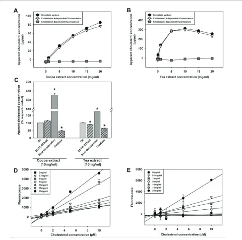
The presence of vegetable derived extracts in a duodenal micelle model gave rise to its apparent cholesterol con-tent using a commercial method based on a cholesterol-oxidase coupled reaction (Figure 1). This kit is also rec-ommended by the manufacturer for the use in food extracts. Different vegetable extracts (in concentration ranging 0 to 20 mg/ml) were analyzed for the potential interference in the cholesterol assay. Cocoa and tea extracts increased the apparent cholesterol concentration (figure 2A and 2B) in a concentration dependent fashion, even in the absence of cholesterol oxidase, the key enzyme in this system. Considering the enzyme-coupled reaction of this method (Figure 1), we analyzed (in com-parison to the complete system), i) the cholesterol inde-pendent fluorescence (system without cholesterol esterase and cholesterol oxidase), ii) the peroxidase activ-ity independent fluorescence and iii) the resorufin inde-pendent fluorescence. Fluorescence found in both cholesterol dependent and independent conditions sug-gested either a generation of vegetable extract derived H2O2 and/or an interference from peroxidase activity present in vegetable extracts.
Phytoesterol interferences were ruled out as ergosterol and other sterols (data not shown) did not offer fluores-cence in the complete system. Metal chelation (EDTA-DTPAC) did not inhibit significantly fluorescence in the complete system in the presence of vegetable extracts except in the case of tea extract (figure 2C). The potential contribution of vegetable derived peroxidases was ruled out by heat denaturation of vegetable extracts, even resulting in the increase of fluorescence (figure 2C).
Finally, the inhibitory activity of catalase [1.11.1.6] (lead-ing to fluorescence decreases to 50%) suggests that there is a vegetable extract dependent H2O2 production which may be derived by the previously described interaction of sample antioxidants with horseradish peroxidase [9]. Moreover, in the presence of vegetable extracts, the fluo-rescence in both cholesterol-dependent and independent reactions offered similar values suggesting the possibility of using the cholesterol independent fluorescence as a blank to account the vegetable extract derived interfer-ences (figures 2A and 2B).
To test this alternative solution, cholesterol standard curves in the presence of vegetable derived extracts were developed (figure 2D and 2E). The slope of the choles-terol concentration-fluorescence linear relationship was inversely related to the concentration of vegetable extract, in a given range (up to 10 mg/ml in cocoa extract). Therefore, it is advisable to develop a cholesterol standard curve with the chosen concentration of vegeta-ble extract in order to ascertain linearity, even in the pres-ence of the cholesterol independent fluorescpres-ence blank. It should be noted that in specific cases the interference of the vegetable extracts precluded the use of cholesterol standard curve even at a lower doses (from 1 mg/ml in tea).
Vegetable extracts induce an apparent decrease in cholesterol incorporation in bile micelles. Chromatographic approach for cholesterol analysis
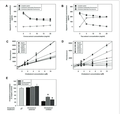
In order to further model duodenal conditions, the extracts were diluted with porcine bile as previously described [8]. The fluorescence was analyzed in choles-terol dependent and independent conditions. Surpris-ingly, the cholesterol independent fluorescence (i.e. vegetable extract derived H2O2) disappeared (cocoa extract) or decreased (tea extract) in this condition (fig-ure 3A and 3B). Nevertheless, a novel interference was detected, since apparent cholesterol concentration decreased. In order to further characterize this new inter-ference, a standard curve using different extract concen-tration was done. The results (figure 3C and 3D) show the same behavior of cholesterol standard curves as in the case of PBS, i.e., the slope of the cholesterol standard curve was reduced when the concentration of food extract was increased.
In order to further characterize those interferences in duodenal-like conditions, we used a chromatographic approach. Figure 3E shows that the cholesterol concen-tration does not change with the addition of cholesterol esterase, probably due to the virtual absence of choles-teryl esters in porcine bile. The addition of cholesterol oxidase in the mixture resulted in a strong decrease in the cholesterol detection because most of the cholesterol was oxidized (Figure 3E). However, the addition of vegetable
derived extracts caused an increase in cholesterol content in those conditions, suggesting an inhibitory activity of those extracts over cholesterol oxidase in the presence of bile.
To further elucidate whether vegetable extracts induced a decrease in the cholesterol in the micellar (bioavailable) phase, cocoa and tea extracts were diluted in bile, incu-bated, centrifuged and filtered to obtain the micellar
tion as previously described [8]. Next, the cholesterol content in this fraction was analyzed both by the enzyme-based assay and by the chromatographic method. The results show that, while porcine bile-vegetable interfer-ences impeded accurate quantification by the enzyme-based assay, chromatographic method allowed it (Figure 4). The results also show that while enzyme-based assay
showed marked differences in the potential for diminish-ing cholesterol absorption between both vegetable extracts, the use of chromatographic assays disclosed similar effects. With these results we concluded that although cholesterol-esterase and cholesterol-oxidase based assays are widely [10-12] used to measure choles-terol, it is necessary to develop artifact controls in order
to offer an accurate measurement in the presence of foodstuffs.
Discussion
There are several methods described to measure choles-terol content in foodstuff using chromatographic approaches [13,14]. However, when it is necessary to ana-lyze a high number of samples, an enzymatic method based on the cholesterol oxidase-peroxidase coupled reaction could be the best option [10-12] if we take into account some interferences reported here.
With the objective of modeling the capability of vegeta-ble extracts decreasing the bioavailavegeta-ble micellar choles-terol, we found different interferences in the measurement of cholesterol using an enzymatic method. In phosphate buffered saline, we evidenced a highly intense cholesterol independent fluorescence that we could attribute partially (50%) to an exogenous H2O2 pro-duction, as in those conditions it was diminished by cata-lase preincubation. The rest of the interference could be explained by a different mechanism recently described in our group [9], showing an interaction of vegetable antiox-idants with peroxidases. Horseradish peroxidase is often used as final step for enzymatic-coupled reactions. Briefly, in the absence of hydrogen peroxide but in aero-bic conditions, the antioxidant compound of the vegeta-ble extracts could reduce the ferric-horseradish peroxidase to ferrous-horseradish peroxidase and then interact with the O2 of the medium producing horserad-ish peroxidase-Compound III. This compound can undergo spontaneous decay to ferriperoxidase with the
generation of O2- which may interact with the antioxidant
producing an antioxidant radical. This antioxidant radical may then react with the Amplex Red® and horseradish
peroxidase and produce resorufin. Thus, all peroxidase based enzymatic methods may show interferences by those vegetable derived compounds. To solve these inter-ferences we propose two different and complementary methods: i) the use of cholesterol independent condition as a blank of the reaction to eliminate the fluorescence when the vegetable extracts are diluted in PBS and ii) the application of cholesterol standard curves including the working concentration of the extract.
For the studies of "in vitro" cholesterol absorption we used porcine bile in order to reproduce a physiologically relevant and a stable source of micellar cholesterol. We first analyze the interference of the extracts diluted in bile and we found a novel interference that decreases the apparent cholesterol concentration. In this case, the use of the cholesterol independent fluorescence is not as use-ful as in the case of buffered saline-based systems because in this latter case the interference is lower. However, the cholesterol and/or cholesterol ester standard curve addi-tion with the extracts could be a good method to reduce the interferences detecting cholesterol at low concentra-tions of vegetable extracts. After incubating the extracts in the presence or in the absence of the enzymes choles-terol esterase and cholescholes-terol oxidase we discover that vegetable extracts can interact directly and/or indirectly decrease the activity of cholesterol oxidase. We cannot exclude that other vegetable derived extracts could inhib-its cholesterol esterase. The mechanisms behind this inhibitory effect are outside the scope of this work, but they may comprise displacement of cholesterol oxidase outside of lipid bilayers needed for efficient catalytic turnover [15] and they may be part of the defensive prop-erties of polyphenols presents in vegetable extracts [16]. In any case, cholesterol oxidase is among the more fre-quently used enzymes and its interference should be accounted when extending the use from clinical chemis-try to food chemischemis-try [17].
Finally, the development of a chromatographic method in order to measure the cholesterol concentration in bio-logical models of food digestion is presented as a solution when the used concentration of foodstuff is high enough to interfere with the standard addition method. Both detection methods were used to test the effect of these extracts in lowering the concentration of potentially bio-available cholesterol. Moreover, it is described that sterol, polyphenols and other nutrients can block the entry of most cholesterol into micelles [8,18-20]. Although there were evidenced a blocking effect of cholesterol entry into micellar phases by both extracts using both methods the magnitude of the change differed. Using the enzymatic method we demostrated that the cholesterol independent
fluorescence in the case of tea extract was very high, quite similar to the fluorescence of complete system. This high fluorescence resulted in a small detection of real choles-terol dependent fluorescence. In the case of cocoa extract the decrease in apparent bioavailability is about 20%. Analyzing the cholesterol concentration using a chro-matographic method both extracts at tested concentra-tions decreased micellar cholesterol about a 50%.
Conclusions
The use of cholesterol-oxidase and/or peroxidase based systems for cholesterol analyses in foodstuffs should be accurately monitored, as important interferences in all the components of the enzymatic chain were evident. The use of adequate controls, standard addition and finally, chromatographic analyses should solve these issues.
Methods
Chemicals
Ethylenediaminetetraacetic acid (EDTA), diethylenetri-aminepentaacetic acid (DTPAC), ergosterol, catalase, chloroform and cholesterol were from Sigma (Sigma-Aldrich, Saint Louis, MO, USA). Acetonitrile, 2-propa-nol, ammonium acetate and formic acid were from Baker (Mallinckrodt Baker, Phillipsburg, NJ, USA). Millex GP filters 0,22 μm and Ultrafree-MC filtres 30,000 from Mil-lipore (MilMil-lipore, Billerica, MA, USA), methanol from Carlo Erba (Carlo Erba, Milano, Italy) and [25,26,26,26,26,27,27,27-2H
7]cholesterol (cholesterol-D7)
from Avanti Polar Lipids (Avanti Polar Lipids Inc, Alabas-ter, AL, USA).
Bile from porcine biliary vesicle was collected from a local abattoir and placed on ice. Immediately after collec-tion, bile was centrifuged at 2000 g at 4°C for 10 min to remove debris. The cholesterol content of the bile was measured (see below) and then the bile was diluted with phosphate buffered saline to obtain a 0.5 mM of choles-terol concentration, as described by Kirana et al [8]. It was aliquoted and stored at -80°C.
Green tea and cocoa vegetable extracts were provided by La Morella Nuts SA (La Morella Nuts, Reus, Spain).
Cholesterol measurement using an enzymatic method The concentration of cholesterol in porcine bile, food extracts and mixes was analyzed by Amplex Red Choles-terol Assay Kit (A12216) based on an enzymatic reaction depicted in figure 1. Briefly, reactions took place in a 96 well plate by the addition of 50 μL of Amplex Red ing solution with 50 μL of assay sample. Five mL of work-ing solution, prepared prior the analysis, contained 75 μL of a 300 μM of Amplex Red reagent and 2 U/mL of HRP. The working solution volume was adjusted to 5 mL with reaction buffer, which contained 25 mM potassium
phos-phate, pH 7.4, 12.5 mM NaCl, 1.25 mM cholic acid and 0.025% Triton X-100. The reactions were incubated for 30 min at 37°C, protected from light. After incubation, fluorescence was measured in a fluorescence microplate reader (Tecan Infinite M200, Männedorf, Switzerland) using excitation wavelength at 560 nm and emission detection at 590 nm.
The samples were suspended with either phosphate buffered saline or porcine bile to obtain the initial desired concentration of nutrient (from 0 to 20 mg/ml). After a dilution in PBS (1:25) cholesterol amount was quantified by fluorescence according the kit instructions.
To model the cholesterol entry into micellar phases and its potential inhibition by vegetable extracts, the method of Kirana et al was used [8]. Briefly, samples suspended in pig's bile at 10 mg/ml were incubated at 37°C for 1 h with continuous shaking at 160 r.p.m. (Unitron, Infors HT, Headquarter, Switzerland). The solution was then centri-fuged at 1000 × g for 10 min, filtered through a 0.22 μm Millex GP and diluted 25 fold. The concentration of cho-lesterol was analyzed by Amplex Red Cholesterol Assay Kit (A12216).
Cholesterol analysis by liquid chromatography coupled to mass-spectrometry
To unequivocally quantify cholesterol in food extract-bile mixtures, cholesterol was extracted using chloro-form:methanol (2:1) as described previously [21,22]. Pre-vious to extraction cholesterol-D7 was added, as an internal standard, to a final concentration of 125 μg/mL. The mixture was vortexed and centrifuged at 4400 × g at room temperature for 15 min. The organic phase was conserved and the procedure was repeated. Combined organic phases were dried in a SpeedVac (Thermo Fisher Scientific, Madrid, Spain) and dissolved with methanol. The samples were filtered in an UltraFree 5 kDa filter (Millipore, Billerica, MA, USA) before liquid chromatog-raphy analysis.
capil-lary voltage was 3500 V with a scan rate of 1 scan/s. N2 was used as a gas nebulizer (Flow: 5 L/min and T = 350°C). The Masshunter Software was used for integra-tion and extracintegra-tion of peak intensities of the cholesterol and cholesterol-D7. The m/z values used for quantifica-tion were: m/z 369.35 [M+H-H2O]+ for cholesterol and
m/z 376.3955 [M+H-H2O]+ for cholesterol-D7.
Interas-say and intraasInteras-say variation < 5%, L.O.Q was 20 pmol, RSQ = 0.98.
Competing interests
The authors declare that they have no competing interests.
Authors' contributions
MJ and JCES performed analytical determinations and designed part of the research. MJB and RP performed statistical analyses. NA, JR and JRM obtained the extract of interest. MJ, NA and MPO conceived the study and participated in its design and coordination and helped to draft the manuscript. All authors read and approved the final manuscript.
Acknowledgements
We thank the support of Saray Arenas-Soria, Laura Arcal, David Argilès and Mer-itxell Martín for excellent technical assistance. Supported by I+D grants from the Spanish Ministry of Education and Science (BFU2009-11879/BFI; AGL2006-12433), the Generalitat of Catalunya (2009SGR-735) and the Spanish Ministry of Health (FIS 08-1843, RD06/0013/0012). Supported by the CENIT program from the Spanish Ministry of Industry and by a consortium of companies led by La Morella Nuts (Reus, Catalonia, Spain) with the following companies: KRAFT, BTSA (Biotecnologías Aplicadas), Selecció Batallé, Industrial Técnica Pecuaria, Neuron BioPharma, Shirota Functional Foods, Grupo Leche Pascual, Innaves. Supported also by the COST B-35 Action.
Author Details
1Institut de Recerca Biomèdica de Lleida-Universitat de Lleida-Parc Científic i Agroalimentari Tecnològic de Lleida (IRBLLEIDA-UdL-PCiTAL). c/Montserrat Roig, 25008 Lleida, Spain and 2La Morella Nuts, SA. Apel.les Mestres, S/N 43006 Reus, Spain
References
1. Poli G, Sottero B, Gargiulo S, Leonarduzzi G: Cholesterol oxidation products in the vascular remodeling due to atherosclerosis. Mol Aspects Med 2009, 30(3):180-189.
2. Temel RE, Rudel LL: Diet effects on atherosclerosis in mice. Curr Drug Targets 2007, 8(11):1150-1160.
3. Brown MS, Goldstein JL: Cholesterol feedback: from Schoenheimer's bottle to Scap's MELADL. J Lipid Res 2009, 50(Suppl):S15-27. 4. Lu K, Lee MH, Patel SB: Dietary cholesterol absorption; more than just
bile. Trends Endocrinol Metab 2001, 12(7):314-320.
5. Nagarajan S, Burris RL, Stewart BW, Wilkerson JE, Badger TM: Dietary soy protein isolate ameliorates atherosclerotic lesions in apolipoprotein E-deficient mice potentially by inhibiting monocyte chemoattractant protein-1 expression. J Nutr 2008, 138(2):332-337.
6. Sutra T, Decorde K, Riss J, Dallas C, Cristol JP, Rouanet JM: A commercial extract of fruits and vegetables, Oxxynea, acts as a powerful antiatherosclerotic supplement in an animal model by reducing cholesterolemia, oxidative stress, and NADPH oxidase expression. J Agric Food Chem 2007, 55(10):4258-4263.
7. Ostlund RE Jr: Phytosterols in human nutrition. Annu Rev Nutr 2002,
22:533-549.
8. Kirana C, Rogers PF, Bennett LE, Abeywardena MY, Patten GS: Naturally derived micelles for rapid in vitro screening of potential cholesterol-lowering bioactives. J Agric Food Chem 2005, 53(11):4623-4627. 9. Serrano J, Jove M, Boada J, Bellmunt MJ, Pamplona R, Portero-Otin M:
Dietary antioxidants interfere with Amplex Red-coupled-fluorescence assays. Biochem Biophys Res Commun 2009, 388(2):443-449.
10. Amundson DM, Zhou M: Fluorometric method for the enzymatic determination of cholesterol. J Biochem Biophys Methods 1999,
38(1):43-52.
11. Mizoguchi T, Edano T, Koshi T: A method of direct measurement for the enzymatic determination of cholesteryl esters. J Lipid Res 2004,
45(2):396-401.
12. Situmorang M, Alexander PW, Hibbert DB: Flow injection potentiometry for enzymatic assay of cholesterol with a tungsten electrode sensor. Talanta 1999, 49(3):639-649.
13. Lin YT, Wu SS, Wu HL: Highly sensitive analysis of cholesterol and sitosterol in foods and human biosamples by liquid chromatography with fluorescence detection. J Chromatogr A 2007, 1156(1-2):280-287. 14. Daneshfar A, Khezeli T, Lotfi HJ: Determination of cholesterol in food
samples using dispersive liquid-liquid microextraction followed by HPLC-UV. J Chromatogr B Analyt Technol Biomed Life Sci 2009,
877(4):456-460.
15. Kreit J, Sampson NS: Cholesterol oxidase: physiological functions. FEBS J 2009, 276(23):6844-6856.
16. Si W, Gong J, Tsao R, Kalab M, Yang R, Yin Y: Bioassay-guided purification and identification of antimicrobial components in Chinese green tea extract. J Chromatogr A 2006, 1125(2):204-210.
17. Pollegioni L, Piubelli L, Molla G: Cholesterol oxidase: biotechnological applications. FEBS J 2009, 276(23):6857-6870.
18. Yasuda A, Natsume M, Sasaki K, Baba S, Nakamura Y, Kanegae M, Nagaoka S: Cacao procyanidins reduce plasma cholesterol and increase fecal steroid excretion in rats fed a high-cholesterol diet. Biofactors 2008,
33(3):211-223.
19. Shibata S, Oda K, Onodera-Masuoka N, Matsubara S, Kikuchi-Hayakawa H, Ishikawa F, Iwabuchi A, Sansawa H: Hypocholesterolemic effect of indigestible fraction of Chlorella regularis in cholesterol-fed rats. J Nutr Sci Vitaminol (Tokyo) 2001, 47(6):373-377.
20. Osada K, Suzuki T, Kawakami Y, Senda M, Kasai A, Sami M, Ohta Y, Kanda T, Ikeda M: Dose-dependent hypocholesterolemic actions of dietary apple polyphenol in rats fed cholesterol. Lipids 2006, 41(2):133-139. 21. Ostrow JD, Murphy NH: Isolation and properties of conjugated bilirubin
from bile. Biochem J 1970, 120(2):311-327.
22. Stark M, Jornvall H, Johansson J: Isolation and characterization of hydrophobic polypeptides in human bile. Eur J Biochem 1999,
266(1):209-214.
23. Laaksonen R, Katajamaa M, Paiva H, Sysi-Aho M, Saarinen L, Junni P, Lutjohann D, Smet J, Van Coster R, Seppanen-Laakso T, Lehtimaki T, Soini J, Oresic M: A systems biology strategy reveals biological pathways and plasma biomarker candidates for potentially toxic statin-induced changes in muscle. PLoS One 2006, 1:e97.
doi: 10.1186/1476-511X-9-65
Cite this article as: Jové et al., When cholesterol is not cholesterol: a note on the enzymatic determination of its concentration in model systems contain-ing vegetable extracts Lipids in Health and Disease 2010, 9:65
Received: 10 May 2010 Accepted: 21 June 2010 Published: 21 June 2010
This article is available from: http://www.lipidworld.com/content/9/1/65 © 2010 Jové et al; licensee BioMed Central Ltd.