R E S E A R C H
Open Access
Induction of virus-specific effector immune
cell response limits virus replication and
severe disease in mice infected with
non-lethal West Nile virus Eg101 strain
Mukesh Kumar
1,2, Kelsey Roe
1,2, Maile O
’
Connell
1,2and Vivek R. Nerurkar
1,2*Abstract
Background:West Nile virus (WNV) is a neurotropic flavivirus that has emerged globally as a significant cause of viral encephalitis in humans. Herein, we investigated the immunological responses induced by two phylogenetically related WNV strains of lineage 1, WNV NY99, and WNV Eg101.
Methods:Eight-week-old C57BL/6J mice were inoculated with WNV NY99 or WNV Eg101 and mortality, virus burden in the periphery and brain, type 1 interferon response, WNV-specific antibodies, leukocyte infiltration, and inflammatory responses were analyzed.
Results:As expected, WNV NY99 infected mice demonstrated high morbidity and mortality, whereas no morbidity and mortality was observed in WNV Eg101 infected mice. Virus titers were comparable in the serum of both WNV NY99 and WNV Eg101 infected mice at day 3 after inoculation; however, at day 6, the virus was cleared from WNV Eg101 infected mice but the virus titer remained high in the WNV NY99 infected mice. Virus was detected in the brains of both WNV NY99 and Eg101 infected mice, albeit significantly higher in the brains of WNV NY99 infected mice. Surprisingly, levels of type 1 interferon and WNV-specific antibodies were significantly higher in the serum and brains of WNV NY99 infected mice. Similarly, protein levels of multiple cytokines and chemokines were significantly higher in the serum and brains of WNV NY99 infected mice. In contrast, we observed significantly higher numbers of innate and adaptive immune cells in the spleens and brains of WNV Eg101 infected mice. Moreover, total number and
percentage of IFN-γand TNF-αproducing WNV-specific CD8+T cells were also significantly high in WNV Eg101 infected
mice.
Conclusions:Our data demonstrate that induction of virus-specific effector immune cell response limits virus
replication and severe WNV disease in Eg101 infected mice. Our data also demonstrate an inverse correlation between leukocyte accumulation and production of pro-inflammatory mediators in WNV-infected mice. Moreover, increased production of pro-inflammatory mediators was associated with high-virus titers and increased mortality in WNV NY99 infected mice.
Keywords:West Nile virus encephalitis, Neuroinflammation, Immune cell migration
* Correspondence:[email protected]
1
Department of Tropical Medicine, Medical Microbiology and Pharmacology, John A. Burns School of Medicine, University of Hawaii at Manoa651 Ilalo Street, Honolulu 96813Hawaii, USA
2Pacific Center for Emerging Infectious Diseases Research, John A. Burns
School of Medicine, University of Hawaii at Manoa, 651 Ilalo Street, Honolulu, Hawaii 96813, USA
Background
West Nile virus (WNV), a neurotropic flavivirus, has emerged as a significant cause of viral encephalitis in the USA [1]. WNV infection in humans is usually asymptom-atic or self-limiting, with a mild febrile illness, but may progress to meningitis, encephalitis, paralysis, and death. Until 1999, WNV was geographically distributed in Africa, the Middle East, western and central Asia, India, and Europe, where it caused sporadic cases of febrile dis-ease and occasional outbreaks of encephalitis in elderly people and in equines [2, 3]. The unexpected emer-gence of WNV in the USA in 1999 was associated with the introduction of the NY99 strain, which is more viru-lent, and resulted in higher incidence of meningoenceph-alitis in humans as compared to the non-virulent strains such as the WNV Eg101 strain [4, 5]. Recent outbreaks of highly virulent WNV strains have also been reported in the Mediterranean basin, southern Europe, and Russia [6, 7]. Although the worldwide incidence of WNV in-fection is increasing, there is no specific treatment or vaccine available for use in humans.
Studies using the well-characterized WNV encephalitis (WNVE) mouse models show that an intact innate and adaptive immune response is required to limit WNV in-fection. Antiviral type I interferon production is essential in suppressing viral titers in the brain and peripheral organs [8]. The induction of WNV-specific immuno-globulins is essential for suppressing viremia and virus dissemination [9]. In the central nervous system (CNS), neurons are the primary target of WNV replication, and virus-associated pathology is characterized by neuronal death, activation of glial cells, and massive in-filtration of leukocytes in the perivascular space and parenchyma. The migration of leukocytes into the brain is essential for controlling WNV infection in the brain [10–12]. WNV also induces a dramatic increase in sev-eral pro-inflammatory cytokines such as tumor necrosis factor alpha (TNF-α) and interleukin (IL)-1β and che-mokines such as CCl2 and CXCL10, which regulate leukocyte trafficking into the brain and neuronal death after infection [11, 13, 14]. The global increase of WNV neuroinvasive disease warrants a greater understanding of the molecular mechanisms associated with virus clearance and neuroinflammation.
The WNV strain Eg101 was isolated from human blood in Egypt in 1950 [15]. NY99 and Eg101 strains of WNV are 95.4 and 99.6 % identical at the nucleotide and amino acid levels, respectively. Both the WNV NY99 and WNV Eg101 strains are classified in same genotypic lineage and belong to same clade 1a of the lineage 1 [5, 16]. Lineage 1 strains are considered emer-ging and associated with outbreaks of neuroinvasive dis-ease [5]. While the WNV NY99 strain is highly pathogenic and implicated in large-scale mortality, the
WNV Eg101 strain is largely non-pathogenic. We have previously demonstrated that mice immunized with both 1000 and 100 plaque-forming units (PFU) of WNV Eg101 demonstrated 100 % protection against both sub-cutaneous and intracranial challenge with a lethal dose (1000 PFU) of the WNV NY99 strain [17]. However, the underlying mechanisms associated with differential patho-genesis of lethal, WNV NY99, and non-lethal, WNV Eg101, strains are largely unknown. We hypothesized that differential host immune response is the critical determin-ant of distinct disease outcome associated with the WNV NY99 and WNV Eg101 strains. In the present study, we examined the immunological changes in the serum and brain of mice during WNV NY99 strain and WNV Eg101 strain infection to understand the underlying immuno-pathological mechanisms leading to severe WNV disease. While both strains were neuroinvasive in adult mice, our results revealed phenomenal differences in the clinical dis-ease, induction of type 1 interferon and WNV-specific antibodies, innate and adaptive immune cell response, and inflammatory response in the periphery and in the brain.
Methods
Ethics statement
This study was specifically approved by the University of Hawaii Institutional Animal Care and Use Committee (IACUC) (protocol number 13–1759), and conducted in strict accordance with guidelines established by the National Institutes of Health and the University of Hawaii IACUC. All animal experiments were con-ducted in consultation with veterinary and animal care staff at the University of Hawaii in animal bio-safety level-3 laboratory, and mice that exhibited se-vere disease were euthanized to limit suffering. The animal suite was maintained at 72 °F, 45 % humidity, and on 12/12-light/dark cycle. Sawdust bedding was provided along with paper towels.
WNV infection experiments
symptoms appear at day 8 after inoculation. Mortality is ob-served between days 8 and 12 after inoculation [19, 20].
Brains were harvested and flash frozen in 2-methylbutane and stored at−80 °C until further processing. Frozen brain tissues were weighed and homogenized in a bullet blender using glass or zirconium oxide beads. WNV titers in the serum and brain homogenates were measured by plaque assay using Vero cells [20, 22]. Alternatively, mice were per-fused with PBS followed by 4 % paraformaldehyde (PFA), and brains were harvested, cryoprotected in 30 % sucrose, and embedded in the optimum cutting temperature (OCT) media [23]. In separate experiments, brains and spleens were harvested at day 8 after inoculation for the isolation of leukocytes and were processed for flow cytometry [23].
Measurement of WNV-specific antibodies
The titers, defined as median fluorescent intensity, of WNV-specific IgM and IgG antibodies were measured in the serum of WNV NY99 and WNV Eg101 infected mice using microsphere immunoassay (MIA) for WNV envelope E protein [17, 24].
Plaque reduction neutralization test (PRNT)
The titers of anti-WNV neutralizing antibodies were mea-sured in the serum of WNV NY99 and WNV Eg101 in-fected mice using PRNT assay as described previously [17]. Serum collected from WNV NY99 and WNV Eg101 in-fected mice were serially diluted from 1:40 to 1:5000, and PRNT was conducted against WNV NY99 and WNV Eg101 viruses, respectively. The highest dilution of serum resulting in 80 % reduction in the number of plaques com-pared to the growth of the virus control was determined as described previously [17, 24, 25].
Measurement of cytokines and chemokines
The levels of cytokines and chemokines were measured in the serum and brain homogenates by multiplex im-munoassay using MILLIPLEX MAP Mouse Cytokine/ Chemokine kit (Millipore) [20, 23].
ELISA
The levels of IFN-αand IFN-βwere measured in the serum and brain homogenates by ELISA, using the VeriKine™ Mouse Interferon-α ELISA Kit and VeriKine™ Mouse Interferon-βELISA Kit (PBL Interferon Source) [20].
Immunohistochemistry
Sagittal sections of 10-μm thicknesses were cut from the hemi-brain tissues frozen in OCT, and whole sagittal tis-sue sections were stained with hematoxylin and eosin (H&E) [23, 26].
Analysis of splenic leukocytes
Splenocytes were harvested from WNV NY99 and WNV Eg101 infected mice on day 8 after inoculation. Cells were counted and washed with 1X fluorescence-activated cell sorter buffer. Cells were stained for CD45, CD3, NK1.1, CD11b, and Ly6c by directly conjugated antibodies (eBiosciences) for 30 min at 4 °C and then fixed with 2 % PFA. Samples were analyzed by multi-color flow cytometry using FACSAria, and data were an-alyzed using FlowJo software [19, 23]. Briefly, after gat-ing live cells by forward and side scatter (FSC and SSC) parameters, CD45+leukocytes were selected. CD45+ leu-kocytes were further divided into CD3+, CD11b+, and NK1.1+ cells. CD11b+ cells were further delineated into subsets of CD11b+ Ly6C+ cells. CD3+ T cells were fur-ther divided into the CD4+ and CD8+subsets. The total numbers of CD4+ T cells, CD8+ T cells, NK1.1+ cells, and CD11b+ Ly6C+ cells from each spleen were deter-mined by multiplying the percentage of these cells by the total number of CD45+ leukocytes harvested as de-scribed previously [23].
In separate experiments, 106splenocytes were stimu-lated with 2 μg/ml of an immunodominant WNV-specific NS4B peptide (SSVWNATTAI) (21st Century Biochemical) for 6 h at 37 °C in the presence of Golgi-Plug (BD Biosciences). Splenocytes were stained with anti-CD8 antibody for 30 min at 4 °C followed by fix-ation and permeabilizfix-ation (BD Biosciences) and stained with anti-IFNγand anti-TNF-αantibodies (eBiosciences) for 30 min at 4 °C. Samples were analyzed by multi-color flow cytometry using FACSAria, and the percent-ages of CD8+T cells that expressed IFNγor TNF-αwere determined using FlowJo software [23].
Flow cytometric analysis of infiltrated immune cells in the brain
Statistical analysis
Log-rank (Mantel-Cox) Test and Gehan-Breslow-Wilcoxon Test were used to analyze survival data. Mann–Whitney test and unpaired Student t test using GraphPad Prism 5.0 was used to calculate p values of difference between viral titers and immune responses, respectively. Differences of p< 0.05 were considered significant.
Results
Morbidity and mortality in mice following WNV NY99 and WNV Eg101 infection
Eight-week-old C57BL/6J mice were inoculated subcuta-neously with 100 PFU of WNV Eg101 or WNV NY99 or PBS (mock) and then monitored daily for clinical signs and mortality. PBS inoculated mice remained healthy throughout the observation period of 21 days. WNV NY99 inoculated mice began to demonstrate clinical signs at day 8 after inoculation and exhibited high (65 %) mortality. In comparison, Eg101 inoculated mice rarely displayed clinical signs of disease and no mortality was observed in these mice (Fig. 1a, b). The difference in mortality between WNV NY99 and WNV Eg101 in-fected mice was statistically significant. All surviving ani-mals were positive for anti-WNV IgG antibodies, confirming virus infection (data not shown).
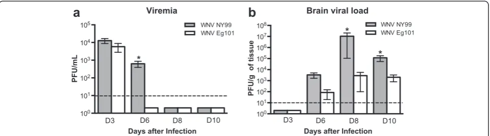
Viral load in the serum and brains of WNV NY99 and WNV Eg101 infected mice
We first examined the WNV titers in the serum and brains of WNV NY99 and WNV Eg101 infected mice by plaque assay. High, but similar virus titers were detected by plaque assay in the serum of both WNV NY99 and WNV Eg101 infected mice at day 3 after inoculation (Fig. 2a). At day 6 after inoculation, virus was cleared from the periphery of all the WNV Eg101 infected mice; however, it remained significantly high in WNV NY99
infected mice. The virus was cleared from the periphery of all WNV NY99 infected mice by day 8 after inocula-tion. WNV was not detected in the brains of both WNV NY99 and WNV Eg101 infected mice at day 3 after in-oculation, which is consistent with the previous studies [20]. WNV was first detected in the brains at day 6 after inoculation and peaked at day 8 after inoculation (Fig. 2b). WNV titers in the brains of WNV NY99 fected mice was slightly higher than WNV Eg101 in-fected mice at day 6 after inoculation, but the difference was not statistically significant. However, virus titers were significantly higher in the brains of WNV NY99 in-fected mice when compared to WNV Eg101 inin-fected mice at both days 8 and 10 after inoculation.
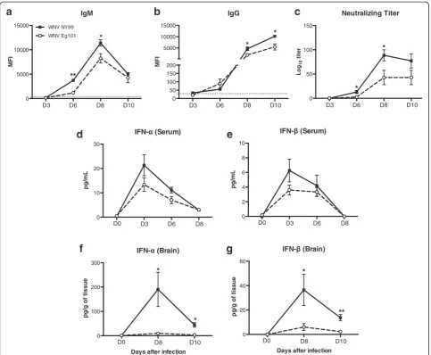
WNV-specific antibodies and type 1 interferon responses in mice following WNV NY99 and WNV Eg101 infection
The titers of WNV-specific IgM and IgG antibodies in the serum of WNV NY99 and WNV Eg101 infected mice were measured using MIA. Infection with both WNV NY99 and WNV Eg101 elicited robust WNV-specific antibody responses. Anti-WNV IgM first ap-peared at day 6 peaked at day 8 and then decreased at day 10 after inoculation (Fig. 3a). Similarly, anti-WNV IgG first appeared at day 6 and gradually increased up to day 10 after inoculation (Fig. 3b). However, titers of both IgM (days 6 and 8) and IgG (days 8 and 10) antibodies were significantly higher in WNV NY99 infected mice than WNV Eg101 infected mice. Similarly, titers of neu-tralizing antibodies were also significantly higher in WNV NY99 infected mice than WNV Eg101 infected mice (Fig. 3c).
We measured the levels of IFN-α and IFN-β in the serum of WNV NY99 and WNV Eg101 infected mice using ELISA. High levels of IFN-α and IFN-β were de-tected in the serum of both WNV NY99 and WNV Eg101 infected mice at day 3 after inoculation, which
0 1 2 3 4
Days after infection
Cl
in
ic
al
S
c
o
re
D8 D9 D10 D11 D12 D13
** **
**
** *
0 5 10 15 20 25
0 20 40 60 80 100
Days after infection
P
e
rcen
t
s
u
rvi
val
WNV Eg101 WNV NY99
**
decreased gradually at days 6 and 8 after inoculation. However, as demonstrated in Fig. 3d, e, we observed a trend of slightly reduced levels of IFN-α and IFN-β in the serum of WNV Eg101 infected mice at days 3 and 6 after inoculation. We also examined IFN-α and IFN-β levels in the brains at days 8 and 10 after inoculation. In the brain, as compared to WNV Eg101 which did not elicit a strong interferon response, WNV NY99 infected mice developed a robust interferon response at day 8 after inoculation. This response further decreased by day 10 after inoculation (Fig. 3f, g).
Peripheral cytokine and chemokine profiles in mice after WNV NY99 and WNV Eg101 infection
It has been demonstrated that WNV-induced expression of chemokines such as CCL2, CCL3, CCL4, CCL5, and CXCL10 are associated with enhanced trafficking of leu-kocytes into the brain [11, 27–29, 30–32]. WNV-induced expression of pro-inflammatory cytokines such as IL-1β and TNF modulate BBB permeability, facilitate leukocyte trafficking into the brain, activate glial cells, and mediate neuronal death after infection [13, 14]. Therefore, we next assessed the protein levels of these cytokines and chemokines in the serum of WNV NY99 and WNV Eg101 infected mice using multiplex immunoassay. Very low levels of these cytokines and chemokines were de-tected in the serum of uninfected mice (day 0). WNV NY99 infection resulted in a dramatic increase in protein levels of multiple cytokines and chemokines at day 3 after inoculation, which further increased slightly at day 6 after inoculation (Fig. 4). We also observed a slight in-crease in the levels of these cytokines and chemokines in WNV Eg101 infected mice at day 3 after inoculation. However, unlike WNV NY99 infected mice, there was no increase in their levels at day 6 after inoculation. Levels of interleukin (IL)-1β, −10, −13, and −15 were
significantly higher in WNV NY99 infected mice than WNV Eg101 infected mice at day 6 after inoculation. Similarly, TNF-α and GM-CSF levels in the serum of WNV NY99 infected mice were significantly higher when compared to WNV Eg101 infected mice at day 6 after inoculation. A similar pattern was also observed for chemokine expression. CCL3 level was signifi-cantly higher in WNV NY99 infected mice at day 6 after inoculation, whereas the level of CXCL10 was significantly higher in WNV NY99 infected mice at both days 3 and 6 after inoculation. The level of CXCL1 was only increased significantly at day 3 after inoculation when compared to WNV Eg101 mice. However, the comparison of levels of serum cytokines and chemokines, such as IL-6, CCL2, and CCL5 be-tween both groups demonstrated no significant differ-ences in their levels at any time point after inoculation (Fig. 4).
Cytokine and chemokine profiles in the brains of WNV NY99 and WNV Eg101 infected mice
We further examined the protein levels of multiple in-flammatory cytokines and chemokines in the mice brain homogenates following WNV NY99 and WNV Eg101 infection. Very low levels of these cytokines and chemo-kines were detected in the brains of uninfected mice (day 0). As expected, levels of multiple cytokines and chemokines increased dramatically in WNV NY99 fected mice brains at day 8 after inoculation, but this in-crease was not observed in the brains of WNV Eg101 infected mice (Fig. 5). Levels of these cytokines and che-mokines decreased slightly in the brains of WNV NY99 infected mice at day 10 after inoculation. In contrast, there was a slight increase in the levels of these inflamma-tory mediators in the brains of WNV Eg101 infected mice at day 10 after inoculation. The levels of IL-1α, IL-5, IL-7,
100
101
102
103
104
105
Days after Infection
PFU/mL
D3 D6
WNV Eg101 WNV NY99
*
D8 D10 10
0
101
102
103
104
105
106
107
108
Days after Infection
PFU/g of tissue
D6 D10
WNV NY99 WNV Eg101
*
*
D8 D3
IFNγ, GM-CSF, CCL5, CXCL2, and CXCL9 were signifi-cantly higher in the brains of WNV NY99 infected mice than WNV Eg101 infected mice at day 8 after inoculation. Similarly, the levels of IL-13, IL-17, G-CSF, MCSF, CCL2, and CCL11 were significantly higher in WNV NY99 in-fected mice at both days 8 and 10 after inoculation when compared to WNV Eg101 mice. However, the levels of IL-6, TNF-α, CCL4, CXCL1, and CXCL5 were increased sig-nificantly only at day 10 after inoculation when compared to WNV Eg101 mice. Whereas, levels of IL-10, IL-12, LIF,
and CCL3 did not differ between WNV NY99 and WNV Eg101 infected mice at any time point after inoculation (Fig. 5).
Leukocyte accumulation in the spleens of WNV NY99 and WNV Eg101 infected mice
Splenocytes were isolated from mock, WNV NY99 and WNV Eg101 infected mice at day 8 after inoculation and were analyzed by flow cytometry (Fig. 6). Cells were stained for CD45, CD3, CD4, CD8, NK1.1, CD11b, and IFN- (Brain)
0 100 200 300
pg/g of tissue
Days after infection
D0 D8 D10
*
*
IFN- (Brain)
0 20 40 60
pg/g of tissue
Days after infection
D0 D8 D10
*
** IFN- (Serum)
0 10 20 30
pg/mL
D0 D3 D6 D8
0 50 100 150 200 5000 10000 15000
MFI
D3 D6 D8 D10
* *
0 5000 10000 15000
MFI
WNV NY99 WNV Eg101
D3 D6 D8 D10
*
**
IFN- (Serum)
0 2 4 6 8 10
pg/mL
D0 D3 D6 D8
d
f
e
g
0 50 100 150
Log
10
titer
D3 D6 D8 D10
* *
Fig. 3WNV-specific IgM and IgG titers, neutralizing antibodies titers, and levels of IFN-αand IFN-βin mice following WNV NY99 and WNV Eg101 infection. Serum was collected from WNV NY99 and WNV Eg101 infected mice at indicated time points and WNV-specific IgM (a) and IgG (b) responses were measured by MIA using WNV E antigen. Results are reported as median fluorescent intensity (MFI) per 100 microspheres. Data are expressed as MFI ± SEM and is representative of two independent experiments (n= 8–16 mice per group). *p< 0.05, **p< 0.001.Horizontal dotted lineindicates the cutoff value.cNeutralizing antibodies titers in the serum of WNV NY99 and WNV Eg101 infected mice. Serum collected from WNV NY99 and WNV Eg101 infected mice at indicated time points was serially diluted from 1:40 to 1:5000 and PRNT was conducted against WNV NY99 and WNV Eg101 viruses, respectively. Data are expressed as mean log10titer ± SEM and is representative of two independent experiments (n= 8–16 mice per group). IFN-αand IFN-βproduction was measured in the serum (d) and (e) collected from WNV NY99 and WNV Eg101 infected mice at indicated time points using mouse IFN-αand mouse IFN-βELISA kits and from brains (f) and (g) harvested from WNV NY99 and WNV Eg101 infected mice at days 6 and 8 after inoculation. The data are expressed as pg/mL of serum or pg/g of brain tissue.Error barsrepresent SEM (n= 6–8 mice per group).
Ly6C markers to analyze both T cells and non-T cell subpopulations. Interestingly, WNV Eg101 infected mice spleens had significantly higher numbers of total leuko-cytes (CD45+ cells), CD4+ T cells (CD45+ CD3+ CD4+), CD8+ T cells (CD45+ CD3+ CD8+), NK cells (CD45+ CD3− NK1.1+), and monocytes/macrophages (CD45+ CD11b+ Ly6c+) than WNV NY99 infected mice spleen (Fig. 6a). Additionally, the percentage of NK cells and monocytes/macrophages in the spleens of WNV Eg101 infected mice were also higher than in WNV NY99 in-fected mice (Fig. 6b). No difference was observed in the percentage of T cells in the spleens of WNV Eg101 and NY99 infected mice.
WNV-specific T cell responses in WNV NY99 and WNV Eg101 infected mice
Splenocytes from mock, WNV NY99, and WNV Eg101 infected mice were harvested at day 8 after inoculation and re-stimulated ex vivo with an immunodominant WNV NS4B peptide, and CD8+T cells were analyzed for the production of intracellular IFNγand TNF-αby flow cytometry. As depicted in Fig. 7a, b, total number and percentage of CD8+IFNγ+cells (3.50 versus 1.65 %) and CD8+ IFNγ+ TNF-α+ cells (2.82 versus 1.45 %) were
significantly higher in WNV Eg101 infected mice than in WNV NY99 infected mice, respectively. We observed no significant difference in the percentage of CD8+ TNF-α+
cells from WNV NY99 and WNV Eg101 in-fected mice after incubation with WNV-specific peptide.
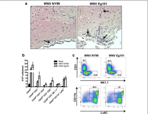
Leukocyte infiltration in the brain after WNV NY99 and WNV Eg101 infection
We initially examined the mice brain tissues for histo-pathological changes following WNV NY99 and WNV Eg101 infection. As expected, H&E staining of brain sec-tions from the WNV NY99 infected mice demonstrated leukocyte infiltration along the meninges at day 8 after inoculation (Fig. 8a). Interestingly, we observed higher numbers of leukocytes in the meninges and parenchyma of WNV Eg101 infected mice. To further verify these re-sults, we isolated leukocytes from the brains of mock, WNV NY99, and WNV Eg101 infected mice at day 8 after inoculation and analyzed by flow cytometry. Con-sistent with our immunohistochemistry results, we ob-served higher numbers of total leukocytes (CD45hicells) in the brains of WNV Eg101 infected mice (Fig. 8b), which was statistically not significant. However, the total number of CD4+ T cells (CD45hiCD3+CD4+) and CD8+ IL-6
0 20 40 60
pg/mL
D0 D3 D6
IL-1
0 10 20 30 40
pg/mL
WNV NY99
WNV Eg101 *
D3 D6
D0
CCL5
0 50 100 150
Days after infection
pg/mL
D3 D6
D0
CXCL10
0 500 1000 1500
Days after infection
pg/mL
D3 D6
* *
D0
CCL2
0 50 100 150 200
pg/mL
D3 D6
D0
TNF-0 5 10 15
pg/mL
D3 D6
*
D0
IL-10
0 50 100 150
pg/mL
D3 D6
*
D0
IL-13
0 50 100 150
pg/mL
D3 D6
*
D0
IL-15
0 50 100 150 200
pg/mL
D3 D6
*
D0
CCL3
0 20 40 60 80 100
Days after infection
pg/mL
D3 D6
**
D0
CXCL1
0 50 100 150
Days after infection
pg/mL
D3 D6
*
D0
GM-CSF
0 20 40 60 80 100
pg/mL
D3 D6
**
D0
G-CSF 0 2000 4000 6000 8000 10000
pg/g of tissue
D8 D10 * ** D0 CCL11 0 500 1000 1500 2000
pg/g of tissue
D8 D10 * * D0 GM-CSF 0 500 1000 1500
pg/g of tissue
D8 D10 ** D0 IFN 0 100 200 300 500 1000 1500
pg/g of tissue
D8 D10 * D0 0 100 200 300 400 500
pg/g of tissue
WNV NY99 WNV Eg101 D8 D10 * D0 0 50 100 150
pg/g of tissue
D8 D10 * D0 0 100 200 300 400 500 2500 5000
pg/g of tissue
D8 D10 * D0 IL-10 0 200 400 600 800
pg/g of tissue
D8 D10 D0 IL-12 (p40) 0 200 400 600 800 1000 1000 1500
pg/g of tissue
D8 D10 D0 LIF 0 50 100 150 200
pg/g of tissue
D8 D10 D0 IL-13 0 200 400 600 800 1000
pg/g of tissue
D8 D10 *** *** D0 CXCL5 0 200 400 600
Days after infection
pg/g of tissue
D8 D10 * D0 IL-17 0 50 100 150
pg/g of tissue
D8 D10 * * D0 CXCL10 0 2000 4000 6000 8000 10000
Days after infection
pg/g of tissue
D8 D10 * D0 CXCL1 0 100 200 300 400 500 1000 2000 3000 4000 5000
pg/g of tissue
D8 D10 * D0 CCL2 0 2000 4000 6000 8000 10000
pg/g of tissue
D8 D10 * *** D0 CCL3 0 500 1000 1500 2000 2000 3000 4000 5000
pg/g of tissue
D8 D10 D0 CCL4 0 100 200 300 400 2000 4000 6000
pg/g of tissue
D8 D10 * D0 CXCL2 0 500 1000 1500 2000
Days after infection
pg/g of tissue
D8 D10 * D0 CXCL9 0 500 1000 1500 2000 3000 4000
Days after infection
pg/g of tissue
D8 D10 * D0 CCL5 0 100 200 300 400 500 750 1000
pg/g of tissue
D8 D10 * D0 TNF-0 50 100 150 200 400 800
pg/g of tissue
D8 D10 * D0 0 50 100 150
pg/g of tissue
D8 D10 * D0 MCSF 0 200 400 600 800 1000
pg/g of tissue
D8 D10
* *
D0
T cells (CD45hi CD3+ CD8+) trafficking into the brains of WNV Eg101 infected mice were significantly higher than in WNV NY99 infected mice. There was no differ-ence between the numbers of NK cells (CD45hi CD3−
NK1.1+) and monocytes/macrophages (CD45hi CD11b+ Ly6c+) in the brains of WNV Eg101 and WNV NY99 in-fected mice. As demonstrated in Fig. 8c, the percentage of T cells in the brains of WNV Eg101 infected mice was higher than in those of WNV NY99 infected mice (43.5 versus 30.7 %). On the other hand, unlike the spleen, the percentage of monocytes/macrophages was lower in the brains of WNV Eg101 infected mice (33 versus 46.4 %). No difference between percentage of NK cells in the brains of WNV Eg101 and WNV NY99 in-fected mice was observed.
Discussion
In this study, NY99 and Eg101, two phylogenetically closely related lineage 1 WNV strains (95.4 and 99.6 % identical at the nucleotide and amino acid level, respect-ively) of contrasting pathogenicity, were compared with regard to induction of clinical disease and immune re-sponses in the periphery and brain of mice. We observed that both viral strains were neuroinvasive and induced a robust anti-WNV immune response when inoculated subcutaneously at similar doses. While the production of type 1 interferon and WNV-specific antibodies were higher in WNV NY99 infected mice, the innate and adaptive cellular immune response was more predomin-ant in WNV Eg101 infected mice. Furthermore, by dem-onstrating that immune cells accumulation was higher in the spleens and brains of mice infected with the non-virulent WNV strain, we suggest that pathogenicity is not directly correlated to the number of immune cells in the spleen and brain. Moreover, our results demonstrat-ing an inverse correlation between the number of immune cells in the brain and the levels of pro-inflammatory mediators indicate that WNV-infected/ activated resident brain cells not infiltrating immune cells are the primary source of these inflammatory me-diators and their increased levels correlate with high brain viral load and severe WNV disease.
Enhanced WNV-specific antibodies, type 1 interferon and inflammatory response in WNV NY99 infected mice
Despite the high degree of homology between the two strains, WNV Eg101 is non-pathogenic in adult mice after peripheral inoculation. WNV Eg101 infected mice rarely displayed clinical signs and no mortality was ob-served in these mice [17]. Similar to our study, it has been demonstrated that peripheral inoculation of the Eg101 strain into MBR/ICR albino Swiss mice rarely re-sulted in fatal illness in mice older than 4 weeks. However, direct intracranial inoculation of WNV Eg101 in same age-matched mice was lethal [33], thereby suggesting the role of peripheral immune response in limiting virus repli-cation, neuroinvasion, and severe WNV disease in WNV Eg101-infected mice. Interestingly, initial virus replication
b
CD45 +
CD45 + CD3
+
CD45 + CD3
+ CD4 +
CD45 + CD3
+ CD8 +
CD45 + CD3
- NK 1.1 +
CD45 + CD11b
+ Ly6C +
0 10 20 30 40 50 50 100 150 200
X1
0
6 c
e
lls
/s
ple
e
n
WNV NY99 WNV Eg101
*
*
*
*
Mock
*
*
WNV Eg101
WNV NY99
NK1.1
Ly6C
CD3
CD11b
21.2 19
8.2 13.9
4.7 9.8
was similar in mice after infection with both WNV NY99 and WNV Eg101 strains as demonstrated by high and comparable viremia at day 3 after inoculation in both the groups. However, viremia in WNV Eg101 infected mice was short-lived than WNV NY99 infected mice, suggest-ing the induction of an early and effective immune re-sponse in these mice. It has been demonstrated that type I interferon and WNV-specific immunoglobulins are rapidly produced following WNV infection and are essential for suppressing viremia and virus dissemination [8, 9]. In this study, we also observed a rapid and robust production of type I interferon and WNV-specific antibodies in both groups. Surprisingly, we observed reduced levels of IFN-α and IFN-β, WNV-specific IgM and IgG antibodies and neutralizing antibodies, in the serum of WNV Eg101 in-fected mice than WNV NY99 inin-fected mice.
Similar to interferon and WNV-specific immunoglob-ulins, WNV-induced pro-inflammatory mediators are also known to protect mice from lethal WNV disease [11, 14, 31]. TNF-α and IL-1β promotes immune cell trafficking into the brain [14, 31]. CXCL10 promotes trafficking of WNV-specific CD8+ T cells via binding to its cognate receptor CXCR3 [11]. Enhanced expression of CCL3, CCL4, and CCL5 by WNV infection leads to CCR5-dependent trafficking of CD4+ and CD8+ T cells, NK cells, and macrophages into the brain [32]. CCL2 is important in the trafficking of inflammatory monocyte into the brain after WNV infection [33]. Interestingly in this study, we observed an enhanced production of key pro-inflammatory cytokines and chemokines in the serum of WNV NY99 infected mice than WNV Eg101 infected mice despite increased mortality observed in
WNV NY99 infected mice. One possible explanation for these differences is increased viral antigen load in WNV NY99 infected mice, leading to enhanced type 1 interferon, antibodies, and inflammatory mediators in these mice.
Increased WNV-specific innate and adaptive immune cell response in WNV Eg101 infected mice
WNV dissemination is also countered by the effector functions of innate and adaptive immune cells. Several studies have demonstrated that immune cells such as monocyte/macrophages and T cells play an indispens-able role in the control of WNV infection [10, 12, 34]. The role of NK cells in regulating tissue tropism to WNV infection or in controlling WNV replication in vivo has not been clearly defined [35, 36]. However, vari-ous in vitro and ex vivo studies have suggested their role in the control of WNV infection by their recognition and elimination of infected cells [37, 38].
Unlike the interferon and inflammatory response, we observed significantly higher numbers of total leuko-cytes, T cells, NK cells, and monocytes/macrophages in the spleens of WNV Eg101 infected mice. Moreover, total number and percentage of IFN-γ and TNF-α pro-ducing WNV-specific CD8 T cells were also significantly higher in WNV Eg101 infected mice. This is surprising as interferon and various inflammatory mediators have been demonstrated to enhance immune cell responses in WNV infection [39–41]. Another interesting finding is the increased percentage of NK cells and monocytes/ macrophages observed in the spleens of WNV Eg101 in-fected mice suggesting a predominant role of innate
X1
0
4 s
ple
noc
y
te
s WNV NY99WNV Eg101
*
Mock
*
Fig. 7T cell responses in the spleens following WNV NY99 and WNV Eg101 infection. Spleens were harvested after extensive perfusion of mock, WNV NY99, and WNV Eg101 infected mice at day 8 after inoculation. Splenocytes were isolated and stimulated ex vivo for 6 h with an immunodominant NS4B peptide as described in the materials and methods. Cells were co-stained for CD8 and intracellular IFN-γand TNF-αand analyzed by flow cytometry. aTotal number of CD8+TNF-α+cells, CD8+IFNγ+cells, and CD8+TNF-α+IFNγ+cells. Data represents the mean ± SEM, representing two independent experiments (n= 6 mice per group) *p< 0.05, ***p< 0.0001.bRepresentative flow cytometry profiles demonstrating percentages of CD8+TNF-α+cells, CD8+IFNγ+cells, and CD8+TNF-α+IFNγ+cells. Data represents two independent experiments (
immune cells in limiting virus replication in these mice. Collectively, these data demonstrate that induc-tion of virus-specific effector immune cell response limits virus replication and severe WNV disease in Eg101 infected mice.
It could be argued that differences in virulence ob-served after infection with WNV NY99 and WNV Eg101 strains in this study are virus dose-dependent. For example, a lower dose (10 PFU) of WNV NY99 could give similar results to those seen with 100 PFU of WNV Eg101, or a higher dose of WNV Eg101 (1000 PFU) could give similar results to those seen with 100 PFU of WNV NY99. However, our previously published data demonstrate that a lower dose (10 PFU) of WNV NY99
results in 37 % mortality when compared to 0 % mortal-ity as observed with 100 PFU of WNV Eg101 [19, 20]. This data clearly demonstrates that a lower dose of WNV NY99 (10 PFU) does not give similar results to those observed with 100 PFU of WNV Eg101. Moreover, our published data demonstrate no significant difference in terms of survival, clinical score, viremia, WNV-specific IgG and IgM antibodies, and neutralizing anti-bodies in mice infected with different doses (100 or 1000 PFU) of WNV Eg101 [17].
WNV entry and replication into the CNS represents an important event in the clinical outcome of WNV dis-ease. Following WNV infection, immune cells, especially T cells and macrophages, travel into the brain and
participate in viral clearance [10, 12, 34]. Moreover, the production of IFN-α and –β is essential in suppressing virus replication in the brain [8]. In this study, we ob-served significantly higher virus titers in the brains of WNV NY99 infected mice when compared to WNV Eg101 infected mice at both days 8 and 10 after inocula-tion. Similar to the periphery, the levels of IFN-αand -β were significantly lower in the brains of WNV Eg101 in-fected mice. Interestingly, similar to the spleen, the total number of immune cells migrating into the brains of WNV Eg101 infected mice was higher than in WNV NY99 infected mice. However, unlike the spleen, we only observed a significantly higher number of T cells in the brains of WNV Eg101 infected mice. No difference was observed between the number of NK cells and mono-cytes/macrophages in the brains of WNV Eg101 and WNV NY99 infected mice. Similarly, we also observed a high percentage of T cells in the brains of WNV Eg101 infected mice. On the other hand, we observed a low percentage of monocytes/macrophages in the brains of WNV Eg101 infected mice. Collectively, these data are consistent with previous observations that virus-specific T cells are the predominant cell types in clearing WNV from the brain.
Absence of functional CD8+ or CD4+ T cells resulted in failure to clear WNV from infected neurons in the CNS [10,12]. It has been demonstrated that increased CD8+ T cells contribute to the immunopathology with high doses of WNV challenge [42]. It is also known that migration of inflammatory monocytes plays a pathogenic role during WNVE [33, 43, 44]. Similarly, immune cell-mediated pathology in the brain has been demonstrated in several neurotropic virus infections such as tick-borne encephalitis virus, Murray Valley encephalitis virus, and Theiler's murine encephalomyelitis virus [45–48]. How-ever, in this study, we did not observe any immunopa-thology associated with increased immune cell migration into the brains of WNV Eg101 infected mice. This may be due to significantly low levels of inflammatory media-tors in the brains of WNV Eg101 infected mice.
Negative correlation between migrating immune cells and associated inflammation
One of the most intriguing findings of our study was the difference in the inflammatory responses observed in the brain. The migration of leukocytes into WNV-infected brain is associated with increased expression of several cytokines and chemokines in the brain. Also, increased expression of cytokines and chemokines in the WNV-infected brain is usually associated with enhanced traf-ficking of leukocytes into the brain [11, 14, 32]. In con-trast, we demonstrate that increased accumulation of leukocytes in the brains of WNV Eg101 (non-virulent strain) infected mice was associated with reduced
production of pro-inflammatory cytokines and chemo-kines. This phenomenon of significantly increased mi-gration of leukocytes in to the brain of WNV Eg101 infected mice despite reduced production of chemokines and cytokines could be due to the higher numbers of WNV-specific primed cells in the periphery of these mice. Lower numbers of infiltrating cells in the brains of WNV NY99 infected mice could be due to different che-mokine receptor expression on the leukocytes leading to differential responsiveness to the chemokines emanating from the brain. Differential virus cytopathic effects of WNV NY99 and WNV Eg101 viruses could also result in difference in leukocytes numbers observed in the brain and spleen. Moreover, we observed a dramatic in-crease in various chemokine and cytokine levels in the brains of WNV NY99 (virulent strain) mice, which cor-relates with high brain viral load detected in these mice. These data indicate that infiltrating immune cells may play a limited role in the production of these inflamma-tory mediators and WNV-infected/activated resident brain cells are the primary source(s) of these inflamma-tory mediators in the brain. However, our data do not rule out the possibility that monocytes/macrophages/ microglial may be less activated in Eg101 infected mice brains and may produce less pro-inflammatory cyto-kines, or the infiltrating immune cell population is sup-pressing/regulating brain cytokine production.
Resident brain cells such as neurons and astrocytes have been associated with producing inflammatory me-diators in several neuroinflammatory scenarios including WNV infection. It has been previously demonstrated by us and others that WNV-infected neurons directly con-tribute to inflammation by secreting various chemokines and cytokines [11, 13]. Similarly, we and others have previously demonstrated that WNV-infected and acti-vated astrocytes can produce high levels of cytokines and chemokines [49, 50]. Recent studies of WNV infec-tion in brain slices cultures demonstrate the ability of the brain cells to produce pro-inflammatory cytokine re-sponse and to activate astrocytes and microglial cells in the absence of infiltrating inflammatory cells and sys-temic immune responses [51, 52].
infected mice limits virus replication and severe WNV disease in these mice.
Conclusions
In conclusion, we demonstrate that effector functions of specific innate and adaptive immune cell subsets protect mice against WNV Eg101 disease. Increased migration of immune cells in the brain is not associated with en-hanced production of pro-inflammatory mediators and associated immunopathology in mice. In contrast, in-creased production of pro-inflammatory mediators was associated with high-virus titers and increased mortality in WNV NY99 infected mice. To our knowledge, this is the first comprehensive study to examine the immuno-logical changes in mice after infection with a naturally non-virulent strain of WNV and will assist to under-stand the underlying immunopathological mechanisms leading to severe WNV disease.
Competing interests
The authors declare that they have no competing interests.
Authors’contributions
MK and VRN designed, analyzed results, and wrote the manuscript. MK, MO, and KR conducted the experiments. All authors have read and approved the final version of the manuscript.
Acknowledgements
This study was partially supported by grants (P20GM103516) from the Centers of Biomedical Research Excellence (COBRE), National Institute of General Medical Sciences, National Institutes of Health (NIH), and Institutional funds. We thank the Histopathology Core of the Research Centers in Minority Institutions Program (G12MD007601), National Institute on Minority Health and Health Disparities, NIH, for assistance with histology. We also thank Ms. Alexandra Gurary and Madhuri Namekar of the COBRE Molecular and Cellular Immunology core for assistance with flow cytometry and antibodies assays, respectively.
Received: 19 May 2015 Accepted: 11 September 2015
References
1. Hayes EB, Gubler DJ. West Nile virus: epidemiology and clinical features of an emerging epidemic in the United States. Annu Rev Med. 2006;57:181–94. 2. Hubalek Z, Halouzka J. West Nile fever—a reemerging mosquito-borne viral
disease in Europe. Emerg Infect Dis. 1999;5:643–50.
3. Donadieu E, Bahuon C, Lowenski S, Zientara S, Coulpier M, Lecollinet S. Differential virulence and pathogenesis of West Nile viruses. Viruses. 2013;5:2856–80.
4. Lanciotti RS, Roehrig JT, Deubel V, Smith J, Parker M, Steele K, et al. Origin of the West Nile virus responsible for an outbreak of encephalitis in the northeastern United States. Science. 1999;286:2333–7.
5. Lanciotti RS, Ebel GD, Deubel V, Kerst AJ, Murri S, Meyer R, et al. Complete genome sequences and phylogenetic analysis of West Nile virus strains isolated from the United States, Europe, and the Middle East. Virology. 2002;298:96–105.
6. Calistri P, Giovannini A, Hubalek Z, Ionescu A, Monaco F, Savini G, et al. Epidemiology of West Nile in Europe and in the Mediterranean basin. Open Virol J. 2010;4:29–37.
7. Rizzo C, Salcuni P, Nicoletti L, Ciufolini MG, Russo F, Masala R, et al. Epidemiological surveillance of West Nile neuroinvasive diseases in Italy, 2008 to 2011. Euro Surveill. 2012;17(20):17.
8. Samuel MA, Diamond MS. Alpha/beta interferon protects against lethal West Nile virus infection by restricting cellular tropism and enhancing neuronal survival. J Virol. 2005;79:13350–61.
9. Diamond MS, Shrestha B, Marri A, Mahan D, Engle M. B cells and antibody play critical roles in the immediate defense of disseminated infection by West Nile encephalitis virus. J Virol. 2003;77:2578–86.
10. Shrestha B, Diamond MS. Role of CD8+ T cells in control of West Nile virus infection. J Virol. 2004;78:8312–21.
11. Klein RS, Lin E, Zhang B, Luster AD, Tollett J, Samuel MA, et al. Neuronal CXCL10 directs CD8+ T-cell recruitment and control of West Nile virus encephalitis. J Virol. 2005;79:11457–66.
12. Sitati EM, Diamond MS. CD4+ T-cell responses are required for clearance of West Nile virus from the central nervous system. J Virol. 2006;80:12060–9. 13. Kumar M, Verma S, Nerurkar VR. Pro-inflammatory cytokines derived from West Nile virus (WNV)-infected SK-N-SH cells mediate neuroinflammatory markers and neuronal death. J Neuroinflammation. 2010;7:73.
14. Shrestha B, Zhang B, Purtha WE, Klein RS, Diamond MS. Tumor necrosis factor alpha protects against lethal West Nile virus infection by promoting trafficking of mononuclear leukocytes into the central nervous system. J Virol. 2008;82:8956–64.
15. Melnick JL, Paul JR, Riordan JT, Barnett VH, Goldblum N, Zabin E. Isolation from human sera in Egypt of a virus apparently identical to West Nile virus. Proc Soc Exp Biol Med. 1951;77:661–5.
16. Shirato K, Kimura T, Mizutani T, Kariwa H, Takashima I. Different chemokine expression in lethal and non-lethal murine West Nile virus infection. J Med Virol. 2004;74:507–13.
17. Kumar M, O'Connell M, Namekar M, Nerurkar VR. Infection with non-lethal West Nile virus Eg101 strain induces immunity that protects mice against the lethal West Nile virus NY99 strain. Viruses. 2014;6:2328–39.
18. Kumar M, Nerurkar VR. Integrated analysis of microRNAs and their disease related targets in the brain of mice infected with West Nile virus. Virology. 2014;452–453:143–51.
19. Kumar M, Roe K, Orillo B, Muruve DA, Nerurkar VR, Gale Jr M, et al. Inflammasome adaptor protein Apoptosis-associated speck-like protein containing CARD (ASC) is critical for the immune response and survival in west Nile virus encephalitis. J Virol. 2013;87:3655–67.
20. Kumar M, Roe K, Nerurkar PV, Namekar M, Orillo B, Verma S, et al. Impaired virus clearance, compromised immune response and increased mortality in type 2 diabetic mice infected with West Nile virus. PLoS One. 2012;7:e44682. 21. Sejvar JJ. Clinical manifestations and outcomes of West Nile virus infection.
Viruses. 2014;6:606–23.
22. Verma S, Lo Y, Chapagain M, Lum S, Kumar M, Gurjav U, et al. West Nile virus infection modulates human brain microvascular endothelial cells tight junction proteins and cell adhesion molecules: Transmigration across the in vitro blood–brain barrier. Virology. 2009;385:425–33.
23. Kumar M, Roe K, Nerurkar PV, Orillo B, Thompson KS, Verma S, et al. Reduced immune cell infiltration and increased pro-inflammatory mediators in the brain of Type 2 diabetic mouse model infected with West Nile virus. J Neuroinflammation. 2014;11:80.
24. Namekar M, Kumar M, O'Connell M, Nerurkar VR. Effect of serum heat-inactivation and dilution on detection of anti-WNV antibodies in mice by West Nile virus E-protein microsphere immunoassay. PLoS One. 2012;7:e45851. 25. Lieberman MM, Nerurkar VR, Luo H, Cropp B, Carrion Jr R, de la Garza M, et
al. Immunogenicity and protective efficacy of a recombinant subunit West Nile virus vaccine in rhesus monkeys. Clin Vaccine Immunol. 2009;16:1332–7. 26. Roe K, Kumar M, Lum S, Orillo B, Nerurkar VR, Verma S. West Nile
virus-induced disruption of the blood–brain barrier in mice is characterized by the degradation of the junctional complex proteins and increase in multiple matrix metalloproteinases. J Gen Virol. 2012;93:1193–203.
27. Zhang B, Chan YK, Lu B, Diamond MS, Klein RS. CXCR3 mediates region-specific antiviral T cell trafficking within the central nervous system during West Nile virus encephalitis. J Immunol.
2008;180(4):2641-9.
28. Lim JK, Glass WG, McDermott DH, Murphy PM. CCR5: no longer a "good for nothing" gene-chemokine control of West Nile virus infection. Trends Immunol. 2006;27(7):308-12.
29. Lim JK, Murphy PM. Chemokine control of West Nile virus infection. Exp Cell Res. 2011;317(5):569-74.
30. Weiner LP, Cole GA, Nathanson N. Experimental encephalitis following peripheral inoculation of West Nile virus in mice of different ages. J Hyg (Lond). 1970;68:435–46.
32. Glass WG, Lim JK, Cholera R, Pletnev AG, Gao JL, Murphy PM. Chemokine receptor CCR5 promotes leukocyte trafficking to the brain and survival in West Nile virus infection. J Exp Med. 2005;202:1087–98.
33. Getts DR, Terry RL, Getts MT, Muller M, Rana S, Shrestha B, et al. Ly6c + "inflammatory monocytes" are microglial precursors recruited in a pathogenic manner in West Nile virus encephalitis. J Exp Med. 2008;205:2319–37.
34. Lim JK, Obara CJ, Rivollier A, Pletnev AG, Kelsall BL, Murphy PM. Chemokine receptor Ccr2 is critical for monocyte accumulation and survival in West Nile virus encephalitis. J Immunol. 2011;186:471–8.
35. Suthar MS, Brassil MM, Blahnik G, McMillan A, Ramos HJ, Proll SC, et al. A systems biology approach reveals that tissue tropism to West Nile virus is regulated by antiviral genes and innate immune cellular processes. PLoS Pathog. 2013;9:e1003168.
36. Wang T, Welte T. Role of natural killer and Gamma-delta T cells in West Nile virus infection. Viruses. 2013;5:2298–310.
37. Zhang M, Daniel S, Huang Y, Chancey C, Huang Q, Lei YF, et al. Anti-West Nile virus activity of in vitro expanded human primary natural killer cells. BMC Immunol. 2010;11:3.
38. Liu Y, Blanden RV, Mullbacher A. Identification of cytolytic lymphocytes in West Nile virus-infected murine central nervous system. J Gen Virol. 1989;70(Pt 3):565–73.
39. Purtha WE, Chachu KA, Virgin HW, Diamond MS. Early B-cell activation after West Nile virus infection requires alpha/beta interferon but not antigen receptor signaling. J Virol. 2008;82:10964–74.
40. Brien JD, Daffis S, Lazear HM, Cho H, Suthar MS, Gale Jr M, et al. Interferon regulatory factor-1 (IRF-1) shapes both innate and CD8(+) T cell immune responses against West Nile virus infection. PLoS Pathog. 2011;7:e1002230. 41. Pinto AK, Daffis S, Brien JD, Gainey MD, Yokoyama WM, Sheehan KC, et al. A temporal role of type I interferon signaling in CD8+ T cell maturation during acute West Nile virus infection. PLoS Pathog. 2011;7:e1002407. 42. Wang Y, Lobigs M, Lee E, Mullbacher A. CD8+ T cells mediate recovery and
immunopathology in West Nile virus encephalitis. J Virol. 2003;77:13323–34. 43. Terry RL, Deffrasnes C, Getts DR, Minten C, van Vreden C, Ashhurst TM, et al.
Defective inflammatory monocyte development in IRF8-deficient mice abrogates migration to the West Nile virus-infected brain. J Innate Immun. 2015;7:102–12.
44. Getts DR, Terry RL, Getts MT, Muller M, Rana S, Deffrasnes C, et al. Targeted blockade in lethal West Nile virus encephalitis indicates a crucial role for very late antigen (VLA)-4-dependent recruitment of nitric oxide-producing macrophages. J Neuroinflammation. 2012;9:246.
45. Ruzek D, Salat J, Palus M, Gritsun TS, Gould EA, Dykova I, et al. CD8+ T-cells mediate immunopathology in tick-borne encephalitis. Virology. 2009;384:1–6. 46. Andrews DM, Matthews VB, Sammels LM, Carrello AC, McMinn PC. The
severity of murray valley encephalitis in mice is linked to neutrophil infiltration and inducible nitric oxide synthase activity in the central nervous system. J Virol. 1999;73:8781–90.
47. King NJ, Getts DR, Getts MT, Rana S, Shrestha B, Kesson AM.
Immunopathology of flavivirus infections. Immunol Cell Biol. 2007;85:33–42. 48. Bennett JL, Elhofy A, Canto MC, Tani M, Ransohoff RM, Karpus WJ. CCL2
transgene expression in the central nervous system directs diffuse infiltration of CD45(high)CD11b(+) monocytes and enhanced Theiler's murine encephalomyelitis virus-induced demyelinating disease. J Neurovirol. 2003;9:623–36.
49. Verma S, Kumar M, Gurjav U, Lum S, Nerurkar VR. Reversal of West Nile virus-induced blood–brain barrier disruption and tight junction proteins degradation by matrix metalloproteinases inhibitor. Virology. 2010;397:130–8. 50. Verma S, Kumar M, Nerurkar VR. Cyclooxygenase-2 inhibitor blocks the
production of West Nile virus-induced neuroinflammatory markers in astrocytes. J Gen Virol. 2011;92:507–15.
51. Quick ED, Leser JS, Clarke P, Tyler KL. Activation of intrinsic immune responses and microglial phagocytosis in an ex vivo spinal cord slice culture model of West Nile virus infection. J Virol. 2014;88:13005–14.
52. Clarke P, Leser JS, Quick ED, Dionne KR, Beckham JD, Tyler KL. Death receptor-mediated apoptotic signaling is activated in the brain following
infection with West Nile virus in the absence of a peripheral immune response. J Virol. 2014;88:1080–9.
53. Shrestha B, Gottlieb D, Diamond MS. Infection and injury of neurons by West Nile encephalitis virus. J Virol. 2003;77:13203–13.
54. van Marle G, Antony J, Ostermann H, Dunham C, Hunt T, Halliday W, et al. West Nile virus-induced neuroinflammation: glial infection and capsid protein-mediated neurovirulence. J Virol. 2007;81:10933–49.
Submit your next manuscript to BioMed Central and take full advantage of:
• Convenient online submission
• Thorough peer review
• No space constraints or color figure charges
• Immediate publication on acceptance
• Inclusion in PubMed, CAS, Scopus and Google Scholar
• Research which is freely available for redistribution