R E S E A R C H
Open Access
TANK-binding kinase 1 (TBK1) modulates
inflammatory hyperalgesia by regulating MAP
kinases and NF-
κ
B dependent genes
Christine V. Möser, Heike Stephan, Katharina Altenrath, Katharina L. Kynast, Otto Q. Russe, Katrin Olbrich,
Gerd Geisslinger and Ellen Niederberger
*Abstract
Background:TANK-binding kinase (TBK1) is a non-canonical IκB kinase (IKK) involved in the regulation of type I interferons and of NF-κB signal transduction. It is activated by viral infections and inflammatory mediators and has therefore been associated with viral diseases, obesity, and rheumatoid arthritis. Its role in pain has not been investigated so far. Due to the important roles of NF-κB, classical IκB Kinases and the IKK-related kinase, IKKε, in inflammatory nociception, we hypothesized that TBK1, which is suggested to form a complex with IKKεunder certain conditions, might also alter the inflammatory nociceptive response.
Methods:We investigated TBK1 expression and regulation in “pain-relevant” tissues of C57BL/6 mice by immunofluorescence, quantitative PCR, and Western blot analysis. Furthermore, nociceptive responses and the underlying signal transduction pathways were assessed using TBK1−/−mice in two models of inflammatory nociception.
Results:Our data show that TBK1 is expressed and regulated in the spinal cord after peripheral nociceptive stimulation and that a deletion of TBK1 alleviated the inflammatory hyperalgesia in mice while motor function and acute nociception were not altered. TBK1-mediated effects are at least partially mediated by regulation of NF-κB dependent COX-2 induction but also by alteration of expression of c-fos via modulation of MAP kinases as shown in the spinal cord of mice and in cell culture experiments.
Conclusion:We suggest that TBK1 exerts pronociceptive effects in inflammatory nociception which are due to both modulation of NF-κB dependent genes and regulation of MAPKs and c-fos. Inhibition of TBK1 might therefore constitute a novel effective tool for analgesic therapy.
Keywords:I-κB Kinases, TBK1, Nociception, Inflammation, c-fos
Background
TANK-binding kinase 1 (TBK1, NAK1, T2K) constitutes an IκB kinase (IKK)-related kinase which has been associated with interferon regulatory factor (IRF)- and nuclear factor (NF)-κB-activation [1]. In this context, it is involved in in-nate immune defense and its dysregulation has been de-scribed in the context of several diseases, particularly inflammatory disorders and cancer. So far, it is not clear which of the TBK1-driven pathways is functionally active in
different diseases. Several publications suggest that TBK1 is able to act in context with its potential binding partner IκB kinase epsilon (IKKε) [2–4]. On the other hand, it is pro-posed to be an independent kinase [5, 6], a suggestion that is favored by constitutive TBK1 expression in many cells while IKKεmust be induced by a variety of stimuli in most cells. To date, it appears that pain, inflammation, and can-cer are associated with IKKε- or TBK1-specific NF-κB pathway activation while IRFs and the IFN pathways are in-volved in pathologies such as rheumatic diseases [7]. The classical NF-κB activation cascade has frequently been asso-ciated with the development of pathophysiological pain (reviewed in [8]). Depletion of NF-κB p50 as well as
* Correspondence:[email protected]
Pharmazentrum Frankfurt/ZAFES, Institut für Klinische Pharmakologie, Klinikum der Goethe-Universität Frankfurt, Theodor Stern Kai 7, 60590 Frankfurt am Main, Germany
inhibition or knock-out of classical IKKs resulted in re-duced nociceptive responses due to a decreased expres-sion of NF-κB-regulated genes [9–12]. We recently showed that IKKεaffects inflammatory nociception also by modulation of NF-κB rather than IRF activity. Dele-tion of IKKε in mice reduced the nociceptive behavior in inflammatory models associated with a loss of inflammation-evoked NF-κB activation and a decreased induction of NF-κB dependent proinflammatory genes
while IRF-3 was not altered. Knock-down of IKKε by
siRNA indicated that impaired phosphorylation of p65 serine 536 might be the underlying molecular mechan-ism for the NF-κB inhibition [13].
The role of TBK1 in nociception has not been investi-gated so far although several data hint at important TBK1 functions, particularly in inflammatory processes. TBK1 modulation has been associated with regulation of inflammatory mediators such as IL-6, TNF-α, or IFN-β [14, 15], and TBK1-deficient cells show strong defects in IFN induction [16]. TBK1-depleted fibroblast-like syno-viocytes revealed reduced capacity for IRF-3-mediated induction of IFN-β and IP-10, suggesting that, particu-larly, TBK1 is involved in the pathogenesis of rheuma-toid arthritis [17]. Furthermore, TBK1 is involved in insulin receptor signaling and might therefore form a link between inflammation and insulin resistance/dia-betes [18]. In this study, we investigated its expression, regulation, and function in the nervous system during inflammatory nociception in two mouse models of in-flammatory hyperalgesia to determine the impact of TBK1 and its potential downstream target genes on pathophysiological pain.
Materials and methods Animals
C57BL/6 mice were obtained from Harlan Winkelmann, Germany at the age of 6–8 weeks. TBK1−/−/TNFR−/−-mice were kindly provided by Prof. Shizuo Akira, Japan. For TBK1 deletion, a 1.5-kbp fragment in exon 9 of the murine
TBK1 gene was replaced with a neomycin-resistant
gene cassette. Mice heterozygous for TBK1 remain alive and healthy. However, TBK1−/− mice have been shown to die around embryonic day 14.5 (E14.5) [16]. Therefore, mice have to be bred on a TNF-receptor knock-out (TNFR−/−) background [19] which prevents premature death [20]. TBK1−/−/TNFR−/− mice were crossed with TBK1+/+/TNFR−/−mice and heterozygous offspring were then mated to generate TBK1+/+/TNFR−/− and TBK1−/−/TNFR−/− littermates. Backcrossing with C57BL/6 wild type mice was performed to generate
TBK1+/+/TNFR+/+ littermates. Genotyping was
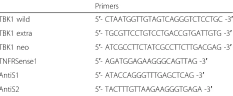
per-formed using the primers in Table 1.
For experiments, we used 2–3-month-old littermate mice. Animals had free access to food and water and were
maintained in climate- and light-controlled rooms (24 ± 0.5 °C, 12/12 dark/light cycle). In all experiments, the eth-ical guidelines for investigations in conscious animals were obeyed and the procedures were approved by the local Ethics Committee for Animal Research (F95/53). All efforts were made to minimize animal suffering and to reduce the number of animals used. All behavioral experiments were assessed by a blinded observer in a dedicated room with restricted sound level and activity.
Western blot analysis
For Western blot analysis, mice were injected with either formalin or zymosan A into the hind paws. Lumbar spinal cords (L4-L6) were dissected out at the indicated time points. Tissues were homogenized in PhosphoSafe Extraction Buffer (Merck, Darmstadt, Germany) with protease inhibitor mixture (1 mM Pefabloc SC Alexis Biochemicals, Lausen, Switzerland) immediately after preparation. To remove cellular debris, extracts were centrifuged at 16.800 ×g for 1 h at 4 °C. The superna-tants were stored at–80 °C. Proteins from tissue homog-enates (15–30μg) were separated electrophoretically by 10 % SDS-PAGE and then transferred onto nitrocellu-lose membranes by wet-blotting. To confirm equal load-ing all blots were stained with Ponceau S red solution. Membranes were blocked for 60 min at room temperature in Odyssey blocking reagent (LI-COR Biosciences, Bad Homburg, Germany) diluted 1:2 in 0.1 M PBS, pH 7.4. Then the blots were incubated overnight at 4 °C with pri-mary antibody against TBK1 (84 kDa), p42 (42 kDa), p-p42 (42 kDa), p44 (44 kDa), p-p44 (44 kDa), JNK (46 kDa), p-JNK (46 kDa), SAPK (54 kDa), p-SAPK (54 kDa), p-p65(Ser536) (65 kDa) (all 1:250, Cell Signaling Technology, Boston, USA) in blocking buffer. After washing three times with 0.1 % Tween 20 in 0.1 M PBS, the blots were incubated for 60 min with an IRDye 800- or IRDye 700-conjugated secondary anti-body (1:5000 in blocking buffer). After rinsing in 0.1 % Tween 20 in 0.1 M PBS, protein-antibody complexes were detected with the Odyssey Infrared Imaging System
(LI-COR, Bad Homburg, Germany). β-Actin (37 kDa),
(1:1200, Sigma) or Hsp90 (90 kDa), (1:500, BD Bioscience) was used as loading control. Densitometric analysis of the
Table 1Primer sequences for genotyping
Primers
TBK1 wild 5′- CTAATGGTTGTAGTCAGGGTCTCCTGC -3′
TBK1 extra 5′- TGCGTTCCTGTCCTGACCGTGATTGTG -3′
TBK1 neo 5′- ATCGCCTTCTATCGCCTTCTTGACGAG -3′
TNFRSense1 5′- AGATGGAGAAGGGCAGTTAG -3′
AntiS1 5′- ATACCAGGGTTTGAGCTCAG -3′
blots was performed with Image Studio Lite Software (LI-COR, Bad Homburg, Germany).
Real-time PCR (Taqman)
RNA was prepared from the lumbar spinal cords and dorsal root ganglia (DRGs) (L4-L6) using TRI reagent according to the standard procedure [13]. Two hundred nanogram of total RNA was used for the reverse transcrip-tion which was performed with Random and Oligo-dT Primers (2:1 ratio) in a Verso cDNA Synthesis Kit (Thermo Scientific, Darmstadt, Germany). Twenty nanogram RNA equivalent was subjected to real-time PCR in an Applied Biosystems sequence detection system AB7500 using the FastStart Universal SYBR Green Master (Rox) System (Roche Diagnostics, Mannheim, Germany). Expression of TBK1, COX-2, iNOS, MMP-9, c-fos, and Akt1 mRNA was assessed related to GAPDH mRNA. The following gene-specific primers were used:
TBK1 FW5′-GAG CCG GGA ACA ACT CAA TA-3′
RV5′-TTG CTT TTG TGG CAT GGT AA-3′
COX-2 FW5′-AGACACTCAGGTAGACATGATCTA
CCCT-3′
RV5′-GGCACCAGACCAAAGACTTCC-3′
iNOS FW5′-TCACCCACACTGTGCCCATCTAC
GA-3′
RV5′-CAGCGGAACCGCTCATTGCCAA
TGG-3′
MMP-9 FW5′-GAAGCCAAACCCTGTGTGTT-3′
RV5′-AGAGTACTGCTTGCCCAGGA-3′
c-fos FW5′-ACCATGATGTTCTCGGGTTTCAA-3′
RV5′-GCTGGTGGAGATGGCTGTCAC-3′
Akt 1 FW5′-GGCTGGCTGCACAAACG-3′
RV5′-GACTCTCGCTGATCCACATCCT-3′
GAPDH FW5′-CAA TGT GTC CGT CGT GGA TCT-3′ RV5′-GTC CTC AGT GTA GCC CAA GAT G-3′
The cycle number at which the fluorescence signal crosses a defined threshold (Ct-value) is proportional to the number of RNA copies present at the start of the PCR. The threshold cycle number for the specific mRNA was standardized by subtracting the Ct-value of GAPDH from the Ct-value of gene-specific amplifications of the same sample. Relative quantitative level of samples was determined by standard 2(-ΔΔCt)calculations and expressed as fold-change in a single reference control sample (un-treated control mice of either genotype).
Immunofluorescence
Mice were perfused intracardially with 0.9 % saline followed by 4 % paraformaldehyde in 0.1 M phosphate-buffered saline (PBS), pH 7.4, under deep ketamine/ xylazine anesthesia. Lumbar spinal cords (L4-L6) were dissected out, post-fixed in 4 % PFA in 0.1 M PBS (pH 7.4),
cryoprotected in 20 % sucrose in 0.1 M PBS overnight at 4 °C and then embedded in Tissue-Tek O.C.T. Compound (Sakura Finetek Europe B.V., Alphen aan den Rijn, Netherlands) frozen on dry ice. Cryostat sections were cut at a thickness of 16μm and stored at−80 °C. For immuno-fluorescence, slices were permeabilized for 15 min with 0.1 M PBS containing 0.1 % Triton-X 100. The sections were then blocked in 3 % BSA in 0.1 M PBS for 1 h to re-duce non-specific binding. After rinsing in 0.1 M PBS, sec-tions were incubated with TBK1 antibody (1:500, Acris) and for co-immunofluorescence with mouse anti-neuronal nuclei (NeuN, 1:1000, Sigma Aldrich), mouse anti-GFAP (1:1000, Chemicon), or CD11b (1:500). Antibodies were dissolved in 0.1 % Tween 20 in 0.1 M PBS and incubated at 4 °C overnight. After rinsing in 0.1 M PBS, sections were incubated for 2 h at room temperature with Alexa Flour 488- or Cy3-conjugated secondary antibodies dissolved in 0.1 % Tween 20 in 0.1 M PBS, rinsed again in 0.1 M PBS and coverslipped with Fluoromount G. Images were cap-tured using an inverted fluorescence microscope (Axio Observer.Z1; Zeiss, Germany) equipped with a mono-chrome CCD camera and AxioVision software (Zeiss, Germany).
Transcription factor analysis
Nuclear extracts from the spinal cord or cell culture were prepared using a commercially available Nuclear Extract Kit following the manufacturer’s instructions (Active Motif, Rixensart, Belgium). These nuclear extracts were then subjected to TransAMTMNF-κB p65, c-fos, and IRF-3 transcription factor ELISAs (Active Motif). Five microgram nuclear protein was allowed to bind to an oligonucleotide-coated plate. NF-κB p65, c-fos, or IRF-3 was detected by incubation with specific primary antibodies and HRP-conjugated secondary antibody. The colorimetric readout was measured photometrically at 450 nm and is propor-tional to transcription factor activity.
Behavioral testing
Littermates were used in all behavioral tests. Animals were habituated to the experimental room and were in-vestigated by observers blinded for the genotype of the animals. Genotyping was done as described above.
Rotarod test
Motor coordination was assessed with a Rotarod Treadmill for mice (Ugo Basile, Comerio, Italy) at a constant rotating speed of 32 rpm. All mice had five training sessions before the day of the experiment. The fall-off latency was averaged from five tests. The cut-off time was 90 s.
Mechanical sensitivity
consisting of a steel rod that is pushed against the plantar surface of the paw with increasing force until the paw is withdrawn (Dynamic Plantar Aesthesiometer, Ugo Basile, Varese, Italy). The maximum force was set at 5 g to prevent tissue damage and the ramp speed was 0.5 g/s. The cut-off time was 20 s. Mice were placed in test cages with a metal grid bottom. After 1-h accommodation time, the paw with-drawal latency was measured. The mean of 4–6 consecutive trials at each time point were used for further analysis.
Hot-plate test
Animals were placed into a Plexiglas cylinder on a heated plate maintained at 52.0 ± 0.2 °C (Ugo Basile), and the latency to jump, shake/flutter a hind paw, or licking the front paws was recorded. Each animal was tested only once since repeated testing in this assay leads to latency changes [21]. The cut-off time was 30 s.
Formalin test
The formalin test was performed as described [22]. Mice were placed on a table top within a Plexiglas cylinder and were allowed to habituate for 30 min. Twenty mi-croliters of a 5 % formaldehyde solution (formalin) was injected subcutaneously into the dorsal surface of the left hind paw. The time spent licking the formalin-injected paw was recorded at 5 min intervals up to 45 min, starting right after formalin injection. Two hours after, formalin injection animals were deeply anes-thetized with isoflurane and then killed by cardiac punc-ture. Lumbar spinal cords and DRGs (L4-L6) were dissected out and immediately frozen in liquid nitrogen and stored at−80 °C until further preparation.
Zymosan A-induced paw inflammation
Paw withdrawal latency in response to mechanical stimulation was assessed as described above. Mice were placed in test cages with a metal grid bottom. After 1 h, baseline paw withdrawal latencies were measured. On the next day, hind paw inflammation was induced by subcutaneous injection of 20μl of a 10 mg/ml zymosan A (Sigma-Aldrich, Munich, Germany) suspension in phosphate-buffered saline (0.1 M, pH 7.4) into the mid plantar region of the left hind paw [23]. The mean of four consecutive trials with 10 s intervals was deter-mined hourly up to 8 h after zymosan A injection and then again after 24 and 48 h. At the end of the observa-tion period (48 h after zymosan A injecobserva-tion), animals were deeply anesthetized with isoflurane and then killed by cardiac puncture. Lumbar spinal cords and DRGs (L4-L6) were dissected out and immediately frozen in li-quid nitrogen and stored at−80 °C until further prepar-ation. Ipsi- and contralateral paws were detached at the ankle. The paw volume was determined by weighing the paws.
Cell culture
RAW264.7 mouse macrophages were cultured and incu-bated in RPMI 1640 medium containing 10 % FCS and 1 % penicillin/streptomycin. Cells were stably transduced with either TBK1-specific shRNA (GIPZ-system, Thermo Scientific, Germany) or negative control virus (NC) and cultivated under puromycin selection. Permanent expres-sion of shRNA resulted in 80 % reduction of the TBK1 protein level, which remained stable throughout the whole incubation period (Additional file 1: Figure S6 A). Bone marrow derived macrophages (BMM) were generated from TNFR−/−and TBK1−/−/TNFR−/−mice. Muscle tissue was removed from the hind legs, and the bones were cut directly beyond and above the joints. The bones were placed into a perforated 0.2 ml tube, which was inserted into a 1.5 ml reaction tube. After centrifugation at 16.800 ×g for 15 s, the bone marrow was resuspended in RPMI 1640 medium supplemented with 10 % FCS, 1 % Pen/Strep, and 20 ng/ml recombinant murine macrophage colony stimulating factor (M-CSF, Peprotech, Hamburg, Germany). Cells were seeded in six well plates and culti-vated and differentiated for 7 days before stimulation.
RAW264.7 and BMM were stimulated with 10 μg/ml
lipopolysaccharide (LPS) for the indicated time points. Protein was isolated using Phophosafe/Pefabloc (Merck/ Novagen, Darmstadt, Germany/Alexis Biochemicals, Lörrach, Germany) according to manufacturer’s instruction.
Data analysis
Statistical evaluation was done with Graph Pad Prism 5.0 for Windows. Data are presented as mean ± SEM. Data were either compared by univariate analysis of
vari-ance (ANOVA) with subsequent t tests employing a
Bonferroni α-correction for multiple comparisons or by student’sttest. For analysis of inflammatory hyperalgesia in the zymosan A-induced paw inflammation, paw with-drawal latencies in response to mechanical stimulation were expressed as the relative difference between the zy-mosan A-treated left and the untreated right hind paw calculated as ΔPWL = (left-right) / right × 100 (mean ± SEM). For analysis of inflammatory hyperalgesia in the zymosan A-induced paw inflammation and the formalin test, repeated measures ANOVA was performed. For all tests, a probability valueP< 0.05 was considered as sta-tistically significant.
Results
Expression and regulation of TBK1 in the spinal cord and the DRGs
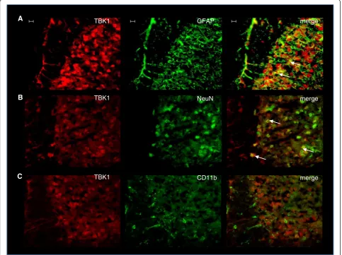
type tissues with markers for neurons, microglia, and astrocytes revealed TBK1 localisation in NeuN-positive neurons and some GFAP-positive astrocytes. CD11b-positive microglial cells showed no co-staining with TBK1 indicating that it is not expressed in this cell type under naïve conditions (Fig. 1). Accordingly, we found low levels of basal TBK1 expression in primary microglia and astro-cytes, which however were upregulated upon inflamma-tory stimulation with a cytokine mix (Additional file 3: Figure S2). To gain an impression whether TBK1 expres-sion in the spinal cord is due to localization in neurons of the terminals of primary nociceptive afferents, immuno-fluorescence images from DRG slides were prepared additionally. TBK1 expression was detectable in small non-myelinated nociceptive neurons and large myelinated non-nociceptive neurons of the DRGs, as assessed by co-staining with IB4 and Neurofilament 200, respect-ively (Additional file 4: Figure S3), indicating its role in
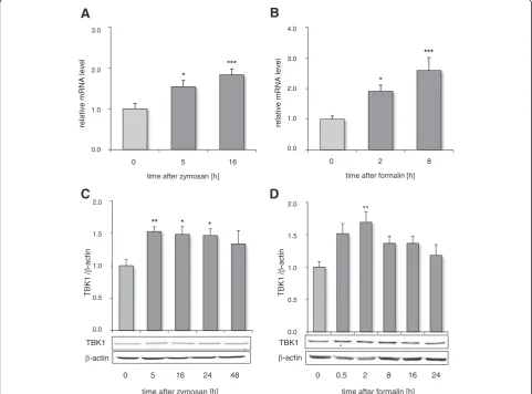
nociceptive transmission as well as general functions in sensory perception in the periphery. Potential regula-tion of spinal TBK1 during nocicepregula-tion was assessed in two different inflammatory models in mice: the forma-lin test which reflects short time tonic nociception as-sociated with mechanisms of central sensitization and the zymosan A-induced paw inflammation model which leads to the formation of paw edema and in-creased sensitivity to nociceptive stimuli that lasts for several days. In both models, we observed an increase in TBK1 mRNA and protein levels in the spinal cord after peripheral noxious stimulation (Fig. 2) suggesting that TBK1 is a modulator of inflammatory nociception and sensitization towards ongoing nociceptive input. In contrast, no TBK1 regulation after inflammatory nocicep-tive stimulation could be observed in DRGs indicating its main function in nociceptive processing in the cen-tral nervous system (Additional file 4: Figure S3).
A
B
C
GFAP
NeuN
CD11b TBK1
TBK1
TBK1
merge
merge
merge
Fig. 1TBK1 expression in the dorsal spinal cord. Representative co-immunofluorescence showing TBK1 protein expression in the dorsal horn of
spinal cord in combination with cell markers for (a) astrocytes (GFAP), (b) neurons (NeuN), and (c) microglia (CD11b). TBK1 was stained with Cy3
(red), cell markers with Alexa Fluor 488 (green). The three images show from left to right: TBK1 alone, cell marker alone, and merged (representative result
TBK1 knock-out is associated with antinociceptive effects in inflammatory models
Genetically modified mice were used to further evaluate TBK1 function in nociceptive processing. Before starting nociceptive tests, the Rotarod Test was performed to assess intact motoric coordination of TBK1−/−/TNFR−/− and TNFR−/− mice, which is an important prerequisite for proper behavioral nociceptive testing. Neither TBK1−/−/TNFR−/− nor TNFR−/− mice showed any dif-ferences in their ability to balance on the rotating rod and remained there until the cut-off time of 90 s indi-cating that the genetic modification does not impair their motor functions. Hot-plate and Dynamic Plantar tests were applied to assess the impact of the genetic modifications on acute thermal and mechanical noci-ception, respectively. The results of both tests showed that there were no significant differences between TBK1−/−/TNFR−/− and TNFR−/− mice indicating that the physiologically important immediate response to
acute noxious thermal and mechanical stimulation is unaffected in the knock-out mice (mean ± SEM; Hot Plate: TNFR−/− 14.7 ± 1.1; TBK1−/−/TNFR−/− 19.4 ± 2.0 (P= 0.101); Dynamic Plantar: TNFR−/− 7.7 ± 0.6; TBK1−/−/TNFR−/− 7.6 ± 0.6). Due to the homology of TBK1 with other IκB kinases and a potential compen-satory regulation of similar proteins in response to sys-temic knock-out of genes, we determined the protein levels of IKKα, IKKβ, and IKKε in spinal cord protein extracts of TBK1−/−/TNFR−/−, TNFR−/−, and wild type mice. The results of the Western blot analysis showed equal protein expression for all kinases in all genotypes tested suggesting that these proteins were not com-pensatory regulated in TBK1 knock-out mice (data not shown).
In the inflammatory nociceptive models, wild type littermates showed typical nociceptive responses. After injection of zymosan A, a strong decrease in paw with-drawal latencies towards mechanical stimulation was
** * *
0 5 16 24 48
time after zymosan [h]
**
2.0
1.5
1.0
0.5
0.0
1
K
B
T/
-a
c
ti
n
-actin TBK1
2.0
1.5
1.0
0.5
0.0
TBK1
/
-actin
0 0.5 2 8 16 24
time after formalin [h] -actin
TBK1
C
D
3.0
2.0
1.0
0.0
0 5 16
time after zymosan [h]
A
N
R
m
e
vit
al
er
lev
el
*** ***
0 2 8
time after formalin [h]
4.0
3.0
2.0
1.0
0.0
*
*
rel
a
ti
v
e
m
R
N
A
lev
e
l
A
B
Fig. 2TBK1 regulation in the spinal cord after peripheral inflammatory stimulation. Time course of the TBK1 mRNA and protein expression in the
lumbar spinal cord after peripheral injection of zymosan A (a,c) and formalin (b,d), respectively (n= 4 mice/group). The Western blots (c,d) show one
representative blot; the diagrams depict the densitometric analysis of all blots (mean ± S.E.M). Univariate ANOVA with Bonferroni post-hoc
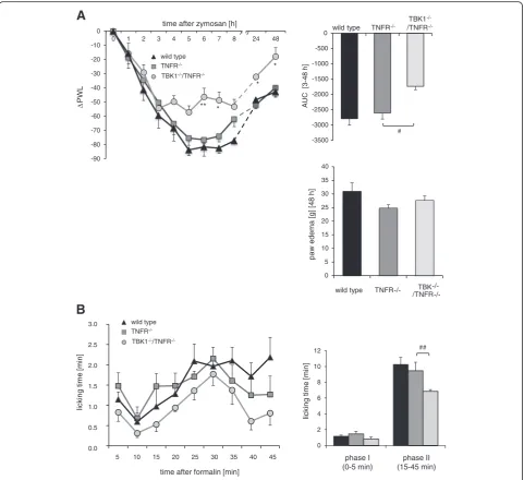
observed which lasted until the end of the experiment at 48 h (Fig. 3a). This effect was already reduced in TNFR−/− animals and further alleviated in TBK1−/−/TNFR−/− mice in the period from 3 h up to 48 h after zymosan A injection. Latencies almost returned to baseline in
TBK1−/−/TNFR−/− mice at 48 h (repeated measures ANOVA, *P< 0.05 (TNFR−/−) and **P< 0.01 (TBK1−/−/ TNFR−/−). To investigate the impact of TBK1 on paw inflammation, the zymosan A-injected paws were weighted at the end of the experiments to determine
A
wild type TNFR
-/-TBK1
-/-/TNFR
-/-24 48
-90 -80 -70 -60 -50 -40 -30 -20 -10 0
0 1 2 3 4 5 6 7 8
2,5 7,5 12,5 17,5 22,5 27,5 32,5 37,5 42,5
** *
*
PW
L
time after zymosan [h]
5 10 15 20 25 30 35 40 45
time after formalin [min]
lick
ing
ti
m
e
[m
in
]
3.0
2.5
2.0
1.5
1.0
0.5
0.0
lick
ing
tim
e
[m
in
]
TNFR -/-TBK1-/-/TNFR
-/-B
wild type TNFR -/-TBK1-/-/TNFR -/-wild type
*
-3500 -3000 -2500 -2000 -1500 -1000 -500 0
#
AUC
[3-48
h]
0 5 10 15 20 25 30 35 40
TBK
wild type TNFR-/-
-/-
/TNFR-/-paw
edema
[g
]
[48 h]
0 2 4 6 8 10 12
phase I (0-5 min)
phase II (15-45 min)
##
Fig. 3Inflammatory hyperalgesia in TNFR−/−and TBK1−/−/TNFR−/−mice.aLeft panel: time course of mechanical hyperalgesia in wild type (black
triangle), TNFR−/−(dark grey square), and TBK1−/−/TNFR−/−(light grey circle) mice after injection of 10 mg/ml (20μl) zymosan A into a hind paw.
The diagram shows the delta paw withdrawal latencies (ΔPWL) in response to mechanical stimulation as assessed with a Dynamic Plantar
Aesthesiometer (n= 6 mice/group), repeated measure ANOVA with Bonferroni post-hoc analysis, *P< 0.05 and **P< 0.01. Upper right panel: comparison of the area under the paw withdrawal latency versus time curve between wild type (black column), TNFR−/−(dark grey column),
the edema volume. Since no significant differences were observed between the genotypes (Fig. 3a), it has to be suggested that TBK1 does not directly influence the inflammatory response in the paw. This assump-tion was further supported by RT-PCR analyses of paw tissue. Although TBK1 in wild type mice and the inflamma-tory cytokines IL-1β, IL-6, and IFN-β1 in wild type, TBK1
−/−
/TNFR−/− and TNFR−/− animals were strongly upregu-lated after inflammatory stimulation, there were no signifi-cant differences in the level of inflammatory cytokines between the genotypes (Additional file 5: Figure S4).
Following injection of formalin, TNFR−/− as well as TBK1−/−/TNFR−/− mice showed the typical biphasic noci-ceptive behavior consisting of licking the injected paw. The first phase (0–5 min) is due to chemical-fiber stimulation of peripheral nociceptors [24]. After a gap phase of ~10 min, the second phase lasts from 15–45 min and corresponds to spinal sensitization. In comparison to wild type mice, we already found an impaired nociceptive response in mice with a deletion of TNFR. However, TBK1−/−/TNFR−/− mice revealed a further decrease in licking of the formalin-injected hind paw which was sig-nificant in the second phase of the formalin assay as com-pared to TNFR−/− mice (Fig. 3b) indicating that TBK1 makes an important contribution to the formalin-induced C-fiber sensitization.
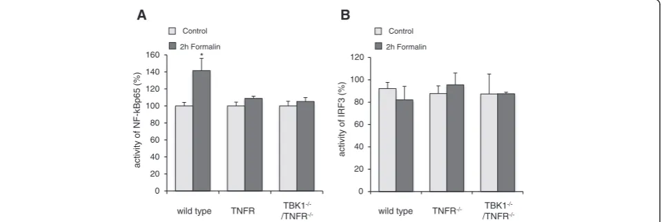
Regulation of NF-κB and IRF3 activity
Since NF-κB as well as IRF3 are important targets of TBK1, we studied the regulation of these transcription factors in the spinal cord of wild type, TBK1−/−/TNFR−/− as well as TNFR−/− mice with and without formalin stimulation (Fig. 4). NF-κB was significantly activated in nuclear ex-tracts of the spinal cord of wild type animals after periph-eral formalin injection while IRF3 activity remained
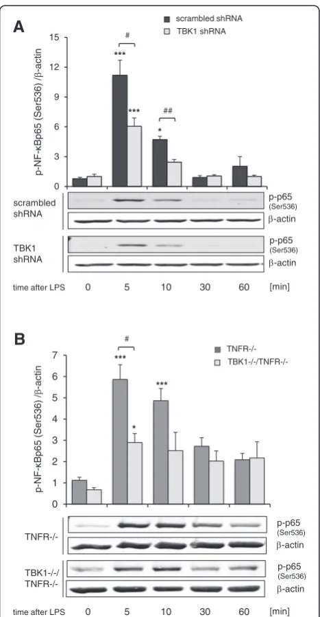
unaltered. In accordance with these data, IRF3 was nei-ther affected in TBK1−/−/TNFR−/− nor in TNFR−/− mice in this study. However, in contrast to the activa-tion of NF-κB in wild type mice, we found no signifi-cant effect of formalin in either TBK1−/−/TNFR−/− or TNFR−/− mice indicating that the knock-out of TNFR already inhibits NF-κB activation which covers possible ef-fects of TBK1. Therefore, cell culture experiments with RAW264.7 mouse macrophages, which were stably trans-duced with TBK1 shRNA were additionally performed. In comparison to control cells, which showed a significant increase in phosphorylated NF-κB p65 (Ser536) 5 and 10 min after stimulation with LPS, knock-down of TBK1 by shRNA significantly decreased this phosphorylation by about 50 % (Fig. 5a). These results are further supported by experiments with primary bone marrow macrophages (BMM) derived from TBK1−/−/TNFR−/− and TNFR−/− mice. The LPS-induced increase of p65 (Ser536)-phos-phorylation after 5 and 10 min stimulation in TNFR−/− BMMs was significantly reduced in BMMs from TBK1−/−/ TNFR−/− mice (Fig. 5b) indicating that TBK1 is involved in stimulus-dependent NF-κB activation which might at least partially contribute to the effects observed in the nociceptive animal models.
Regulation of potential TBK1 downstream targets
Further experiments were performed to clarify which signal transduction pathways participate in the TBK1-mediated nociception in the spinal cord. Since NF-κB activation was only partially inhibited by downregulation of TBK1 in RAW264.7 macrophages or BMMs from TBK1−/−/TNFR−/− mice, we suggested that TBK1 down-stream targets are not limited to the NF-κB cascade and analyzed a panel of NF-κB dependent as well as inde-pendent “pain-relevant” genes. The NF-κB dependent
acti
v
ity
of
N
F
-)
%(
5
6
p
B
k
Control
2h Formalin
Control
2h Formalin
acti
v
ity
of
IR
F3 (%)
A B
0 20 40 60 80 100 120 140 160
wild type TNFR TBK1
-/-/TNFR
-/-0 20 40 60 80 100 120
wild type TNFR-/- TBK1
-/-/TNFR
-/-*
Fig. 4Regulation of NF-κB and IRF3 activation in the spinal cord.ap65 andbIRF3 transcription factor activity as assessed by TransAM transcription
factor ELISA. Nuclear extracts of the spinal cord of wild type, TNFR−/−and TBK1−/−/TNFR−/−mice were prepared from control mice and mice 2 h after
injection of formalin into the hind paws (n= 3 mice/group); bright columns = control; dark columns = 2 h formalin, *P< 0.05, statistically
genes cyclooxygenase-2 (COX-2), inducible nitric oxide synthase (iNOS), and matrix metalloproteinase-9 (MMP-9) showed the expected upregulation in the spinal cord after peripheral formalin injection in wild type mice. iNOS and MMP-9 induction were repressed in both TBK1−/−/ TNFR−/−and TNFR−/−mice in a similar manner, whereas
COX-2 regulation was normal in TNFR−/− mice and
significantly decreased in TBK1−/−/TNFR−/− animals.
For the NF-κB-independent genes c-fos and Akt1, a
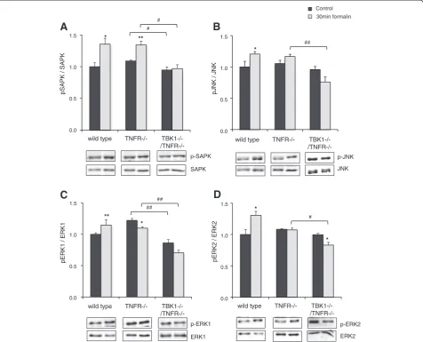
formalin-induced increase in the spinal cord was ob-served in wild type mice. The upregulation of Akt1 was inhibited in both TBK1−/−/TNFR−/− and TNFR−/− mice, whereas c-fos modulation was significantly repressed in TBK1−/−/TNFR−/− animals but remained unaltered in TNFR−/− mice (Fig. 6). These data indicate that COX-2 and c-fos strongly contribute to the TBK1-mediated nociception. The role of COX-2 in TBK1 signaling in nociception was further confirmed by experiments with the COX-2 selective inhibitor celecoxib. While cele-coxib treatment was able to further reduce the antino-ciceptive effects of the TNFR-deletion in a significant manner, the reduction in TBK1−/−/TNFR−/− mice was only marginal thus further supporting the hypothesis that COX-2 is a TBK1 downstream target (Additional file 6: Figure S5). The strong regulation of c-fos prompted us to investigate the regulation of MAP kinases which are involved in the control of c-fos expression [25, 26] and have been repeatedly associated with nociceptive re-sponses [27–29]. As expected, we found a significant in-crease in ERK 1/2, SAPK, and JNK activity in the spinal cord of wild type mice after injection of formalin into the paw as assessed by Western blot with phospho-specific antibodies. The formalin-induced phosphorylation of ERK 1/2 and JNK was already inhibited in TNFR−/− mice, but in TBK1−/−/TNFR−/− mice, we even found a decrease in their phosphorylation to below baseline level. The increase in p-SAPK levels was similar in wild type and TNFR−/− mice but significantly inhibited in TBK1−/−/TNFR−/−mice (Fig. 7). These results indicate that TBK1 contributes to c-fos activation in inflammatory nociception by regulation of MAP kinase activation. This is supported by cell culture experiments with TBK1 depleted RAW264.7 macrophages which showed a 50 % reduced c-fos activation in compari-son to control cells in which activated c-fos was strongly increased 60 min after LPS stimulation (Additional file 1: Figure S6 B).
Discussion
The classical I-κB kinase complex consisting of the two catalytical subunits IKKα and βand the regulatory sub-unit IKKγ (NEMO) has been suggested to be an essen-tial player in inflammatory NF-κB activation [30] and also contributes to inflammatory nociception [10, 31]. In a recent study, we found that the IKK-related IKKε is
0 3 6 9 12 15
0 5 10 30 60
***
#
*** ##
p
-N
F
-/ )
6
3
5r
e
S(
5
6
p
B-act
in
-actin p-p65
(Ser536)
-actin p-p65
(Ser536)
scrambled shRNA
TBK1 shRNA
[min]
time after LPS
scrambled shRNA
TBK1 shRNA
A
*
0 1 2 3 4 5 6 7
B
0 5 10 30 60
p
-NF
-/ )
6
3
5r
e
S(
5
6
p
B-act
in
TNFR-/-TBK1-/-/
TNFR-/-[min]
time after LPS
TNFR-/-
TBK1-/-/TNFR-/-***
#
***
*
-actin p-p65
(Ser536)
-actin p-p65
(Ser536)
Fig. 5Regulation of NF-κB in RAW264.7 and primary bone marrow
macrophages.aWestern blot showing serine 536 phosphorylation
of p65 in LPS-stimulated RAW264.7 macrophages stably transduced
with scrambled (black columns) or TBK1-specific shRNA (light grey
columns), (n= 4). The blots show a representative result; the diagram
shows the densitometric analysis of all experiments.bWestern blot
showing serine 536 phosphorylation of p65 in LPS-stimulated BMMs
derived from TNFR−/−(dark grey columns) and TBK1−/−/TNFR−/−(light
grey columns) mice, (n= 4). The blots show a representative result, the diagram shows the densitometric analysis of all experiments. Univariate
ANOVA with Bonferroni post-hoc analysis *P< 0.05 and ***P< 0.001
statistically significant differences compared with unstimulated
cells. Student’sttest#P< 0.05 and##P< 0.01 significant mean difference
between scrambled shRNA and TBK1 shRNA or TNFR−/−and
also involved in processing of inflammatory pain by regulating NF-κB activity and the expression of NF-κB dependent genes [13]. TBK1 constitutes another
non-canonical IKK which might form a complex with IKKε
and is involved in regulation of antiviral responses by modulating IFN Type I and also in inflammatory pro-cesses by regulating NF-κB signaling (reviewed in [7, 32, 33]). The aim of this study was to clarify whether TBK1 is also involved in inflammatory nociceptive processing. We observed TBK1 expression in“pain-relevant”cells in the spinal cord and the DRGs. After peripheral inflam-matory nociceptive stimulation, TBK1 was upregulated in the spinal cord but not in the DRGs indicating its main role in the central nervous system. Effects in the formalin model could be detected at earlier time points compared to the zymosan A model which might reflect the fact that the formalin model constitutes a more acute inflammatory nociceptive model leading to early effects while zymosan A-induced paw inflammation pro-vides more information about persistent hyperalgesia and later regulation [23]. TBK1 knock-out mice were used to assess inflammatory hyperalgesia in the two dif-ferent mouse models after confirming that similar ki-nases such as IKKα,β, andεdo not compensate for the TBK1 deletion. Due to embryonic lethality of complete TBK1 knock-out mice as a result of liver degeneration and apoptosis [16, 34], the TBK1−/− mice had to be bred
under a TNFα-receptor knock-out and were compared
to TNF-α receptor knock-out mice (TNFR−/−) as con-trols. A knock-out of TNFR in mice has already been re-peatedly associated with reduced nociceptive responses in models of inflammatory, cancer, and neuropathic pain [35–37] and might thus cover antinociceptive effects
mediated by TBK1 knock-down. Accordingly, we showed that the TNFR knock-out already ameliorates nociception in the formalin test and in the zymosan A-induced paw inflammation. Nevertheless, in TBK1/TNFR double-knock-out mice, this effect was significantly enhanced indicating that TBK1 contributes to inflammatory hyperalgesia. These results are in accordance with studies showing anti-inflammatory effects of TBK1 inhibition in fibroblast-like synoviocytes by reducing the expression of the proinflam-matory protein IP-10 [17]. Furthermore, TBK1 is involved in the inflammatory response in obesity and hypertension and contributes to phosphorylation of the insulin receptor thus impairing its function and supporting insulin resist-ance [18]. Treatment of obese mice with the TBK1/IKKε inhibitor amlexanox led to weight loss and improved insu-lin sensitivity by elevation of energy expenditure [38, 39], which further confirms that TBK1 contributes to proin-flammatory reactions. Our concern was to investigate whether TBK1 effects in nociception are mediated via
NF-κB signaling possibly in concert with IKKε or via other pathways. In a recent study, we used BX795 as a pharma-cological inhibitor of IKKεand TBK1 and showed that the combined inhibition of both kinases led to a stronger re-duction of the noxious response in comparison to single IKKε knock-out [13]. Together, with the results of this study, it seems that inhibition of both kinases by BX795 leads to an addition of the effects of IKKε- and TBK1-mediated analgesia which might be due to synergistic modulation of the same target genes or to regulation of different targets, respectively. For IKKε, we showed that a complete inhibition of NF-κB p65 phosphorylation at Ser 536 was responsible for inhibition of NF-κB dependent target genes, such as COX-2, iNOS, and MMP-9, while
time after formalin[[2h]
COX-2 iNOS MMP-9 c-fos Akt1
wild type
TNFR
-/-TBK1-/-/TNFR
-/-******
***
##
******
**
* *
#
**
*
3.5
3.0
2.5
2.0
1.5
1.0
0.5
e
tiv
al
erA
N
R
m
lev
el
#
Control
Fig. 6Regulation of NF-κB dependent and independent genes in the spinal cord. Quantitative RT-PCR (Taqman) of cyclooxygenase-2 (COX-2), inducible NO-synthase (iNOS), matrix metalloproteinase-9 (MMP-9), c-fos, and Akt1 mRNA in the spinal cord of naïve mice and 2 h after formalin injection into a hind
paw. GAPDH-RNA was used as reference control gene (n= 4 mice/group) and each Taqman-PCR was performed twice in triplicate; black columns = wild
type; dark grey columns = TNFR−/−, light grey columns = TBK1−/−/TNFR−/−, Student’sttest *P< 0.05, **P< 0.01, and ***P< 0.001 in comparison to untreated
NF-κB independent targets were not affected [13]. The re-sults of this study revealed that a knock-down of TBK1 also inhibits NF-κB activation by decreasing phosphoryl-ation of p65 Ser536 but to a lesser extent than that shown for IKKεinhibition. From these data and published studies which hint towards divergent functional roles for TBK1 and IKKε [5, 6], we postulated that TBK1 might affect other target genes in addition. Indeed, we found that the NF-κB-independent marker of neural activity, c-fos, was also differentially regulated in TBK1 knock-out mice in comparison to TNFR−/−and wild type mice in association with a decreased activation of the MAP kinases ERK1/2 and SAPK/JNK. These MAPKs have been frequently asso-ciated with nociceptive responses [27–29] and inhibition of their activation might contribute to antinociceptive effects in
TBK1 knock-out mice. A relationship between TBK1 and MAPKs has already been found in a study with TBK1−/− mouse embryonic fibroblasts (MEFs) which showed reduced TNFα-induced MAPK activation in comparison to TBK1+/+ cells [40]. In contrast, TBK1 was not involved in MAPK activation stimulated by double stranded DNA [41] or TLR-3 activation [42]. A recent study has further shown that
NF-κB activation is capable of participation in c-fos induction, but only in concert with ERK-mediated fos induction [43], a mechanism which might also have contributed to TBK1 sig-naling in this study and supports the increase in c-fos. IRF3, which has been frequently described as a prominent TBK1 target, seems to play only a minor role in nociception, since it was not affected by nociceptive stimulation or by deletion of TBK1.
1.5
1.0
0.5
0.0
Control 30min formalin
wild type TNFR-/-
TBK1-/-
/TNFR-/-*
##
p-JNK
JNK
1.5
1.0
0.5
0.0
wild type TNFR-/-
TBK1-/-
/TNFR-/-K
P
A
S
pK
P
A
S
/
*
#
** #
p-SAPK
SAPK
**
##
* ## 1.5
1.0
0.5
0.0
wild type TNFR-/-
TBK1-/-
/TNFR-/-1
K
R
E
/
1
K
R
E
p
p-ERK1
ERK1
pJNK
/ JNK
1.5
1.0
0.5
0.0
wild type TNFR-/-
TBK1-/-
/TNFR-/-p-ERK2
ERK2
pERK2 /
ERK2
*
#
*
A
B
C
D
Fig. 7Regulation of MAPK activity in the spinal cord of wild type, TNFR−/−and TBK1−/−/TNFR−/−mice. Western blots showing the activation of
SAPK (a), JNK (b), ERK1 (c), and ERK 2 (d) in different genotypes as assessed by Western blot analysis with phospho-specific antibodies. The blots
were normalized to the respective total protein levels and additionally against the loading control HSP90 orβ-actin. The blots show a representative
result, the diagram shows the densitometric analysis of three independent experiments (n= 3 mice/group); dark columns = control; bright columns = 30 min
formalin. Student’sttest *P< 0.05 and **P< 0.01 significant mean difference between untreated and formalin–injected mice.#P< 0.05 and
##
Conclusions
This study shows that TBK1, similar to IKKε, is involved in inflammatory nociceptive processing in the spinal cord and might therefore constitute a promising target for pharmacological intervention in pain. Our data
indi-cate that, on the one hand, TBK1 and IKKε work
to-gether in a complex to inhibit NF-κB signal transduction while, on the other hand, TBK1 additionally exerts IKKε-independent actions by the so far undescribed in vivo regulation of MAP kinases and c-fos. Neverthe-less, the embryonic lethality of a complete TBK1 knock-out raises concerns that inhibition of TBK1 in humans would be associated with severe unwanted side effects. A possible solution to this problem might be tissue-specific modulation of TBK1 or drugs with combined IKKε/TBK1 inhibitory potency which can be adminis-tered at low doses.
Additional information
Additional methods and figure legends are provided in Additional file 7.
Additional files
Additional file 1: Figure S6.Stable downregulation of TBK1 is associated with decreased LPS-induced c-fos activity. (A) Western blot showing TBK expression in RAW264.7 macrophages stably transduced with scrambled or TBK1-specific shRNA during the time course of LPS-incubation. The blots show a representative result, the diagram shows the densitometric analysis of four independent experiments. (B) c-fos transcription factor activity in nuclear extracts of RAW264.7 cells stably transduced with scrambled shRNA or TBK1 shRNA, respectively, as assessed by
TransAM transcription factor ELISA (n= 3); black columns = scrambled shRNA;
light grey columns = TBK1 shRNA, Univariate ANOVA with Bonferroni post-hoc
analysis *P< 0.05 and ***P< 0.001 in comparison to untreated control,
#
P< 0.05, significant mean difference between scrambled shRNA and
TBK1 shRNA.
Additional file 2: Figure S1.Antibody specificity. Western Blot analysis (A) and immunofluorescence ((B): dorsal spinal cord, (C): DRG) of TBK1 in different mouse genotypes to confirm specificity of the antibody. Scale
Bar: 10μm.
Additional file 3: Figure S2.Regulation of TBK1 in primary immune cells after inflammatory stimulation. Regulation of TBK1 mRNA in primary
astrocytes (A) and microglia (B) after stimulation with a cytokine mix (TNF-α,
5 ng/ml; IL-1β, 1 ng/ml; LPS, 1μg/ml) for 6 and 24 h, respectively, analyzed by
quantitative RT-PCR.n= 3 independent incubations/cell type. Univariate
ANOVA with Bonferroni post-hoc analysis *P< 0.05, and*** P< 0.001.
Additional file 4: Figure S3.TBK1expression and regulation in the dorsal root ganglia. (A) Representative co-immunofluorescence showing TBK1 expression in the dorsal root ganglia (one of 3 independent experiment,
n= 3 mice/group) in combination with cell markers of non-myelinated
nociceptive afferents (IB4) (A) and large myelinated non-nociceptive neurons (NF200) (B), respectively. TBK1 was stained with Cy-3 (red), cell markers with Alexa Fluor 488 (green). The images show (from left to right side): TBK1 alone, cell marker alone, and merged (representative
result from 3 independent experiments). Scale Bar: 20μm. (C, D) Time
course of the TBK1 mRNA expression in the dorsal root ganglia after
peripheral injection of zymosan A (C) and formalin (D), respectively, (n= 3
mice/group).
Additional file 5: Figure S4.Regulation of TBK1 and inflammatory cytokines in the paw. (A) TBK1 mRNA expression in the contra- and ipsilateral
paws of wild type mice 48 h after zymosan injection. Student’sttest
***P < 0.001significant mean difference between contra- and ipsilateral paws
(n= 3 mice/group). (B-D) Regulation of inflammatory cytokines (B: IL-1β, C: IL-6,
D: IFN-β1) in the contra- and ipsilateral paws of wild type (black columns),
TNFR−/−(dark grey columns) and TBK1−/−/TNFR−/−mice (light grey columns)
48 h after zymosan injection. Univariate ANOVA with Bonferroni
post-hoc analysis, ***P < 0.001significant mean difference compared
to contralateral paw (n= 3 mice/group).
Additional file 6: Figure S5.Effects of celecoxib on zymosan-induced hyperalgesia in mice with different genotypes. Time course of mechanical
hyperalgesia in wild type (A), TNFR−/−mice (B) and TBK1−/−/TNFR−/−mice
(C) with and without administration of celecoxib (10 mg/kg BW, p.o.) 30 min prior to zymosan A injection. The diagram shows the delta paw
withdrawal latencies (ΔPWL) in response to mechanical stimulation as
assessed with a Dynamic Plantar Aesthesiometer (n= 6 mice/group (D)). Comparison of the area under the paw withdrawal latency versus time curve between wild type (black column), TNFR−/−(dark grey column) and TBK1−/−/TNFR−/−(light grey column) mice 3 to 7 h after zymosan A
injection. Univariate ANOVA with Bonferroni post-hoc analysis, *P < 0.05 significant mean difference between celecoxib/zymosan treated groups and zymosan treated controls.#P< 0.05 significant mean difference between the genotypes.
Additional file 7:Additional methods and figure legends.
Competing interests
The authors declare that they have no competing interests.
Authors’contributions
CVM planned the experiments and performed animal, biochemical, and molecular biological analyses. HIS performed biochemical and molecular biological experiments. KA performed animal experiments and immunohistochemical analyses. KLK was involved in the primary cell culture experiments. OQR performed Western blot analyses. KO was involved in animal tissue preparation. GG participated in the design of the study and editing of the manuscript. EN conceived and designed the study, co-ordinated subprojects, analyzed data, and wrote the manuscript. All authors have read and approved the final manuscript.
Acknowledgements
The authors would like to thank Prof. Shizuo Akira and Osama Takeuchi for
kindly providing TBK1−/−/TNFR−/−mice and Prof. Simone Fulda for kindly
providing viral packaging plasmids. Furthermore, the authors would like to thank Julia Häusler and Annett Häussler for excellent technical assistance and Prof. Michael Parnham for carefully reading and correcting the manuscript. The work was supported by the Deutsche Forschungsgemeinschaft (NI 705/
2-1 and Graduate School“Biologicals”GRK 1172) and a grant of the Medical
Faculty from the Johann Wolfgang Goethe-Universität Frankfurt to Christine Möser (Nachwuchsforscher). Further support was provided by the Else Kröner-Fresenius Foundation (EKFS), Research Training Group Translational
Research Innovation—Pharma (TRIP).
Received: 18 December 2014 Accepted: 5 May 2015
References
1. Fitzgerald KA, McWhirter SM, Faia KL, Rowe DC, Latz E, Golenbock DT, et al.
IKKepsilon and TBK1 are essential components of the IRF3 signaling
pathway. Nat Immunol. 2003;4:491–6.
2. Peters RT, Liao SM, Maniatis T. IKKepsilon is part of a novel PMA-inducible
IkappaB kinase complex. Mol Cell. 2000;5:513–22.
3. Shimada T, Kawai T, Takeda K, Matsumoto M, Inoue J, Tatsumi Y, et al. IKK-i,
a novel lipopolysaccharide-inducible kinase that is related to IkappaB kinases.
Int Immunol. 1999;11:1357–62.
4. Pomerantz JL, Baltimore D. NF-kappaB activation by a signaling complex
containing TRAF2, TANK and TBK1, a novel IKK-related kinase. Embo J.
1999;18:6694–704.
5. Tenoever BR, Ng SL, Chua MA, McWhirter SM, Garcia-Sastre A, Maniatis T.
Multiple functions of the IKK-related kinase IKKepsilon in interferon-mediated
6. Ng SL, Friedman BA, Schmid S, Gertz J, Myers RM, Tenoever BR, et al. IkappaB kinase epsilon (IKK(epsilon)) regulates the balance between type I
and type II interferon responses. Proc Natl Acad Sci U S A. 2011;108:21170–5.
7. Niederberger E, Moser C, Kynast K, Geisslinger G: The non-canonical IkappaB
kinases IKKepsilon and TBK1 as potential targets for the development of novel therapeutic drugs. Curr Mol Med 2013,;13(7):1089-97
8. Niederberger E, Geisslinger G. The IKK-NF-kappaB pathway: a source for
novel molecular drug targets in pain therapy? Faseb J. 2008;22:3432–42.
9. Niederberger E, Schmidtko A, Gao W, Kuhlein H, Ehnert C, Geisslinger G.
Impaired acute and inflammatory nociception in mice lacking the p50
subunit of NF-kappaB. Eur J Pharmacol. 2007;559:55–60.
10. Tegeder I, Niederberger E, Schmidt R, Kunz S, Guhring H, Ritzeler O, et al.
Specific inhibition of IkappaB kinase reduces hyperalgesia in inflammatory
and neuropathic pain models in rats. J Neurosci. 2004;24:1637–45.
11. Ebersberger A, Buchmann M, Ritzeler O, Michaelis M, Schaible HG. The role
of spinal nuclear factor-kappa B in spinal hyperexcitability. Neuroreport.
2006;17:1615–8.
12. Bockhart V, Constantin CE, Haussler A, Wijnvoord N, Kanngiesser M, Myrczek
T, et al. Inhibitor kappaB Kinase beta deficiency in primary nociceptive
neurons increases TRP channel sensitivity. J Neurosci. 2009;29:12919–29.
13. Moser CV, Kynast K, Baatz K, Russe OQ, Ferreiros N, Costiuk H, et al. The
protein kinase IKKepsilon is a potential target for the treatment of
inflammatory hyperalgesia. J Immunol. 2011;187:2617–25.
14. Lee JK, Kim SY, Kim YS, Lee WH, Hwang DH, Lee JY. Suppression of the
TRIF-dependent signaling pathway of Toll-like receptors by luteolin. Biochem
Pharmacol. 2009;77:1391–400.
15. Xie XH, Zang N, Li SM, Wang LJ, Deng Y, He Y, et al. Resveratrol inhibits
respiratory syncytial virus-induced IL-6 production, decreases viral replication, and downregulates TRIF expression in airway epithelial cells. Inflammation.
2012;35:1392–401.
16. Hemmi H, Takeuchi O, Sato S, Yamamoto M, Kaisho T, Sanjo H, et al. The
roles of two IkappaB kinase-related kinases in lipopolysaccharide and double
stranded RNA signaling and viral infection. J Exp Med. 2004;199:1641–50.
17. Hammaker D, Boyle DL, Firestein GS: Synoviocyte innate immune responses:
TANK-binding kinase-1 as a potential therapeutic target in rheumatoid arthritis. Rheumatology (Oxford) 2012,;51(4):610-8
18. Munoz MC, Giani JF, Mayer MA, Toblli JE, Turyn D, Dominici FP.
TANK-binding kinase 1 mediates phosphorylation of insulin receptor at serine residue 994: a potential link between inflammation and insulin resistance.
J Endocrinol. 2009;201:185–97.
19. Pfeffer K, Matsuyama T, Kundig TM, Wakeham A, Kishihara K, Shahinian A,
et al. Mice deficient for the 55 kd tumor necrosis factor receptor are resistant to endotoxic shock, yet succumb to L. monocytogenes infection.
Cell. 1993;73:457–67.
20. Perry AK, Chow EK, Goodnough JB, Yeh WC, Cheng G. Differential
requirement for TANK-binding kinase-1 in type I interferon responses to
toll-like receptor activation and viral infection. J Exp Med. 2004;199:1651–8.
21. Mogil JS, Wilson SG, Bon K, Lee SE, Chung K, Raber P, et al. Heritability of
nociception II.‘Types’of nociception revealed by genetic correlation
analysis. Pain. 1999;80:83–93.
22. Dubuisson D, Dennis SG. The formalin test: a quantitative study of the
analgesic effects of morphine, meperidine, and brain stem stimulation in
rats and cats. Pain. 1977;4:161–74.
23. Meller ST, Gebhart GF. Intraplantar zymosan as a reliable, quantifiable model
of thermal and mechanical hyperalgesia in the rat. Eur J Pain. 1997;1:43–52.
24. Hunskaar S, Hole K. The formalin test in mice: dissociation between
inflammatory and non-inflammatory pain. Pain. 1987;30:103–14.
25. Kominato Y, Tachibana T, Dai Y, Tsujino H, Maruo S, Noguchi K. Changes in
phosphorylation of ERK and Fos expression in dorsal horn neurons following noxious stimulation in a rat model of neuritis of the nerve root.
Brain Res. 2003;967:89–97.
26. Sgambato V, Pages C, Rogard M, Besson MJ, Caboche J. Extracellular
signal-regulated kinase (ERK) controls immediate early gene induction on
corticostriatal stimulation. J Neurosci. 1998;18:8814–25.
27. Coste O, Moser CV, Sisignano M, Kynast KL, Minden A, Geisslinger G, et al.
The p21-activated kinase PAK 5 is involved in formalin-induced nociception through regulation of MAP-kinase signaling and formalin-specific receptors.
Behav Brain Res. 2012;234:121–8.
28. Russe OQ, Moser CV, Kynast KL, King TS, Stephan H, Geisslinger G, et al.
Activation of the AMP-activated protein kinase reduces inflammatory
nociception. J Pain. 2013;14:1330–40.
29. Ji RR, Gereau RW, Malcangio M, Strichartz GR. MAP kinase and pain. Brain
Res Rev. 2009;60:135–48.
30. Karin M, Yamamoto Y, Wang QM. The IKK NF-kappa B system: a treasure
trove for drug development. Nat Rev Drug Discov. 2004;3:17–26.
31. Kanngiesser M, Haussler A, Myrczek T, Kusener N, Lim HY, Geisslinger G,
et al. Inhibitor kappa B kinase beta dependent cytokine upregulation in nociceptive neurons contributes to nociceptive hypersensitivity after sciatic
nerve injury. J Pain. 2012;13:485–97.
32. Kawai T, Akira S. Signaling to NF-kappaB by Toll-like receptors. Trends Mol
Med. 2007;13:460–9.
33. Hacker H, Karin M. Regulation and function of IKK and IKK-related kinases.
Sci STKE. 2006;2006:re13.
34. Bonnard M, Mirtsos C, Suzuki S, Graham K, Huang J, Ng M, et al. Deficiency
of T2K leads to apoptotic liver degeneration and impaired NF-kappaB-dependent
gene transcription. Embo J. 2000;19:4976–85.
35. Geis C, Graulich M, Wissmann A, Hagenacker T, Thomale J, Sommer C, et al.
Evoked pain behavior and spinal glia activation is dependent on tumor necrosis factor receptor 1 and 2 in a mouse model of bone cancer pain.
Neuroscience. 2010;169:463–74.
36. Zhang L, Berta T, Xu ZZ, Liu T, Park JY, Ji RR. TNF-alpha contributes to spinal
cord synaptic plasticity and inflammatory pain: distinct role of TNF receptor
subtypes 1 and 2. Pain. 2011;152:419–27.
37. Vogel C, Stallforth S, Sommer C. Altered pain behavior and regeneration
after nerve injury in TNF receptor deficient mice. J Peripher Nerv Syst.
2006;11:294–303.
38. Reilly SM, Chiang SH, Decker SJ, Chang L, Uhm M, Larsen MJ, et al. An inhibitor of
the protein kinases TBK1 and IKK-varepsilon improves obesity-related metabolic
dysfunctions in mice. Nat Med. 2013;19:313–21.
39. Mowers J, Uhm M, Reilly SM, Simon J, Leto D, Chiang SH, et al.
Inflammation produces catecholamine resistance in obesity via activation of PDE3B by the protein kinases IKK{varepsilon} and TBK1. Elife. 2013;2, e01119.
40. Delhase M, Kim SY, Lee H, Naiki-Ito A, Chen Y, Ahn ER, et al. TANK-binding
kinase 1 (TBK1) controls cell survival through PAI-2/serpinB2 and transglutaminase
2. Proc Natl Acad Sci U S A. 2012;109:E177–86.
41. Abe T, Barber GN. Cytosolic-DNA-Mediated, STING-Dependent Proinflammatory
Gene Induction Necessitates Canonical NF-kappaB Activation through TBK1.
J Virol. 2014;88:5328–41.
42. Bruni D, Sebastia J, Dunne S, Schroder M, Butler MP. A novel IRAK1-IKKepsilon
signaling axis limits the activation of TAK1-IKKbeta downstream of TLR3.
J Immunol. 2013;190:2844–56.
43. Tu YC, Huang DY, Shiah SG, Wang JS, Lin WW. Regulation of c-Fos gene
expression by NF-kappaB: a p65 homodimer binding site in mouse embryonic fibroblasts but not human HEK293 cells. PLoS One. 2013;8, e84062.
Submit your next manuscript to BioMed Central and take full advantage of:
• Convenient online submission
• Thorough peer review
• No space constraints or color figure charges
• Immediate publication on acceptance
• Inclusion in PubMed, CAS, Scopus and Google Scholar
• Research which is freely available for redistribution