Volume 15, Issue 2, Spring 2016
Editorial in Charge
Hossein Pakdaman, M.D.
Professor of Neurology, Shahid Beheshti
University of Medical Sciences,
Tehran, Iran
Editor-in-Chief
Shahriar Nafissi, M.D.
Associate Professor of Neurology,
Neurology Department, Tehran University of
Medical Sciences, Tehran, Iran
Deputy Editor
Farzad Fatehi, M.D.
Assistant Professor of Neurology, Neurology Department, Tehran University of
Medical Sciences, Tehran, Iran
Section Editors
Headache: Mansooreh Togha
, M.D.
,
Tehran
University of Medical Sciences
, Tehran, Iran
Multiple Sclerosis: Mohammad Ali Sahraian
,
M.D., Neurology Department,
Tehran University of
Medical Sciences
, Tehran, Iran
Stroke: Afshin Borhanin Haghighi
, M.D., Shiraz
University of Medical Sciences, Shiraz, Iran
Movement Disorders: Mohammad Rohani
, M.D.,
Iran University of Medical Sciences, Tehran, Iran
Associate Editors
Shahin Akhondzadeh,
Pharm.D., Ph.D, Tehran
University of Medical Sciences, Tehran, Iran
Majid Ghafarpour,
M.D., Tehran University of
Medical Sciences, Tehran, Iran
Massoud Nabavi,
M.D., Shahed University of
Medical Sciences, Tehran, Iran
Scientific Assistant Editor
Ali Amini-Harandi
, M.D., Shahid Beheshti University of Medical Sciences, Tehran, Iran
Editorial Board
Shahram Attarian
, M.D., Centre de Référence des
Maladies Neuromusculaires et de la SLA, France
Mahmoud R. Azarpazhooh
, M.D., Mashhad
University of Medical Sciences, Mashhad, Iran
Keivan Basiri
, M.D., Isfahan University of Medical
Sciences, Isfahan, Iran
Ahmad R. Dehpour
, Pharm.D., Ph.D., Tehran
University of Medical Sciences, Tehran, Iran
Masoud Etemadifar
, M.D., Isfahan University of
Medical Sciences, Isfahan, Iran
Kavian Ghandehari
, M.D., Mashhad University of
Medical Sciences, Mashhad, Iran
Kurosh Gharagozli
, M.D., Shahid Beheshti
University of Medical Sciences, Tehran, Iran
Mohammad H. Harirchian
, M.D., Tehran University
of Medical Sciences, Tehran, Iran
Payam Kabiri
, M.D., Ph.D., Tehran University of
Medical Sciences, Tehran, Iran
Hossein Kalani
, M.D., Shahid Beheshti University of
Medical Sciences, Tehran, Iran
Jamshid Lotfi
, M.D., Tehran University of Medical
Sciences, Tehran, Iran
Alireza Minagar
, M.D., Louisiana State University
Health Sciences Center, USA
Ali Moghtaderi
, M.D., Zahedan University of
Medical Sciences, Zahedan, Iran
Mahmood Motamedi
, M.D., Tehran University of
Medical Sciences, Tehran, Iran
Alireza Nikseresht
, M.D., Shiraz University of
Medical Sciences, Shiraz, Iran
Abdolmohamad M. Rostami
, M.D., Thomas
Jefferson University Hospitals, USA
Mohammad Saadatnia
, M.D., Isfahan University of
Medical Sciences, Isfahan, Iran
Mohammad K. Salajegheh
, M.D., Brigham and
Women's Hospital and Harvard Medical School, USA
Gholam A. Shahidi
, M.D., Tehran University of
Medical Sciences, Tehran, Iran
Vahid Shaygannejad
, M.D., Isfahan University of
Medical Sciences, Isfahan, Iran
Akbar Soltanzadeh
, M.D., Tehran University of
Medical Sciences, Tehran, Iran
Amir A. Zamani
, M.D., Brigham and Women's
Hospital and Harvard Medical School, USA
Babak Zamani
, M.D., Tehran University of Medical
Sciences, Tehran, Iran
Secretary:
Samaneh Bahraminejad, BSc
Email: [email protected]
http://ijnl.tums.ac.ir
Copy Edit, Layout Edit, Proof Reading and Design: Farzanegan Radandish Co. Postal Code: 81465-1798, Isfahan, Iran; Telefax: +98 311 6686302
www.farzaneganco.ir; Email: [email protected]
Indexed in
PubMed, PubMed Central, Academic Keys,
Cite Factor (Directory Indexing of International Research Journals),
Directory of Open Access Journals (DOAJ), Directory of Research Journal Indexing (DRJI), Ebsco,
Electronic Journals Library,
Google Scholar, InfoBase Index,
Islamic World Science Citation Center (ISC), LocatorPlus,
Scientific Information Database (SID), Ulrichsweb Global Serials Directory, Universal Impact Factor,
WorldCat
Iranian Journal of Neurology © 2016
II Iran J Neurol 2016; 15(2)
http://ijnl.tums.ac.ir 3 April
Aim and Scope
Iranian Journal of Neurology
INFORMATION FOR AUTHORS
The submission file is in Microsoft Word document file format.
The Iranian Journal of Neurology is dedicated to the Iranian Neurological Association. The journal is a peer- reviewed journal published quarterly and publishes neurological experiences in basic or clinical fields in
English Language. The Iranian Journal of Neurology aims to publish manuscripts of a high scientific quality representing original clinical, diagnostic or experimental works or observations in neurological sciences. Papers in
English are welcomed, particularly those which bring novel information and researches in clinical or basic fields from the neurological disorders. All received manuscripts coving the scope of the journal will be evaluated by properly competent referees.
Submission Cover Letter:
Submissions should be accompanied by a cover letter including a declaration by the first author on behalf of the others to the effect that
(1) The paper has not been published to date (except for abstracts of conference materials).
(2) The paper has not been accepted for publication elsewhere.
(3) All persons listed as the authors have read it and approved it for publication. The cover letters should be submitted in section "Comments for the Editor".
Articles must be written in accurate scientific English appropriate for publication. The articles are subject to review and editing; however, the authors are responsible for the correctness the manuscript's English language.
The articles must be submitted only online: ijnl.tums.ac.ir
Policies
The Editorial Board reserves the right to reject a paper without seeking reviewers’ opinion provied the content or the form of the paper does not meet minimum acceptance criteria or if the subject of the paper is beyond the aims and scope of the journal.
Everyone listed as the author of a paper is responsible for the reliability and completeness of data presented in the paper.
Do not submit papers that copy fully or partially previously published papers.
Indicate that this submission is ready to be considered by this journal by checking off the following:
The submission has not been previously published, nor is it before another journal for consideration (or an explanation has been provided in Comments to the Editor).
Where available, URLs for the references have been provided.
The text is double-spaced; uses an Arial 12-point font; and all illustrations, figures, and tables are placed within the text at the appropriate points, rather than at the end.
The text adheres to the stylistic and bibliographic requirements outlined in the Author Guidelines, which is found in About the Journal.
If the Editorial Board is not notified in advance and the paper is found to have been copied during editorial work, the paper shall be rejected.
We expect that all studies reported in the journal conform to the requirements of the Declaration of Helsinki (1989). Information on the consent of a relevant ethics committee to perform the trial and the informed consent of the patients to participate in the trial should be given in the Material and methods section of each paper in which diagnostic or therapeutic intervention does not follow from the standard procedure. Authors of case reports must not disclose personal data of patients described.
Manuscripts
The journal publishes: Original Article Review Article Case Report
Short Communication Clinical Notes Editorial Letters to Editor Neurological Images Neurological Videos Iranian Neurological Events Clinical Quiz
Details
Original and review papers: The maximum length of original and review papers (including tables and figures materials) is 3000 words.
Case reports: Case reports will be accepted only as Letter to the Editor.
Short communications: The maximum word number of short communications should be below 1200 words with maximum one table or figure and 10 references. The manuscript should be structured including introduction, materials and methods, results, discussion, and conclusion with a structured abstracts as original articles.
Iranian Journal of Neurology © 2016
Iran J Neurol 2016; 15(2) III
http://ijnl.tums.ac.ir 3 April neurological images or videos are welcome. They should
be maximally 400 words with legends without abstract and unstructured. The videos should be uploaded as supplementary files.
Letter to the Editor: May concern short scientific reports and comments. The maximum number of words should be below 800 words with maximum 5 references, no abstract, no table or figure, and unstructured.
Clinical notes: Refer to important interesting observations which are imperative for reminders in clinical practice. The maximum number is 1000 words with maximum 5 references, 1 table and 1 figure with no abstract.
Iranian neurological events: Include the brief description of major regional events (congresses or seminar) implemented in Iran.
Structure of Articles
Manuscripts should be submitted in 12 points, Arial font, with double line spacing and sufficient margins of 2.5 cm.
The text should not be formatted.
Each section of the paper should begin on a new page
The manuscript must include:
Page 1: Title Page
Page 2: Abstract and Key Words
Page 3 and subsequent pages: manuscript body including Introduction, Materials and Methods, Results, Discussion, Conclusion, References, Tables, Figures
1. Title page:
Title page should contain paper title, full names of authors, authors’ place of work, full name and address of the corresponding author (including e-mail address and telephone number), given in that order.
2. Abstract page:
The length of the abstract should be at least 200 and not more than 250 words for original papers and not more than 150 words for review papers and case reports. Abstracts of original papers should be structured to include the background, methods, results and conclusion.
Below the abstract authors should provide between three and six keywords conforming to Medical Subject Headings (Index Medicus).
3. Page three and subsequent pages of the original paper and short communication should include the text arranged in the following order (for other mansucript type, see above):
1. Introduction: The introduction should be as concise as possible and introduce the context of the paper to the reader; the paper should clearly state the research hypothesis and the objective of the study.
2. Materials and Methods: Description of the studied population or material should be detailed and include all information necessary to assess the reliability of results obtained in the study and/or allow the experiment to be repeated by other researchers; the section related to statistical analysis should have information on
applied statistical tests and programs.
3. Results: Present results directly related to the topic of the paper only; tables and/or figures are recommended.
4. Discussion
5. Conclusions: These should be brief, follow directly from results presented above and correspond to the aim of the paper outlined in the introduction.
6. Acknowledgements: Should comprise information on sources of funding (grant numbers); acknowledgements should concern those who made a significant contribution to the paper, but who did not meet the criteria to be listed as authors.
7. References: References should be listed in the order quoted in the paper. Please cite source and major papers that offer interested readers an opportunity to obtain more detailed information. Avoid citing review papers and conference reports, if they are not the only materials on a given topic.
References
In the paper references should be given in superscripts
with no space between the comma and the consecutive number.
Authors are advised to carefully verify citation details. Give names of first six authors; if there are more authors, add “et al.“. Use Index Medicus abbreviations for journal titles. Then mention the volume and the issue of the journal.
The recommended style for journal references is as follows:
[Reference number][Authors]. [Article title]. [Journal Name] [Year of publication]; [volume](issue): [Pages range].
For Journal Example:
1. Janghorbani M, Amini M, Willett WC, Mehdi Gouya M, Delavari A, Alikhani S, et al. First nationwide survey of prevalence of overweight, underweight, and abdominal obesity in Iranian adults. Obesity (Silver Spring) 2007; 15(11): 2797-808.
For Books Example:
2. Ropper AH, Brown RJ. Adams and Victors principles of neurology. 8th ed. New York, NY: McGraw Hill Professional; 2005. p. 271.
Tables: Each table should be placed on a separate page. Tables should be numbered with Arabic numerals in the order in which they appear in the text. Authors should indicate the position of tables in the paper. Titles and headings of tables should be given in English. Information given in tables should not be repeated in the body of the text. Explanations concerning tables, e.g. full names of abbreviations should be given in footers below tables and should be consecutively marked: “*”,“**”,“***” etc.
Figures: Figures and photographs should be numbered with Arabic numerals and attached as separate printouts (in the electronic version, as separate files). Figures should be saved in one of the following formats: .jpg.
Iranian Journal of Neurology © 2016
IV Iran J Neurol 2016; 15(2)
http://ijnl.tums.ac.ir 3 April
of 300 dpi and in the .tif or .jpg format. Figures and photographs are placed in the paper in the form delivered, so they must be prepared carefully. Please indicate where they should be placed in the text.
Abbreviations should be always clarified when used for the first time in the text (including the abstract). Abbreviations should not be used in paper titles, unless in exceptional circumstances.
Review process: All papers submitted for publication in the journal are assessed by two independent reviewers with the mutual anonymity rule as to the names of
reviewers and authors observed.
Plagiarism policy: According to the plagiarism policy of Iranian Journal of Neurology, plagiarism is defined as a paper which replicates another publication with as a minimum 25% resemblance and devoid of citation.
In any time the evidence of plagiarism is detected, the manuscript will be withdrawn and the author will be sanctioned from publishing papers permanently.
Iranian Journal of Neurology © 2016
Iran J Neurol 2016; 15(2) V
http://ijnl.tums.ac.ir 3 April
Table of Contents
Original Article(s)
The efficacy of the ketogenic diet on motor functions in Parkinson’s disease: A
rat model
Sheida Shaafi, Safa Najmi, Hamed Aliasgharpour, Javad Mahmoudi, Saeed Sadigh-Etemad,
Mahdi Farhoudi, Negar Baniasadi ... 63-69
Diagnostic value of median nerve ultrasonography for screening of carpal tunnel
syndrome in
hypothyroid patients: A
cross-sectional study
Masoud Mehrpour, Zahra Mirzaasgari, Mohammad Rohani, Mahdi Safdarian ... 70-74
The assessment of proinflammatory cytokines in the patients with the history of
cerebral venous sinus thrombosis
Farnaz Akbari, Askar Ghorbani, Farzad Fatehi ... 75-79
Association between Ala379Val polymorphism of lipoprotein-associated phospholipase
A2 and migraine without
aura in Iranian population
Faraidoon
Haghdoost,
Mahsa
Gharzi,
Farough
Faez,
Elinaz
Hosseinzadeh,
Mohamadhasan Tajaddini, Laleh Rafiei, Fatemeh Asgari, Mahboobeh Banihashemi,
Samaneh Sadat Masjedi, Alireza Zandifar, Shaghayegh Haghjooy-Javanmard ... 80-84
Review Article(s)
Amyotrophic lateral sclerosis mimic syndromes
Majid Ghasemi ... 85-91
Short Communication(s)
A case-series study of cerebral venous thrombosis in women using short course oral
contraceptive
Payam Khomand, Kambiz Hassanzadeh ………92-95
Assessment of cerebral venous sinus thrombosis using T2*-weighted gradient echo magnetic
resonance imaging sequences
Fatemeh Bidar, Fariborz Faeghi, Askar Ghorbani ………..96-99
Psychogenic nonepileptic seizures in adult neurology clinics in southern Iran: A survey of
neurologists
Ali Asadi-Pooya ………..100-102
Clinical Note(s)
Progressive ataxia due to alpha-tocopherol deficiency in Pakistan
Salman Mansoor, Arsalan Ahmad ……….……...103-105
Cryptococcal meningitis in a human immunodeficiency virus-negative patient with
rheumatoid arthritis
Samaneh Haghighi, Maral Seyed Ahadi, Abdorreza Naser Moghadasi …..………106-108
Influenza vaccination in patients with multiple sclerosis is possible with some considerations
Iranian Journal of Neurology © 2016 Corresponding Author: Safa Najmi
Email: [email protected] Email: [email protected]
http://ijnl.tums.ac.ir 3 April
Original Paper
Iran J Neurol 2016; 15(2): 63-69
The efficacy of the ketogenic diet on
motor functions in Parkinson’s
disease: A rat model
Sheida Shaafi1, Safa Najmi1, Hamed Aliasgharpour1, Javad Mahmoudi2, Saeed Sadigh-Etemad2,
Mahdi Farhoudi2, Negar Baniasadi3
1 Department of Neurology, School of Medicine, Tabriz University of Medical Sciences, Tabriz, Iran 2 Neuroscience Research Center, Tabriz University of Medical Sciences, Tabriz, Iran
3Department of Internal Medicine, School of Medicine, Tabriz University of Medical Sciences, Tabriz, Iran
Keywords
Parkinson’s Disease; Pramipexole; Ketogenic Diet
Abstract
Background: The ketogenic diet (KD), high in fat and low in carbohydrate and protein, provides sufficient protein but insufficient carbohydrates for all the metabolic needs of the body. KD has been known as a therapeutic manner intractable epilepsy. In recent years, the effectiveness of KD drew attention to the treatment of some other disorders such as Parkinson’s disease (PD). This study has evaluated the efficacy of KD on motor function in Parkinsonian model of rat and compared it with pramipexole.
Methods: A total of 56 male Wistar rats weighing 200-240 g between 12 and 14 weeks of age were randomized in seven 8-rat groups as follows: Control group; sham-operated group; KD group; Parkinsonian control group; KD-Parkinsonian group; pramipexole-Parkinsonian group; and KD-pramipexole-Parkinsonian group. The results of bar test, beam traversal task test, and cylinder task test were compared between the groups.
Results: The mean number of ketone bodies had increased significantly in the rats blood after KD. Regarding the results of the triad tests, no statistically
significant difference was found between the controls and the sham-operated group. Among the Parkinsonian rats, better results were found in KD groups compared to the non-KD group. The KD enhanced the effect of pramipexole for motor function but did not reach a statistically significant level.
Conclusion: The KD reinforced the motor function in Parkinsonian rats in our study. When the diet was combined with pramipexole, the effectiveness of the drug increased in enhancing motor function.
Introduction
Parkinson’s disease (PD) is the second most common neurodegenerative disease that affects almost 1% of individuals over 60 years of age. First described in 1920,1 the ketogenic diet (KD) comprises fat
(80-90%), carbohydrate, and protein and
metabolically induces a fasting-like condition.2,3 Some studies have shown the usefulness of this diet in PD.2,4,5
Studies on experimental models of Modeling Parkinson’s disease in primates (MPTP)-induced Parkinsonism have shown that a restriction in glucose intake bolsters resistance of the cells located in the substantia nigra against neurotoxic effects of MPTP and prevents the progression of symptoms
Iranian Journal
of Neurology
64 Iran J Neurol 2016; 15(2) Shaafi et al.
http://ijnl.tums.ac.ir 3 April associated with PD 2.5
Another study, by Vanitallie et al.6 on PD patients, shown that the KD for almost 1 month could significantly lessen symptoms and consequently the unified PD rating scale score. Although there is not a consensus on the mechanism underlying the effect of the KD on cerebral pathologies, it seems that the efficacy of ketone bodies in this regard stem from an enhancement of mitochondrial functions and a decrease in oxidative stress7,8 the prevention of the excitotoxicity due to a neurotransmission escalation of excitatory amino acids,8 and fending off inflammatory processes and apoptosis.9
Pramipexole is a non-ergo dopamine receptor agonist with high affinity toward D2 and D3 dopamine receptors, which has been used for symptomatic treatment of PD in recent years. Noting the mechanism of PD in which there is a decrease in cerebral dopamine levels after degradation of neurons in the substantia nigra, this medication can ameliorate PD motor symptoms through exerting dopamine agonist effects and binding to its receptor.10
Because available treatments in PD are usually along with diminished symptoms without affecting progression of the disease and at the same time the potential of causing motor fluctuations, it is essential to use new therapeutic strategies in this regard.1
Because there is no available study regarding the effects of the KD on motor symptoms in PD using rat model, this study seeks to examine the effect of this regimen on motor symptoms in rats with 6-hydroxydopamine (6-OHDA)-induced PD and to evaluate the efficacy of this regimen alone or in combination with pramipexole.
Materials and Methods
In this experimental study, a total of 56 Wistar rats weighing 200-240 g and aged 12-14 weeks were randomized in seven 8-rat groups including controls on a regular diet, sham surgical group, controls on the KD, rats with 6-OHDA-induced PD on a regular diet (negative controls), rats with 6-OHDA-induced PD on the KD for 25 days, rats with 6-OHDA-induced PD on a regular diet and pramipexole for 14 days, and rats with 6-OHDA-induced PD on the KD combined with pramipexole for 14 days.
This study was performed at Tabriz
Neuroscience Laboratory (Iran) from July 2014 to September 2015. The exclusion criteria were an occurrence of any disease during the study period; the expiration of the rats due to intracerebral injections, and no documentation of ketonemia in rats after being on consecutive days of the KD.
This research was conducted in accordance with the latest ethical regulations for laboratory animal
studies issued by Tabriz University of Medical Sciences in terms of providing an appropriate environment for animals, free access to water, and using painless stereotaxic injections.
Rats were first underwent a period of training for a standard bar test, beam traversal task test, and cylinder task test and then randomized in the aforementioned groups. To induce ketonemia, a KD containing medium chain triglycerides (MCTs) oils accounting for a total of 50% of the required calories plus a regular diet for the remaining calories was administered. The MCTs oil was administered orally through standard gavage feeding tubes.
To ensure induced ketonemia, serum levels of beta-hydroxybutyrate (BHB), as an index for the production of ketone bodies, were measured in both groups including rats on the KD and regular diets. In case of documenting a statistically significant difference between the two groups, an induced ketonemia was confirmed in rats on the KD.11,12
In the present experiment, serum levels of ketone bodies were measured using biochemical kits of BHB at baseline and on the day 14 after being on the KD.
To induce experimental Parkinsonism, 11 days after the commencement of the KD and regular diets an intranigral injection of 6-OHDA (12 µg in 2 µl normal saline and 0.2% ascorbic acid) was carried out. 30 minutes before, this injection desipramine (25 mg/kg) was injected into the peritoneal cavity to preclude a possible reabsorption of 6-OHDA back into the noradrenergic neurons and subsequent injuries. In the sham surgical group, 6-OHDA was replaced with normal saline.
Finally, 14 days after induction of Parkinsonism the rats underwent a standard bar test, beam traversal task test, and cylinder task test.
Bar test
As illustrated in figure 1 to perform a standard bar test, the forearms of the animal were placed on a bar (9 mm in diameter) fixed at the height of 9 cm away from the platform of the testing device. The duration of maintaining this position (catalepsy time) was documented. In case of any exploratory head movements or displacement of one or both forearms, the test was considered terminated.
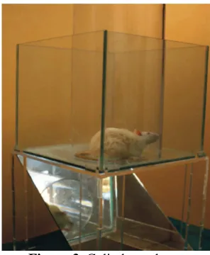
Beam traversal task test
As illustrated in figure 2, 5 cm wide and 1 m long wooden bridge fixed 50 cm above the ground was used. The animal was placed on one end of the bridge and released. The time needed for total crossing the bridge was documented.
Cylinder task test
Efficacy of the ketogenic diet in PD Iran J Neurol 2016; 15(2) 65
http://ijnl.tums.ac.ir 3 April inside a seven-through glass cylinder, and the total
number of forearm contacts with the wall was
documented within a 10-minute period.
Accordingly, the final score was calculated using the following formula.13
Figure 1. Bar test
Figure 2. Beam traversal task test
Figure 3. Cylinder task test
Score = 100 × (Number of forearm contacts on the lesion side + 1/2 of total forearm contacts)/total forearm contacts.
The KD was started from the 1st day of experiment and pramipexole (0.3 mg/kg once a day) was administered on the day 12 for 14 consecutive
days in relevant groups.
The data were presented as mean ± standard error (SE) of the mean. The SPSS software (version 16, SPSS Inc., Chicago, IL, USA) was used. The one-way ANOVA test coupled with the Tukey post-hoc analysis was employed for comparisons. Levels of serum ketone bodies were compared between groups using the Wilcoxon test. A P < 0.050 was considered statistically significant.
Results
The mean level of serum ketone bodies in the group on the KD was 0.25 ± 0.03 mmol/L (range: 0.21-0.28) at baseline and 1.83 ± 0.17 mmol/L (range: 1.60-2.10) on the day 14. According to the results of Wilcoxon test, the mean serum level of ketone bodies increased significantly on the day 14 compared to that at baseline (P = 0.010).
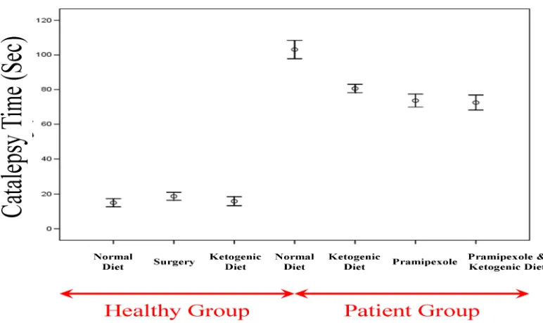
Results of the bar test (catalepsy time)
Results of the bar test in different groups were as follows (Figure 4): The control group on a regular diet: 15.00 ± 1.18 seconds (range: 10-20); the surgical sham group: 18.75 ± 1.15 seconds (range: 15-25); the control group on the KD: 15.88 ± 1.30 seconds (range: 10-21); the PD group on a regular diet: 1 03.13 ± 2.65 seconds (range: 95-115); the PD group on the KD: 80.63 ± 1.21 seconds (range: 75-85); the PD group on a diet with pramipexole: 73.63 ± 1.87 seconds (range: 67-80); and the PD group on the KD with pramipexole: 72.50 ± 2.17 seconds (range: 65-80).
66 Iran J Neurol 2016; 15(2) Shaafi et al.
http://ijnl.tums.ac.ir 3 April
Figure 4. The mean results of the bar test in different study groups
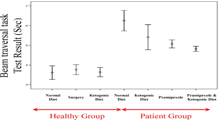
Results of the beam traversal task test
Results of the beam traversal task test in different groups were as follows (Figure 5): The control group on a regular diet: 3.61 ± 0.17 seconds (range: 3-4); the sham surgical group: 3.76 ± 0.13 seconds (range: 3-4); the control group on the KD: 3.64 ± 0.12 seconds (range: 3-4); the PD group on a regular diet: 6.25 ± 0.26 seconds (range: 5-8); the PD group on the KD: 5.41 ± 0.32 seconds (range: 4-7); the PD group on a diet with pramipexole: 5.08 ± 0.10 seconds (range: 5-6); and the PD group on the KD with pramipexole: 4.83 ± 0.06 seconds (range: 4.5-5).
There was not a significant difference between the controls on a regular diet and the sham surgical group in terms of the mean results of the beam traversal task test (P = 0.990); nor in the comparison between the controls on a regular diet and the controls on the KD (P = 0.990). Comparing
unaffected and PD groups, however, showed that the mean results of the beam traversal task test were significantly lower in the former (P < 0.001 for all paired comparisons). Comparing the PD group on a regular diet with the three remaining PD groups showed that the mean results of the beam traversal task test were significantly higher in the former (P = 0.040 in comparison with the PD group on the KD and P < 0.001 for the other two comparisons). Comparing the PD group on the KD and the PD group on a regular diet with pramipexole showed no significant difference between the two groups as to the results of the beam traversal task test (P = 0.860). A similar finding was found in comparisons between the PD group on the KD and the PD group on the KD with pramipexole (P = 0.300), and between the PD group on a regular diet with pramipexole and the PD group on the KD with pramipexole (P = 0.960).
Efficacy of the ketogenic diet in PD Iran J Neurol 2016; 15(2) 67
http://ijnl.tums.ac.ir 3 April
Figure 6. The mean results of the cylinder task test in different study groups
Results of the cylinder task test
Results of the cylinder task test in different groups are as follows (Figure 6): the control group on a regular diet: 48.38 ± 1.50% (range: 42-55); the sham surgical group: 50.38 ± 1.48% (range: 45-56); the control group on the KD: 49.88 ± 1.68% (range: 42-56); the PD group on a regular diet: 71.63 ± 1.86% (range: 62-80); the PD group on the KD: 64.13 ± 1.53% (range: 58-70); the PD group on a diet with pramipexole: 60.63 ± 1.10% (range: 57-65); and the PD group on the KD with pramipexole: 58.00 ± 1.48% (range: 52-65). Comparing the controls on a regular diet and the sham surgical group showed no significant difference between the two groups in terms of the mean results of the cylinder task test (P = 0.970). Similarly, no significant difference was found in the comparison between the controls on a regular diet and the controls on the KD (P = 0.990). Comparing the unaffected and PD groups showed significantly lower mean results of the cylinder task test in the former (P < 0.001 for all paired comparisons). In addition, comparing the PD group on a regular diet with the remaining three PD groups showed significantly lower mean results of the cylinder task test in the former (P = 0.020 in comparison with the PD group on the KD and P < 0.001 for the other two comparisons). There was no significant difference between the PD group on the KD and the PD group on a regular diet and pramipexole in terms of the results of the cylinder task test (P = 0.670). There was also no significant difference between the PD group on the KD and the PD group on the KD and pramipexole in this regard (P = 0.090). Finally, there was no significant
difference between the PD group on a regular diet with pramipexole and the PD group on the KD with pramipexole regarding the results of the cylinder task test (P = 0.890).
Discussion
Nutritional and metabolic therapeutic strategies have been tried in a wide range of neurologic diseases such as epilepsy, headache, neurotrauma, Alzheimer’s disease, sleep disorders, brain cancer, autism, pain, multiple sclerosis (MS), and PD. The related incentive possibly originates from the
ineffectiveness of available pharmacologic
treatments in many of such diseases, as well as a
general inclination toward more natural
products.14
The present experimental study on rats showed that the KD is significantly effective in promoting motor functions in PD animals and at the same time, it may enhance the therapeutic effects of concomitantly administered pramipexole, albeit in an insignificant fashion.
In accordance with these findings, using a standard scale, Vanitallie et al.6 showed that the KD was able to exert beneficial effects in 5 out of 7 PD patients.
One of the major limitations in the current study was using a small sample size that was unable to exclude the placebo effect. In a study by Tieu et al.,5 the injection of BHB acid also effectively prevented
MTPT-induced neurodegenerative and aging
68 Iran J Neurol 2016; 15(2) Shaafi et al.
http://ijnl.tums.ac.ir 3 April nigra against 6-OHDA-induced neurotoxicity. In a
study by Yang and Cheng,16 the anti-inflammatory effects of ketone bodies were confirmed using a model of MPTP-induced neurotoxicity.
In a similar experiment by Beckett et al.17 using a rat model of Alzheimer’s disease, in conformity with our findings the KD managed to significantly improve some aspects of motor functions. In this study, all aspects of motor functions of the examined animals were not influenced equally. Although the underlying cause of this finding is not clear, it seems that various muscular groups are affected unequally by the KD (for example the quadriceps muscles vs. the paw flexors).
In another series by Brownlow et al.,18 there was also a considerable improvement in motor functions in Alzheimeric rats using the KD. Ruskin et al.19 also showed that the administration of the KD could increase motor capabilities of animals with induced Huntington’s disease (HD).
Although the exact neuroprotective property of the KD is not known, some hypotheses have been suggested. The two major aspects in treating with the KD is an increase in the production rate of ketone bodies in the liver and a decrease in serum glucose levels. An increased level of ketone bodies is assumed to be related to fatty acid oxidation. Certain polyunsaturated fatty acids such as arachidonic acid
(AA), docosahexaenoic acid (DHA), and
eicosapentaenoic acid (EPA) may regulate the stimulatory properties of the neural sheaths through inhibiting voltage-dependent calcium and sodium channels, decreasing inflammation and inducing mitochondrial uncoupling proteins that could lead to production of reactive oxygen species (ROS).14
Ketone bodies themselves may also bear
neuroprotective properties.2
This effect develops through increasing the levels of adenosine triphosphate and decreasing ROS production via enhancing nicotinamide adenine dinucleotide hydrogen (NADH) oxidation and preventing mitochondrial permeability change.
Along with other enhanced bioenergetics
pathways, ketone bodies are able to stimulate mitochondrial biogenesis and stabilize synaptic functions. The second major biochemical feature of ketone bodies is diminishing glycolytic flow. This condition is the main feature in calorie intake limitation that could elongate the life of various species including primates. It seems that the observed neuroprotective effect is due to a
diminished incidence of the brain-derived
neurotrophic factor and its principle receptor the tyrosine kinase B, an improved mitochondrial
function, diminished oxidative stress, a
compromised activity of pro-apoptotic factors, and the prevention of inflammatory mediators such as interleukins and tumor necrosis factor-alpha.14
Possible mechanisms involved in the
effectiveness of the KD in relieving PD symptoms could be summarized as follows:
•Providing an efficacious source of energy,
which is capable of preventing local
hypometabolism in the brain •Diminished oxidative injury
•Increasing mitochondrial biogenesis pathways •Exploiting ketone capacities to bypass a failure in the Complex I activity.20
Noh et al.21 showed that one of principle
mechanisms of the KD in preventing
neurodegeneration was via impeding neural apoptosis by caspase-3.
The early pathophysiology of PD is an excitotoxic degeneration of the dopaminergic neurons located in the substantia nigra. On the basis of some findings, ketone bodies may bypass defects in mitochondrial Complex I activities, which are possibly involved in the pathogenesis of PD.6 Therefore, it seems that mitochondrial abnormalities play a pivotal role in this regard.22
Kashiwaya et al. used an analog of heroin to
destroy dopaminergic cells. The suggested
mechanism involved in this process was the blockade of a mitochondrial NADH dehydrogenase multi-enzyme complex.4 Accordingly, much of previous studies on the role of the KD in PD have been focused on this aspect of the disease (i.e., mitochondrial abnormalities).23-27 For example, in a study by Kashiwaya et al.4 on an animal model of PD induced by MPTP, the administration of BHB reduced mitochondrial respiration cycle lesions, which are usually induced by the used toxin.
In a study by Kim et al. the protective effects of ketone bodies against mitochondrial respiratory complex lesions developed by inhibitors of complex I (rotenone) and II (exogenous 3-nitropropionic acid) were examined. They suggested their findings as potential neuroprotective mechanisms of ketone bodies in PD.7
Efficacy of the ketogenic diet in PD Iran J Neurol 2016; 15(2) 69
http://ijnl.tums.ac.ir 3 April Conclusion
According to the findings of the present study, since the KD is effective in improving motor function in PD rats further human studied are recommended in this regard.
Conflict of Interests
The authors declare no conflict of interest in this study.
Acknowledgments
The authors would like to sincerely thanks to the
staff of laboratory of NSRC and Research Council in Tabriz University of Medical Sciences for providing financial and emotional assistance in this project.
How to cite this article: Shaafi Sh, Najmi S, Aliasgharpour H, Mahmoudi J, Sadigh-Etemad S, Farhoudi M, et al. The efficacy of the ketogenic diet on motor functions in Parkinson’s disease: A rat model. Iran J Neurol 2016; 15(2): 63-9.
References
1. Mahmoudi J, Mohajjel NA, Reyhani-Rad S,
Samini M. Fluoxetine improves the effect of levodopa on 6-hydroxy dopamine-induced motor impairments in rats. Adv Pharm Bull 2012; 2(2): 149-55.
2. Gasior M, Rogawski MA, Hartman AL.
Neuroprotective and disease-modifying
effects of the ketogenic diet. Behav Pharmacol 2006; 17(5-6): 431-9.
3. Hartman AL, Gasior M, Vining EP, Rogawski
MA. The neuropharmacology of the ketogenic diet. Pediatr Neurol 2007; 36(5): 281-92.
4. Kashiwaya Y, Takeshima T, Mori N,
Nakashima K, Clarke K, Veech RL. D-beta-hydroxybutyrate protects neurons in models of Alzheimer's and Parkinson's disease. Proc Natl Acad Sci USA 2000; 97(10): 5440-4.
5. Tieu K, Perier C, Caspersen C, Teismann P,
Wu DC, Yan SD, et al.
D-beta-hydroxybutyrate rescues mitochondrial
respiration and mitigates features of Parkinson disease. J Clin Invest 2003; 112(6): 892-901.
6. Vanitallie TB, Nonas C, di Rocco A, Boyar
K, Hyams K, Heymsfield SB. Treatment of
Parkinson disease with diet-induced
hyperketonemia: a feasibility study.
Neurology 2005; 64(4): 728-30.
7. Kim DY, Davis LM, Sullivan PG, Maalouf
M, Simeone TA, van Brederode J, et al. Ketone bodies are protective against oxidative stress in neocortical neurons. J Neurochem 2007; 101(5): 1316-26.
8. Maalouf M, Sullivan PG, Davis L, Kim DY,
Rho JM. Ketones inhibit mitochondrial production of reactive oxygen species production following glutamate excitotoxicity
by increasing NADH oxidation.
Neuroscience 2007; 145(1): 256-64.
9. Maalouf M, Rho JM, Mattson MP. The
neuroprotective properties of calorie
restriction, the ketogenic diet, and ketone bodies. Brain Res Rev 2009; 59(2): 293-315.
10. Silberstein S, Marmura M, Muntner N.
Essential neuropharmacology: the
prescriber's guide. Cambridge, UK:
Cambridge University Press; 2010.
11. Zhao Q, Stafstrom CE, Fu DD, Hu Y,
Holmes GL. Detrimental effects of the ketogenic diet on cognitive function in rats. Pediatr Res 2004; 55(3): 498-506. 12. Streijger F, Plunet WT, Lee JH, Liu J, Lam
CK, Park S, et al. Ketogenic diet improves forelimb motor function after spinal cord injury in rodents. PLoS One 2013; 8(11): e78765.
13. Soleman S, Yip P, Leasure JL, Moon L.
Sustained sensorimotor impairments after
endothelin-1 induced focal cerebral
ischemia (stroke) in aged rats. Exp Neurol 2010; 222(1): 13-24.
14. Stafstrom CE, Rho JM. The ketogenic diet
as a treatment paradigm for diverse neurological disorders. Front Pharmacol 2012; 3: 59.
15. Cheng B, Yang X, An L, Gao B, Liu X, Liu
S. Ketogenic diet protects dopaminergic neurons against 6-OHDA neurotoxicity via up-regulating glutathione in a rat model of Parkinson's disease. Brain Res 2009; 1286: 25-31.
16. Yang X, Cheng B. Neuroprotective and
anti-inflammatory activities of ketogenic diet on MPTP-induced neurotoxicity. J Mol Neurosci 2010; 42(2): 145-53.
17. Beckett TL, Studzinski CM, Keller JN, Paul
Murphy M, Niedowicz DM. A ketogenic diet improves motor performance but does not affect beta-amyloid levels in a mouse model of Alzheimer's disease. Brain Res 2013; 1505: 61-7.
18. Brownlow ML, Benner L, D'Agostino D,
Gordon MN, Morgan D. Ketogenic diet improves motor performance but not cognition in two mouse models of Alzheimer's pathology. PLoS One 2013; 8(9): e75713.
19. Ruskin DN, Ross JL, Kawamura M, Ruiz
TL, Geiger JD, Masino SA. A ketogenic
diet delays weight loss and does not impair working memory or motor function in the R6/2 1J mouse model of Huntington's disease. Physiol Behav 2011; 103(5): 501-7.
20. Paoli A, Bianco A, Damiani E, Bosco G.
Ketogenic diet in neuromuscular and
neurodegenerative diseases. BioMed
Research International 2014; 2014: 10.
21. Noh HS, Kim YS, Choi WS.
Neuroprotective effects of the ketogenic diet. Epilepsia 2008; 49(Suppl 8): 120-3. 22. Camilleri A, Vassallo N. The centrality of
mitochondria in the pathogenesis and treatment of Parkinson's disease. CNS Neurosci Ther 2014; 20(7): 591-602.
23. Gano LB, Patel M, Rho JM. Ketogenic diets,
mitochondria, and neurological diseases. J Lipid Res 2014; 55(11): 2211-28.
24. Valero T. Mitochondrial biogenesis:
pharmacological approaches. Curr Pharm Des 2014; 20(35): 5507-9.
25. Henderson ST. Ketone bodies as a
therapeutic for Alzheimer's disease.
Neurotherapeutics 2008; 5(3): 470-80.
26. Studzinski CM, MacKay WA, Beckett TL,
Henderson ST, Murphy MP, Sullivan PG, et al. Induction of ketosis may improve mitochondrial function and decrease steady-state amyloid-beta precursor protein (APP) levels in the aged dog. Brain Res 2008; 1226: 209-17.
27. Veech RL. The therapeutic implications of
ketone bodies: the effects of ketone bodies
in pathological conditions: ketosis,
ketogenic diet, redox states, insulin
resistance, and mitochondrial metabolism. Prostaglandins Leukot Essent Fatty Acids 2004; 70(3): 309-19.
the upper extremities.2 The recent American Academy of Orthopaedic Surgeons Clinical Guidelines define CTS as asymptomatic compression neuropathy, which is characterized by decreased median nerve function at the level of wrist accompanying with physiologically increased pressure in the CT. With a prevalence of about 50 per 1000 subjects, the incidence of CTS has been estimated 1-3 per 1000 cases per year in the United States.3 Current literature reports a higher prevalence of hypothyroidism and diabetes in patients with CTS. The prevalence of CTS is estimated to be 5.8% in women and 0.6% in men, in the general population4 while its prevalence in hypothyroid patients is reported about 28.5%.1 A retrospective review of patients who underwent surgery for CTS over a 3-year period by Vashishtha et al.5 in the UK showed that CTS is associated with thyroid dysfunction and diabetes.
The British Society for Surgery of the Hand advises screening CTS patients for thyroid and glucose dysfunction before surgery.5 Considering the high prevalence of CTS in hypothyroidism, early diagnosis of this disorder to prevent nerve changes is very important. CTS is characterized by typical anatomic changes, the most probable swelling of the median nerve in the proximal part of the CT.6,7 The diagnosis is usually clinical using Tinel’s sign and Phalen’s maneuver and is confirmed by nerve conduction study (NCS).8 Although NCS is the method most frequently used in practice to confirm a clinical diagnosis of CTS, it is known to be painful or unpleasant for patients, and false negatives and false positives occur even if the most sensitive methods are used.2 In addition, NCS does not provide anatomical information about the nerve or its surroundings that could help in determining its
etiology.9 Recently, using high-frequency
ultrasonography (US) for CTS has emerged as an alternative confirmatory test with a sensitivity of 44-95% and a specificity of about 57-100%.10 Various studies1,11-13 suggested different cut-off points to have the best sensitivity and specificity for median nerve cross-sectional area (CSA) asthe most reliable finding for the diagnosis of CTS.14
Yet, no study has been conducted to evaluate the diagnostic value of high-frequency US in the diagnosis of CTS in patients with hypothyroidism. To determine whether these findings are reliable and can be used to establish the diagnosis, we aimed to evaluate US as an alternative procedure for diagnosis of subclinical CTS in hypothyroid patients in a cross-sectional study.
This study was approved by the Local Ethics
Committee of the Firoozgar Clinical Research Development Center, Iran University of Medical Sciences, Tehran, Iran. Informed consent was taken from the patients before the diagnostic procedures. No invasive method was used in this study and all the diagnostic procedures were harmless. The patients’ information remains confidential and would be used only for analytical study.
Between April 2013 and November 2014, from the patients with the diagnosis of hypothyroidism referring to the institute of endocrinology and metabolism of Firoozgar Hospital, those who met our inclusion criteria entered the study. The patients with CTS or injection, wrist fractures, median nerve neuropathy and cervical radiculopathy, or polyneuropathy were excluded. In addition, the patients with diseases and conditions associated with CTS including pregnancy, rheumatoid arthritis (RA), diabetes mellitus (DM), renal failure, gout, tenosynovitis, tumors, ganglion cysts, amyloidosis, and median nerve with two or more branches were also excluded from the study. Bilateral wrist US at the CT inlet was done for the included patients using a MyLab™40 - Esaote with an 18 MHz ultrasound probe by an expert in the neurology clinic to measure the median nerve CSA.
To compare the US measurements with the standard values, NCS was done for all patients as the gold standard test. Other variables such as age, body mass index (BMI), disease duration, wrist circumference, and median nerve CSA were recorded for each patient in the data collection form. The patients were divided into two groups of subclinical CTS and the control group according to the clinical Tinel’s sign and Phalen’s tests, confirmed by NCS. The median nerve CSA at the forearm CT inlet was assessed by an expert investigator blinded to the clinical and NCS data.
The subjects were seated facing the examiner with their arms extended, their wrists on a flat surface, their forearms supine, and their fingers semi-extended. The transverse US of the median nerve was performed at the inlet of the CT. The pisiform bone was used as an anatomical landmark to measure the median nerve CSA at the CT inlet, by
tracing a continuous line within the
hyperechogenicity boundary of the nerve.
test evaluations. Quantitative variables expressed as mean ± standard deviation (SD) and frequency was used for qualitative data.
A total number of 76 patients (152 wrists bilaterally) were recruited in the study including 70 women and 6 men. The mean age of the subjects was 43.1 ± 13.8 years (range from 19 to 81) with a mean BMI of 27.5 ± 3.8 kg/m2 (minimum 20 and maximum 45). The mean duration of disorder was 7.5 ± 3.3 years (from 2 to 15). The average wrists surface area was 20.0 ± 1.7 cm2 (minimum 16 and maximum of 24), and the average of median nerve CSA at the CT inlet was 7.6 ± 1.9 mm2 (minimum of 4 and maximum of 15). About 31 wrists (20.0%) had CTS diagnostic criteria in the NCS, which were considered as sub-clinical CTS. The median nerve CSA was more than 9 mm2 in 14 cases (9.0%) (Figure 1). The mean median nerve CSA at the tunnel inlet was 9.96 mm2 (SD: 2.2) for the CTS affected wrists and 7.08 mm2 (SD: 1.38) for the normal wrists (P < 0.05). The average of median nerve CSA was 8 mm2 in left and 7.34 mm2 in right wrists. The same parameter was 8 mm2 in males and 7.6 mm2 in females (The difference was not significant). The average of median nerve CSA seemed also not to be associated with age, BMI, wrist circumference and duration of the disease. According to the NCS findings, 31 wrists (20.4%) were diagnosed as CTS while 19 wrists (12.5%) were diagnosed as CTS by the US. The sensitivity and specificity of US in the diagnosis of CTS was 45.0 and 95.8%, respectively, with a CSA cutoff point of 9.8 mm2. Positive and negative predictive values of US were 73.7 and 87.2%, respectively, with a test accuracy of 85.5%.
Although CTS is clinically diagnosed, moderate sensitivity and specificity have been reported for
clinical symptoms15 and false negative and positives results for NCS.16,17 A some of the CTS symptoms such as paresthesia may appear before nerve fiber changes, which can justify the false negative results of NCS. According to different studies, NCS has a sensitivity of about 56-85% and a specificity of 94% or more in the diagnosis of CTS.8 Although semi-invasive, NCS is the gold standard diagnostic procedure for CTS, while comparing to US, the electrical stimuli may not be pleasant for the patient.3,4
In our study, the median nerve US had a sensitivity and specificity of 45% and 95.8% for diagnosis of CTS in patients with hypothyroid. The US measurements of median nerves were found to be increased significantly in patients with CTS when compared with controls, particularly in terms of CSA. As a result, high-frequency US can be used to confirm the diagnosis and due to high positive predictive value, it can be a suitable test to evaluate CTS in the majority of patients with clinical suspicion of CTS, reserving NCS only for with negative US findings. Current literature approves that patients with CTS have a higher prevalence of hypothyroidism and screening helps diagnosing new cases of this condition in this selected group.17 A random-effects meta-analysis of the studies not controlling their estimates for any confounder confirmed an association between CTS and hypothyroidism.18
In an interesting study by Kolovos and Tsiotas19 to establish US examination as a method with at least of the same accuracy with electrodiagnostic (EDX) study, 60 healthy individuals and 30 patients suffering from CTS were scanned. The authors suggested that ratios over the value 1.0 could be considered as a definite diagnosis of CTS. While, ratios up to 0.79 would be surely refers to a healthy wrist and the intermediate ratios between 0.79 and 1.0 refers to a gray zone, which is practically considered healthy.
Recent studies have demonstrated advantages of US in the diagnosis of CTS; however, its role is limited due to lack of adequate data relative to EDX testing. The wide variations of sensitivities and specificities reported in the literature, on the other hand, prevent meaningful analysis of US as either screening or confirmatory test in the diagnosis of CTS.2 The meta-analysis by Fowler et al. on 19 articles with a total sample size of 3131 wrists reported the sensitivity and specificity of US for the diagnosis of CTS, 77.6 and 86.8%, respectively.2 The study by Kwon et al.20 also showed a sensitivity of 66.0% and specificity of 63.0% for median nerve CSA of 10.7 mm2 for the US, while the sensitivity and specificity of NCS were 78.0 and 83.0%, respectively. In another study by Mohammadi et al.,21 the median nerve CSA at the CT inlet and outlet were examined bilaterally in 82 patients with electrophysiologically confirmed CTS to determine whether high-resolution US can be considered as an alternative diagnostic method to NCS in grading the severity of CT. The differences of the median nerve CSA in mild, moderate and severe CTS were not statistically significant either in the CT inlet or in outlet. According to this study, US of the median nerve CSA had a diagnostic value to confirm or exclude CTS, but could not be used for grading of its severity. In contrast, Azami et al.22 prospectively to examined individuals with EDX proven CTS and healthy control subjects to determine the diagnostic value of US compared with NCS. With a sensitivity and specificity of 99.2 and 88.3%, CSA at the tunnel inlet with a threshold of 9.15 mm2 had the best diagnostic accuracy. There was reported a significant difference in the median nerve CSA in mild, moderate and severe CTS in this study. In another cross-sectional case-control study, Ghasemi et al.23 assessed findings in US in correlation with severity of CTS. According to EDX, patients were classified as mild, moderate, and severe CTS and high-resolution US for CSA measurement at the tunnel inlet was performed for all patients. The mean of the CSA in mild, moderate and severe CTS wrists were 0.12 cm2 in, 0.15 and 0.19 cm2, respectively. A significant correlation between the median nerve CSA and the severity of CTS impelled the authors claiming that US may serve as a complementary and reliable method in assessing the severity of CTS.
Yazdchi et al.13 also studied the sensitivity and specificity of median nerve US in diagnosis of CTS in 90 Iranian patients with clinically suspected CTS. The median nerve CSA at the three levels of the CT could fairly differentiate severe CTS from other cases. Their results suggested that median nerve US
cannot replace the NCS because of an overall low sensitivity and specificity.13 Dejaco et al.24 prospectively studied 135 consecutive patients with diagnosis of CTS in order to compare US measurement of median nerve CSA. They measured CSA using US at five different levels at forearm and wrist. The US of median nerve swelling revealed a good reliability with an intraclass correlation coefficient of 0.90, allowing a fairly reliable diagnosis of CTS.
Kim et al.25 studied 187 patients, to determine the criteria for US measurement of the median nerve CSA and differential diagnosis of patients with CTS with or without diabetic polyneuropathy. All the CSAs in this study were larger in the diabetic polyneuropathy group compared with those in the control group. The cutoff value for the CSA at the wrist that yielded the highest sensitivity and specificity was 11.6 mm2. They concluded that diagnosing the comorbidity of CTS with diabetic polyneuropathy could be done according to the median nerve CSA at the wrist and the wrist-forearm ratio. In another similar study, Kanikannan et al.26 compared the diagnostic accuracy of high-resolution US and electrophysiology in the diagnosis of CTS in patients with CTS and CTS associated with peripheral neuropathy. High-resolution US showed a good correlation with EDX studies in all grades of CTS in these patients with the sensitivity, specificity, positive predictive value and negative predictive values of 76.4, 72.7, 89.5 and 68.0 percent, respectively. They claimed that US can be used as a complementary screening tool to EDX.
Although US is an operator-dependent procedure and should be done by or under supervision of an expert, it may be preferred by patients since it is painless and easily accessible. The high-resolution US allows direct imaging of the involved nerves, in addition to documentation of nerve shape changes that occur in compressive syndromes. US can also diagnose a spectrum of entrapment causes such as tenosynovitis, ganglia, soft-tissue tumors, bone and joint abnormalities, and anomalous muscles. According to our study, US may not replace EDX testing as the most sensitive and specific diagnostic test for CTS diagnosis in hypothyroid patients, but it can be used as the firstline confirmatory test as its accuracy was detected 85.5% in our study.
The authors declare no conflict of interest in this study.
We acknowledge all patients that have participated in this study.
1. Cakir M, Samanci N, Balci N, Balci MK. Musculoskeletal manifestations in patients with thyroid disease. Clin Endocrinol (Oxf) 2003; 59(2): 162-7.
2. Fowler JR, Gaughan JP, Ilyas AM. The sensitivity and specificity of ultrasound for the diagnosis of carpal tunnel syndrome: a meta-analysis. Clin Orthop Relat Res 2011; 469(4): 1089-94.
3. Keith MW, Masear V, Chung KC, Maupin K, Andary M, Amadio PC, et al. American Academy of Orthopaedic Surgeons Clinical Practice Guideline on diagnosis of carpal tunnel syndrome. J Bone Joint Surg Am 2009; 91(10): 2478-9.
4. de Krom MC, Knipschild PG, Kester AD, Thijs CT, Boekkooi PF, Spaans F. Carpal tunnel syndrome: prevalence in the general population. J Clin Epidemiol 1992; 45(4): 373-6.
5. Vashishtha M, Varghese B, Mosley F, Kadakia A, de Jager W. Screening for thyroid dysfunction and diabetes in patients with carpal tunnel syndrome. Surgeon 2014. 6. Buchberger W, Judmaier W, Birbamer G, Lener M, Schmidauer C. Carpal tunnel syndrome: diagnosis with high-resolution sonography. AJR Am J Roentgenol 1992; 159(4): 793-8.
7. Martinoli C, Bianchi S, Gandolfo N, Valle M, Simonetti S, Derchi LE. US of nerve entrapments in osteofibrous tunnels of the upper and lower limbs. Radiographics 2000; 20 Spec No: S199-S213.
8. Jablecki CK, Andary MT, Floeter MK, Miller RG, Quartly CA, Vennix MJ, et al. Practice parameter: Electrodiagnostic studies in carpal tunnel syndrome. Report of
the American Association of
Electrodiagnostic Medicine, American Academy of Neurology, and the American Academy of Physical Medicine and Rehabilitation. Neurology 2002; 58(11): 1589-92.
9. Abrishamchi F, Zaki B, Basiri K, Ghasemi M, Mohaghegh M. A comparison of the ultrasonographic median nerve cross-sectional area at the wrist and the wrist-to-forearm ratio in carpal tunnel syndrome. J Res Med Sci 2014; 19(12): 1113-7. 10. Seror P. Sonography and electrodiagnosis in
carpal tunnel syndrome diagnosis, an analysis of the literature. Eur J Radiol 2008; 67(1): 146-52.
11. Mohammadi A, Afshar A, Etemadi A, Masoudi S, Baghizadeh A. Diagnostic value of cross-sectional area of median nerve in grading severity of carpal tunnel syndrome. Arch Iran Med 2010; 13(6): 516-21. 12. Ashraf AR, Jali R, Moghtaderi AR, Yazdani
AH. The diagnostic value of
ultrasonography in patients with electrophysiologicaly confirmed carpal tunnel syndrome. Electromyogr Clin Neurophysiol 2009; 49(1): 3-8.
13. Yazdchi M, Tarzemani MK, Mikaeili H, Ayromlu H, Ebadi H. Sensitivity and specificity of median nerve ultrasonography in diagnosis of carpal tunnel syndrome. Int J Gen Med 2012; 5: 99-103.
14. Sarria L, Cabada T, Cozcolluela R, Martinez-Berganza T, Garcia S. Carpal tunnel syndrome: usefulness of sonography. Eur Radiol 2000; 10(12): 1920-5.
15. Katz JN, Simmons BP. Clinical practice. Carpal tunnel syndrome. N Engl J Med 2002; 346(23): 1807-12.
16. Hamanaka I, Okutsu I, Shimizu K, Takatori Y, Ninomiya S. Evaluation of carpal canal pressure in carpal tunnel syndrome. J Hand Surg Am 1995; 20(5): 848-54.
17. Ferry S, Silman AJ, Pritchard T, Keenan J, Croft P. The association between different patterns of hand symptoms and objective evidence of median nerve compression: a community-based survey. Arthritis Rheum 1998; 41(4): 720-4.
18. Shiri R. Hypothyroidism and carpal tunnel
syndrome: a meta-analysis. Muscle Nerve 2014; 50(6): 879-83.
19. Kolovos S, Tsiotas D. Ultrasonographic diagnosis of carpal tunnel syndrome: introducing a new approach. Eur J Orthop Surg Traumatol 2016; 26(2): 167-75. 20. Kwon BC, Jung KI, Baek GH. Comparison
of sonography and electrodiagnostic testing in the diagnosis of carpal tunnel syndrome. J Hand Surg Am 2008; 33(1): 65-71.
21. Mohammadi A, Ghasemi-Rad M,
Mladkova-Suchy N, Ansari S. Correlation between the severity of carpal tunnel syndrome and color Doppler sonography findings. AJR Am J Roentgenol 2012; 198(2): W181-W184.
22. Azami A, Maleki N, Anari H, Iranparvar Alamdari M, Kalantarhormozi M, Tavosi Z. The diagnostic value of ultrasound compared with nerve conduction velocity in carpal tunnel syndrome. Int J Rheum Dis 2014; 17(6): 612-20.
23. Ghasemi M, Abrishamchi F, Basiri K, Meamar R, Rezvani M. Can we define severity of carpal tunnel syndrome by ultrasound? Adv Biomed Res 2015; 4: 138. 24. Dejaco C, Stradner M, Zauner D, Seel W,
Simmet NE, Klammer A, et al. Ultrasound for diagnosis of carpal tunnel syndrome: comparison of different methods to determine median nerve volume and value of power Doppler sonography. Ann Rheum Dis 2013; 72(12): 1934-9.
25. Kim LN, Kwon HK, Moon HI, Pyun SB, Lee HJ. Sonography of the median nerve in carpal tunnel syndrome with diabetic neuropathy. Am J Phys Med Rehabil 2014; 93(10): 897-907.
Classic risk factors for venous thrombosis are divided into two main groups of acquired factors such as immobilization, surgery and cancers, and genetic risk factors, like activated protein C resistance, and deficiencies of protein C or S and antithrombin.1 Evidence is accumulating that venous thromboembolism is not limited to coagulation system and immune system seems to be involved in formation and resolution of thrombus.2,3