Short Report
Simultaneous Detection of
Helicobacter
Genus and
Helicobacter
pylori
Species using a Multiplex PCR Method
Shohreh Farshad
*, Manoochehr Rasouli and Abdolvahab Alborzi
Professor Alborzi Clinical Microbiology Research Center, Shiraz University of Medical Sciences, Shiraz, Iran
Received 12 June 20032; revised 12 November 2003; accepted 30 December 2003
ABSTRACT
In order to improve simultaneous detection and identification of Helicobacter genus in general and
Helicobacter pylori specifically and reduce the number of amplifications needed, we established a multiplex PCR. In this study, two pairs of primers: Hcom1 and Hcom2 specific for Helicobacter genus, Hicd1 and Hicd2 specific for Helicobacter pylori species were used. To determine the sensitivity of our multiplex PCR, the lower limits of DNA detection from pure culture were established using phenol-chloroform method. To evaluate the specificity of our protocol, we tested DNA extracted from various Gram-negative and -positive bacteria. A study was subsequently undertaken on stomach tissue samples from 18 patients to evaluate this protocol for detection of Helicobacter in tissue samples. In our optimized PCR, two fragments of 389- and 1200-bp were produced using Hcom1-Hcom2 and Hicd1-Hicd2 primers, respectively. Amplification of Helicobacter pylori genomic DNA was achieved at concentration as low as 0.03 pg equivalent to 150 bacteria. No DNA amplification of other Gram- positive and -negative bacteria was seen. Twelve specimens, positive in culture and rapid urease test for Helicobacterpylori were positive for DNA of this organism using this multiplex PCR. Our results demonstrate that this protocol represents a specific and sensitive assay for simultaneous detection of
Helicobacter genus members, in general, and Helicobacter pylori species, specifically, in clinical samples yielding no false-positive. Iran. Biomed. J. 8 (4): 205-209, 2004
Keywords: Helicobacter, Helicobacterpylori, Multiplex PCR
INTRODUCTION
H
elicobacter is a microaerophilic Gram-negative spiral-shaped microbe that was first described in 1983 [1]. It has been cited as the most prevalent bacterial pathogen of human.
Helicobacter species have been isolated from
various animal species in association with or without gastric ulcers [2-5]. Thereafter in 1984,
Helicobacter pylori, the most prevalent species of
Helicobacter genus, was described [6]. It is strongly
associated with gastroduodenal diseases including chronic and active gastritis, duodenal ulcer, peptic ulcer and gastric mucosa-associated lymphoid tissue [7]. A chronic H. pylori infection is also associated with an increasing risk for the patient to develop gastric adenicarcinoma later in life [8]. H. cinaedi
and H. fennelliae can cause gastoenteritis and
protocolitis with septicemia in homosexual men. H.
cinaedi also causes recurrent cellulitis with fever
and bacteremia in immuno-compromised patients [9]. During the past few years, it has been reported that H. pylori and other Helicobacter species can cause some kinds of extradigestive diseases in liver and gallbladder of human [4, 10, 11]. There are some other reports that discuss about the relationship of Helicobacter to diabet and anemiae diseases [12-15]. Therefore, it seems that
Helicobacter organisms can cause a wide rage of
diseases. In addition, one disease can be caused by two different Helicobacter species, as H. heilmannii has recently been identified in human gastric pathology [16, 17]. Hence, it is valuable to make plans for researches on the basis of detectiong
Helicobacter species in any disease suspect to be related to these organisms. Histopathological examinations and urease tests performed on gastric biopsis, urea breath tests, gastric biopsy culturs and serology are techniques used to diagnose H. pylori infections. However, these methods have the disadvantages of being time-consuming (except the breath test) and giving false-negative and -positive results [18,19].
In the last decade, PCR has become an important tool to identify a number of fastidious organisms
such as Helicobacter. Molecular assays are
inherently valuable because detection can be achieved by enhanced sensitivity and specificity, and it is not diminished by non-viable organisms. The aim of this study was to establish a sensitive and specific multiplex PCR according to two pairs of primers based on sequence specific for genus and species to identify Helicobacter genus gene in general and H. pylori species gene in particular. Ultimately, It was our aim to adapt this method for direct detection from clinical specimens.
MATERIALS AND METHODS
Clinical samples and culture. From 18 patients who underwent endoscopy for upper gastro-intestinal complaints, 2 gastric biopsies were taken. For each patient, one biopsy was placed in a sterile
bottle and stored at -70°C until subsequent
preparation for PCR. Another biopsy sample was used for rapid urease test, gram staining and culture
on Brucella agar supplemented with 10% lysed
horse blood and antibiotics of Trimethoprim, amphotericin B and nalidixic acid. The cultures were incubated in a microaerophilic atmosphere in gaspak jars at 37°C for 5-7 days. The isolates were identified as H. pylori by their morphology upon gram staining and by positive oxidase, catalase and rapid urease tests.
Positive controls. Three confirmed H. pylori isolates from gastric biopsies were used as positive controls to determine the PCR in vitro assay sensitivity and test sensitivity and to validate this multiplex PCR protocol.
DNA extraction from bacteria. Genomic DNA from positive controls was extracted using three methods. The bacterial suspensions used in all three methods were made in normal saline and had the same bacterial concentration.
A) Phenol-chloroform method. Briefly, 350 µl of extraction buffer (100 mM NaCl, 10 mM Tris-HCL [pH 8.0], 25 mM EDTA, 0.5% SDS) was added to 100 µl of bacterial suspension in a 1.5 ml Eppendorf tube. To each tube, 3 µl of proteinase K (20 mg/ml) (MBI, Fermentas, USA) was added. The tubes were then incubated in a hot block at 56°C for 120 min and afterwards kept in lab temperature overnight for cell lysis completion. After centrifugation, the supernatant was transferred to new tube. DNA was isolated from supernatant by a sequence of procedures including extraction via phenol chloroform, precipitation with ethanol and dissolution in sterile double distilled water, respectively.
B) Sonication method. The bacterial suspension was sonicated for 2 periods of 5 min. This crude lysate was used as template in the PCR.
C) Boiling method. The bacterial suspension was incubated in a hot block at 95°C for 60 min and then was used in the PCR.
In each method, nucleic acid concentration was determined spectrophotometricaly. Each genomic DNA (10 µl) obtained from above protocols was used as the template in the PCR.
DNA extraction from tissue. In a 1.5-ml Eppendorf tube, 1.2 ml digestion buffer (NaCl 100 mM, Tris-HCl 10 mM [pH 8.0], EDTA 25 mM, SDS 0.5%) and 10 µl proteinase K were added to 100 mg of homogenized stomach tissue and the mixture was vortexed and incubated in a hot block at 56°C for 120 min. These lysates were then kept at 37°C for 4-5 days while shaking. Thereafter, all tissue lysates were processed using the phenol-chloroform method to purify genomic DNA for PCR.
Primer preparation. Two sets of primers (TIB MoLBId, Syntheselabor, Berlin, Germany) were used in this protocol. A pair of primers (Hcom1 and Hcom2) on the basis of the 16 s rRNA gene sequence of Helicobacter genus, as described by Choi et al. [20], was with the sequences of 5- - GTA
AAG GCT CAC CAA GGC TAT-3- and 5-CCA
CCT ACC TCT CCC ACA CTC-3-. The second set
of primers (Hicd1 and Hicd2), based on an isocitrate dehydrogenase gene sequence of H. pylori species, as originally described by Argyros et al. [21], were with the sequences of 5--ATG GCT TAC
AAC CCT AAA ATT TTA CAA AAG CC-3- and
5- -TCA CAT GTT TTC AAT CAT CAC GC-3-.
Optimization of multiplex PCR. To optimize the condition, in which two sets of primers could amplify their specific target, a broad range of each reagent was used. The optimized reaction was performed in a volume of 50 µl, comprising 50 pm
each primer, 10 µl of chromosomal DNA from
positive control, 2 U of Taq DNA polymerase, 0.2 mM deoxynucleoside triphosphates, and 2 mM MgCl2 in a thermal cycler gradient (Eppendorf,
Germany) using a broad range of annealing temperature according to the annealing temperature of each pair of primers. The cycle profiles were set as follows: initial denaturation at 94°C for 5 min denaturation at 94°C for 1 min, primer annealing at 55°C for 2 min, and 2 min extention at 72°C. The samples were amplified for 30 cycles followed by 10 min at 72°C. PCR products were analyzed by electrophoresis of a 10-µl aliquot using a 1.5% (wt/vol) agarose gel and the sizes of the PCR products were estimated by comparison with 100-bp DNA size markers (MBI, Fermentas, USA). Negative control reactions with distilled water were performed with each batch of amplification to exclude the possibility of contamination.
Sensitivity assay. A determined concentration of DNA extracted from pure H. pylori was serial diluted (dilution from 10-1 to 10-6) and optimized
multiplex PCR was done on these dilutions. According to the genomic DNA, molecular weight
of H. pylori [22] sensitivity of this PCR assay was
determined based on the highest dilution of DNA in which the primers could amplify their specific sequences.
Specificity assay. The specificity of optimized multiplex PCR was tested using 9 non-Helicobacter control bacteria including: E. coli, Enterobacter sp.,
Klebsiella sp., Shigella sp., Proteus mirabilis,
Salmonella typhi, Streptococcus viridance,
Campylobacter sp. and Heamophilus influenza.
Identification of Helicobacter in clinical samples. To evaluate this protocol in identification
of Helicobacter in clinical, this multiplex PCR was
performed on DNA extracts from stomach samples of 18 patients who underwent endoscopy.
RESULTS
Development of multiplex PCR. Our optimized multiplex PCR assay successfully amplified two
fragments of the expected sizes of 389- and 1200-bp from a DNA preparation of H. pylori positive control, using Hcom1-Hcom2 and Hicd1-Hicd2 primers, respectively (Fig. 1).
Sensitivity. The sensitivity of this assay has been tested by serial dilution of genomic DNA from H.
pylori positive control. The 389- and 1200-bp
fragments were amplified by this multiplex PCR assay from a minimum of 0.03 pg of DNA from H.
pylori equivalent to 150 organisms.
The comparison of three DNA extraction techniques of boiling, sonication and phenol chloroform showed the same sensitivity.
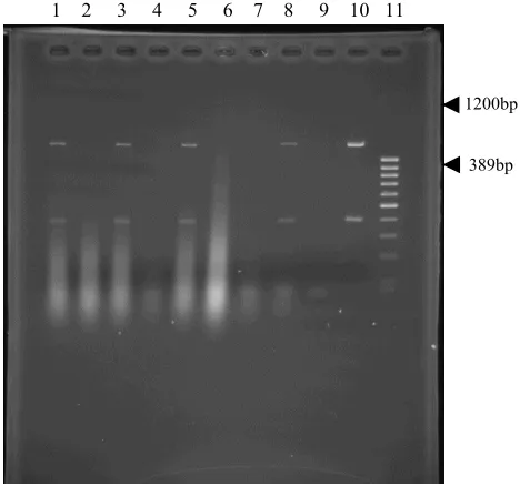
1 2 3 4 5 6 7 8 9 10 11
1200bp
389bp
Fig. 1. Multiplex PCR amplification of 16s RNA region (389 bp) and isocitrate dehydrogenase gene (1200 bp) of H. pylori in tissue samples: (1, 3, 5, 8) positive samples, (2, 4, 6, 7) negative samples, (9) negative control, (10) positive control, (11) molecular marker.
Specificity. Our multiplex PCR proved to be very specific for Helicobacter genus and H. pylori species specific genes and did not result in false-positive with any of the other bacterial species. Therefore, the specificity for this protocol was 100%.
Clinical application. Of 18 stomach samples examined, 12 (66%) positive in culture, gram staining, rapid urease, oxidase and catalase test for
H. pylori were positive for DNA of this organism
using this protocol. In clinical samples, it shows the sensitivity of 100% for this protocol.
DISCUSSION
Helicobacter genus members are fastidious and
very slow growing microorganisms [9]. The development of highly sensitive and specific PCR assays has alleviated problems typically associated with identification of microorganisms like
Helicobacter that are found in low densities in
tissue, difficult to culture or serologically similar. Because H. pylori is considered as the most prevalent species of Helicobacter, in most diseases
suspicious to be produred by Helicobacter
organisms diagnosis is on the basis of isolation of
H. pylori, while different species can cause similar
diseases [9-11]. According to these data, we established a multiplex PCR to identify
Helicobacter members in general and H. pylori
species in particular. In multiplex PCR, in addition to reduction of the preparatory steps of PCR, less materials are used. To prevent false-negative results, the development of efficient DNA purification methods is necessary to isolate genetic material from cellular substances found to inhibit DNA polymerase activity during the PCR. In our study, comparison of three DNA extraction protocols that included boiling, sonication and phenol chloroform showed that all of them have the same sensitivity. It seems that boiling is a cost effective, fast and safe way to extract DNA from pure H. pylori organisms.
To identify H. pylori species, we chose Hicd1-Hicd2 primers according to the experience of Argyros et al. [21]. Using a wide range of bacterial species, including all Helicobacter species, he showed that PCR based upon the highly specific icd genes primers represents a specific and sensitive method for detection of H. pylori. His screening experiments showed that use of these primers doesn’t result in false-positive amplifications with
C. jejuni and E. feacalis that appear when
species-specific protein antigen primers, described by Makristathis et al. [23] are used. On the other hand, Nilssin et al. [10] and Choi et al. [20] showed that the bacterial DNA extracts from non-Helicobacter species did not react with any primer specific for
Helicobacter genus 16s rRNA sequence. The
primers of Hcom1 and Hcom2 used in this multiplex PCR to identify Helicobacter genus also displayed good specificity when tested against several bacteria close to Helicobacter members. The minimum DNA concentration of pure H. pylori for a positive multiplex PCR result was 0.03 pg, approximately equivalent to 150 bacteria, which is comparable to other reported sensitiviy [21, 24]. To evaluate the potency of this protocol for clinical
application, we used PCR on 18 samples from stomach of patients undertaken endoscopy. H.
pylori was identified in 12 samples (66%) using
culture, gram staining, rapid urease test, catalase and oxidase. All these 12 samples were positive for DNA of H. pylori using multiplex PCR. Therefore, this PCR didn’t have any false-negative results and showed the sensitivity of 100%.
As today the number of diseases that can be caused by different species of Helicobacter are increasing, considering the possibility of the presence of other species other than H. pylori can help physicians to treat the patients precisely. Hence, using a multiplex PCR instead of a regular PCR as a molecular method to diagnose the causative agent in diseases related to Helicobacter family members is a valuable way. In conclusion, according to our results, this multiplex PCR represents a specific and sensitive assay for detection of Helicobacter genus members in general and H. pylori species in particular. In the next step, we will test this protocol on different extradigestive samples to evaluate its potency for clinical application in diagnosis of possible role of
Helicobacter organisms in some of extradigesitive
diseases which are suspecious to be caused by Helicobacter.
AKNOWLEDGEMENTS
This study was supported by Prof. Alborzi Clinical Microbiology Research Center, Shiraz, Iran grant # 81-3.
REFERENCES
1. Mobley, H.L.T., Mendz, G.L. (2001) Helicobacter pylori: physiology and genetics. ASM Press. Washington DC. USA.
2. Cantet, F., Magras, C., Marais, A., Federichi, M. and Megraud, F. (1999) Helicobacter species colonizing pig stomach: Molecular characterization and determination of prevalence. Appl. Environ. Microbiol. 65: 4672-4676.
3. De Groote, D., Van Doora, L.J., Ducatells, R., Verschauuren, A., Haesebrouck, F., Quint, W.G.V., Jalava, K. and Vandamme, P. (1999) Candidatus Helicobacter suis, a gastric Helicobacter from pigs and its phylogenetic relatedness to other gastrospirilla. Int. J. Syst. Bacteriol. 49: 1769-1777. 4. Fox, J.G., Yan, L., Dawbirst, F.E., Paster, B.J.,
Shames, B., Murphy, J.C., Hayward, A., Belcher, J.C. and Mendes, E.N. (1995) Helicobacter bilis sp. nov., a novel helicobacter isolated from bile, livers
and intestines of aged, inbred mouse strain. J. Clin. Microbiol. 33: 445-454.
5. Simmons, J.H., Riley, L.K, Besch-Williford, C. and Franklin, C.L. (2000) Helicobacter mesocricetorum sp. nov., a novel helicobacter isolated from the feces of Syrian hamsters. J. Clin. Microbiol. 38: 1811-1817.
6. Marshall, B.J. and Warren, J.R. (1984) Unidentified curved bacilli in the stomach of patients with gastritis and peptic ulceration. Lancet i: 1311-1314. 7. Mc Gowan, C.C., Cover, T.L. and Blaser, M.J.
(1996) Helicobacter pylori and gastric acid: Biological and therapeutic implications. Gastroenterology 110: 926-938.
8. Parsonnet, J., Friedman, G.D., Vandersteen, D.P., Chung, Y., Vogelman, J.H., Orentreich, N. and Sibley, R.K. (1991) Helicobacter pylori infection and the risk of gastric carcinoma. N. Engl. J. Med. 16: 1127-1131.
9. Murray, P.R., Rosenthal, K.S., Kobayashi, G.S. and Pfaller, M.A. (2002) Medical Microbiology. Mosby, Inc. A Harcourt Health Sciences Company, USA. 10. Nilsson, H.O., Taneera, J., Castedal, M., Glatz, E.,
Olsson, R. and Wadstrom, T. (2000) Identification
of Helicobacter pylori and other Helicobacter
species by PCR, hybridization and partial DNA sequencing in human liver samples from patients with primary sclerosing cholangitis or primary biliany cirrhosis. J. Clin. Microbiol. 38 (3): 1072-1076.
11. Fox, J.G., Drolet, R., Higgins, R., Messier, S., Yan, L., Coleman, B.E., Paster, B.J. and Dewhirst, F.E. (1996) Helicobacter canis isolated from a dog liver with multifocal necrotizing hepatitis. J. Clin. Microbiol. 34: 2479-2482.
12. Richy, F. and Megraud, F. (2003) Helicobacter pylori infection as a cause of extra-digestive diseases: myth or reality? Gastroenterol. Clin. Biol. 27 (3 pt 2): 459-466.
13. Bcrabino, A. (2002) Helicobacter pylori related iron deficiency anemia: a review. Helicobacter 7 (2): 71-75.
14. Sykora, J., Cerna, Z., Hejda, V., Varvarovska, J. and Stozicky, F. (2002) Hypergastrinemia associated with Helicobacter pylori infection and sideropenic anemia in a 15-year-old girl. Cas. Lek. Cesk. 22; 141 (23): 739-741.
15. Sugiyama, T., Tsuchida, M., Yokota, K., Shimodan, M. and Asaka, M. (2002) Improvement of long standing iron deficiency anemia in adults after
eradication of Helicobacter pylori infection. Intern. Med. 41 (6): 491-494.
16. Helimann, K.L. and Borchard, F. (1991) Gastritis due to shaped bacteria other than Helicobacter pylori: Clinical, histological, and ultra structural findings. Gut 32: 137-140.
17. Yeomans, N.D. (1996) Helicobacter heilmannii (formerly Gastrospirillum): association with pig and human gastric pathology. Gastroenterology 111: 244-259.
18. Weiss, J., Mecca, J., Da Silva, E. and Gassner, D. (1994) Comparison of PCR and other diagnostic techniques for detection of Helicobacter pylori in dyspeptic patients. J. Clin. Microbiol. 32: 1663-1668.
19. Fabre, R., Sobhani, I., Laurent-Puig, P., Hedef, N., Yazigi, N., Vissnzaine, C., Rodde, I., Potet, F., Mignon, M., Etienne, J.P. and Braquet, M. (1994) Polymeraze chain reaction assay for the detection of Helicobacter pylori in gastric biopsy specimens: Comparison with culture, rapid urease test and histopathological testes. Gut 35: 905-908.
20. Choi, Y.K, Han, J. and Hand Joo, H.S. (2001) Identification of novel helicobacter species in pig stomachs by PCR and partial sequencing. J. Clin. Microbiol. 39 (9): 3311-3315.
21. Argyros, F.C., Ghosh, M., Huang, L., Masubuchi, N., Cave, D.R. and Grubel, P. (2000) Evaluation of a PCR primer based on the isocitrate dehydrogenase gene for detection of Helicobacter pylori in feces. J. Clin. Microbiol. 38 (10): 3755-3758.
22. Goosen, C., Theron, J., Ntsala, M., Maree, F.F., Olckers, A., Botha, S.J., Lastovica, A.J. and Van der Merwe, S.W. (2002) evaluation of a novel heminested PCR assay based on the phosphoglucosamine mutase gene for detection of Helicobacter pylori in saliva and dental plaque. J. Clin. Microbiol. 40 (1): 205-209.
23. Makristathis, A., Pasching, E., Schutze, K., Wimmer, M., Rotter, M.L. and Iiirschi, A.M. (1998) Detection of Helicobacter pylori in stool specimens by PCR and antigen enzyme immunoassay. J. Clin. Microbiol. 36: 2772-2774.
24. McAvin, J.C., Reilly, P.A., Roudabush, R.M., Barnes, W.J., Salmen, A., Jackson, G.W., Beninga, K.K., Astorga, A., McCleskdy, F.K., Huff, W.B., Niemeyer, D. and Lohman, K.L. (2001) Sensitive and specific method for rapid identification of streptococcus pneumonia using real-time fluorescence PCR. J. Clin. Microbiol. 39 (10): 3446-3451.