Monte Carlo-based optimization of a gamma probe system for sentinel
lymph node mapping
Azadeh Nikoogoftar
1,2, Mojtaba Shamsaie
1, Navid Zeraatkar
3, Mohammad Reza Ay
2,41
Faculty of Nuclear Engineering and Physics, Amirkabir University of Technology, Tehran, Iran
2Research Centre for Molecular and Cellular Imaging, Tehran University of Medical Sciences, Tehran, Iran
3
Department of Radiology, University of Massachusetts Medical School, Worcester, MA, USA
4Department of Medical Physics and Biomedical Engineering, Tehran University of Medical Sciences,
Tehran, Iran
(Received 10 June 2018, Revised 14 October 2018, Accepted 17 October 2018)
ABSTRACT
Introduction: Sentinel lymph node biopsy (SLNB) is a standard surgical technique to identify sentinel lymph node (SLN) for the staging of early breast cancer. Nowadays, two methods are used for the identification of SLN: blue dye method aiding visually and radioactive dye using gamma detector. A wide range of gamma probe systems with different design and performance are used in intra-operative surgery. The performance of the probes is evaluated by some parameters such as sensitivity, spatial resolution, angular resolution, and shielding efficiency.
Methods: In this study, we simulated a gamma probe system, SURGEOGUIDE II based on CsI(Tl) scintillator, a silicon photomultiplier (SiPM), and a tungsten collimator, using the MCNP4C Monte Carlo (MC) method and comparing with experimental measurement. Finally we modeled a series of probe with various crystal material, crystal length, and collimator hole length to evaluate the sensitivity and the spatial resolution in order to propose the optimal configuration.
Results: The sensitivity of the system was measured as 2040 cps/MBq in 30 mm distance from the source. The spatial resolution and angular resolution were 43 mm and 70𝑜 at the same distance, respectively. Sensitivity at 30 mm distance from the probe head was the highest for BGO crystal and was the lowest for NaI crystals. The sensitivity and spatial resolution have also been changed by increasing the length of the crystal to a certain amount and then remained constant.
Conclusion: The results showed that the best choice for crystal was CdTe and CsI and the best length for CsI crystal in this type of the systems was 10 mm long. Also, based on the specific application, special probe should be designed taking the length of the collimator hole into consideration.
Key words: Gamma probe; Sentinel lymph node; Monte Carlo; Simulation
Iran J Nucl Med 2019;27(1):8-14 Published: January, 2019 http://irjnm.tums.ac.ir
Corresponding author:Dr. Mohammad Reza. Ay, Department of Medical Physics and Biomedical Engineering, Tehran University of Medical Sciences, Tehran, Iran. E-mail: mohammadreza_ay tums.ac.ir
O
rigi
na
l A
rticl
Ir
an
J
N
u
cl
Me
d
20
1
9
,
V
ol
27
,
No
1
(
S
er
ial
N
o
52
)
h
tt
p:
//
ir
jn
m
.tu
m
s.a
c.ir
Jan
u
ar
y,
20
19
9
INTRODUCTION
Many types of cancers can spread through the lymphatic system, and the first place affected is the first lymph node receiving lymphatic drainage from the tumor called sentinel lymph node (SLN). After introducing the SLN in the early 1990s, identification and evaluation of it has been of great importance [1]. Sentinel lymph node biopsy (SLNB) is a standard surgical technique to identify the SLN in patients without clinical evidence of metastasis in early-stage cancers [2]. The SLNB is a minimally invasive technique that prevents unnecessary lymph node dissection in 50-75%of patients. Complete lymph node dissection is often associated with post-operative morbidities such as edema, numb and limitation in arm movement, and chronic pains [3, 4]. The most common approach of the SLNB involves the injection of a low dose of a radioactive material, mostly technetium-99m (99mTc), and blue dye near the site of
the tumor. The radioactive-labeled material is transported by the lymph vessels to the lymph nodes where it is trapped. After an appropriate delay for uptake, the surgeon uses a radioactive detector (gamma probe), or looks for the dye, to find the SLN. Then, the SLN is removed and sent to the laboratory to be examined under a microscope by a pathologist. Based on the pathology results, the surgeon decides to perform an Axillary lymph node dissection(ALND) or not [5, 6].
In 1950, the first “sentinel node” was sent to the pathologist and at the same time a gamma detector was used in surgery for localization of SLN. Since then, various types of gamma probes were released with differences in the appearance and performance [7-9]. Intra-operative gamma ray detector (gamma probe) is specifically designed to identify the location of SLN in the body. Often, the detector is connected to a console that displays the detection count rate together with an audible signal proportional to the count rate [10]. Most available gamma probes are different in terms of the size of the crystal and the collimator resulting in different basic performance parameters such as sensitivity, spatial resolution, angular resolution, and shielding effectiveness [11].
Monte Carlo (MC) is a computerized mathematical technique of simulation based on statistical methods employing random numbers. MC can be used for simulation of photons and charged particles transport in various materials. MC method has been well known in the field of medical physics over the last fifty years. Now, this method is the gold standard for modeling of many physical processes that are difficult or impossible to assess by experimental measurements or analytical approaches [12]. In medical physics, the MC method is one of the most usable tools for modeling any complex activity and attenuation distributions in the field of nuclear medicine, dose
calculation in radiotherapy, diagnostic radiology, and radiation protection [13]. Growing number of scientific papers related to MC in medical physics studies [14-17] indicates the increasing use of it in modeling of the systems.
The purpose of the present study is the optimization of a gamma probe system. First, we used a MC code to model our system and then compared the MC results with the experimental ones. Next, we modeled various crystal lengths, collimator sizes, and different scintillator crystal materials to evaluate the gamma probe performance by measurement of some important parameters like sensitivity and spatial resolution aiming to find an optimal structure with high sensitivity with respect to spatial resolution and suitable shielding for SLN mapping [18].
METHODS Gamma probe system
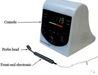
SURGEOGUIDE II [19, 20] is a compact gamma detector used during cancer surgery providing an audio output signal proportional to the number of radiation detected in time. The detection rate is also displayed.
The SURGEOGUIDE II is composed of two main parts: probe that is the gamma detector of the system and console including the control and display unit buttons. The probe head (tip) contains an internal collimator, crystal, and photodiode. The front-end electronic board is placed in the cylindrical hand-piece of the probe. The sensitive part of the probe is a cesium iodide thallium-activated [CsI(Tl)] scintillator crystal shaped as a cylinder with diameter of 7.2 mm and 10 mm in length in contact with a silicon photomultiplier (SiPM) having an active area of 6 × 6 mm2.The scintillator and the SiPM are enclosed in a tungsten collimator/shield. The probe is equipped with a single hole tungsten collimator with a 7.8 mm hole diameter and 2.5 mm length. A thin coating of stainless steel covers the outer surface of the probe. An overview of the appearance of the gamma probe is shown in Figure 1.
Ir
an
J
N
u
cl
Me
d
20
1
9
,
V
ol
27
,
No
1
(
S
er
ial
N
o
52
)
h
tt
p:
//
ir
jn
m
.tu
m
s.a
c.ir
Jan
u
ar
y,
20
19
10
Monte Carlo simulation
Modeling of the gamma probe system was performed using MCNP 4C code. MCNP4C is the first major release of MCNP since version 4B. It was released in 2000 providing unique features such as macrobodies, cumulative tallies, and superimposed importance mesh. MCNP 4C is easy-to-use, flexible, and scalable capable of converting a complex model to basic events and interactions [21].
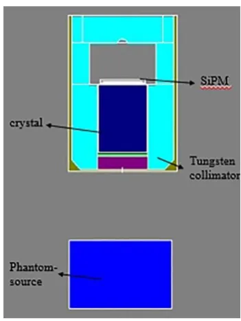
The geometry specification of the gamma probe detection subsection was described accurately in the MC simulation file including tungsten collimator/shield, CsI(Tl) crystal, and SiPM and Co-57 source embedded in a phantom. In Figure 2, a snapshot of the gamma probe geometry and a Co-57 source, simulated using the MCNP 4C code, is shown. To get the desired result from the simulation, in the data card, we used F8 tally with a number of histories equal 107and cut off energy was set to 20 keV.
Fig 2. A snapshot of the gamma probe geometry and a Co-57 source, simulated using the MCNP 4C code.
Validation of the MC model
In this work, performance evaluation of the gamma probe whether for measurement set-up or data processing methods was performed according to NEMANU3-2004 standards [18]. The radionuclide used for all tests was a 7 mm Co-57 source with an activity of 96 kBq.
For validation of the MC model, two main parameters, sensitivity and spatial resolution, which have great influence on the gamma probe performance during surgery [22] were examined in both experimental and
simulation measurements. The simulation results were then compared with the experimental tests.
Sensitivity in air: Due to the small amount of the radiopharmaceutical injected to the patient and slight accumulation in the target organ, the sensitivity of a gamma probe system is evaluated. A point source of Co-57, with 96 kBq activity, was placed along the central axis of the probe at distance of 10mm, 30 mm, and 50mm from the probe tip. Because scatter from the surrounding material can change the recorded sensitivity, we must ensure that the probe to-source axis is at least 50 mm far from the surrounding material.
The sensitivity of the probe in the air is defined as the counts recorded per unit of radioactivity and is expressed in counts per second per MBq.
Spatial and angular resolution: Better spatial resolution is favorable to accurate localization of the SLN from the other sources of activity close to it e.g. other hot nodes or the injection site. To measure the spatial resolution, the point source was positioned at 30mm distance from the detector and the sensitivity was measured at varying lateral distances from the source in the range of -50mm to +50 mm. To improve the accuracy, the measurements were performed using 2.5-mm steps in the range of ±15 mm and using 5-mm steps for other locations. The response function is expected to be a Gaussian distribution; the spatial resolution was hence reported as full-width at half-maximum (FWHM) of the fitted Gaussian.
Angular resolution was determined by measuring the sensitivity of the probe at 30 mm distance from the tip between the central axis of the probe and the hypothetical line connecting the source and the center of the probe tip ranged from -90° to +90°. Five-degree intervals were used for the angular range of ±25 degree while for larger angles, 10 degree intervals were applied. The FWHM of the fitted Gaussian was reported as the angular resolution.
Shielding efficiency: Shielding of the head of the probe is important to prevent radiation from unintended locations such as injection site or tumor. But, it should be noted that a thicker shield increases the weight of the probe while surgeons tend to use thin and light-weight probes [23, 24].
For measurement of shielding, the Co-57 source was positioned in full contact with outer surface of the head of the probe being moved slowly around to reach the highest count rate. Percentage shielding effectiveness (SE%) was then calculated as:
SE (%) = SensitivityAXIS−SensitivityLEAK
SensitivityAXIS (1)
where SensitivityAXIS and SensitivityLEAK are the
Ir
an
J
N
u
cl
Me
d
20
1
9
,
V
ol
27
,
No
1
(
S
er
ial
N
o
52
)
h
tt
p:
//
ir
jn
m
.tu
m
s.a
c.ir
Jan
u
ar
y,
20
19
11
the highest count in contact with the lateral surface of the probe, respectively. Same set-up was used for MC simulation for all above mentioned parameters and finally, for validation, the experimental results were compared with the ones of simulation.
System optimization
The scintillator crystal is the main component of a gamma probe having a significant impact on the sensitivity and the spatial resolution of the system. For the purpose of optimization, we focused on the optimization of the crystal features based on the two mentioned parameters. MC method was used to simulate different lengths and various types of scintillator crystal. For this, we simulated different lengths of CsI crystal, from 2mm to 15mm, and calculated sensitivity and spatial resolution. For sensitivity simulation, 1mm step was used for the crystal length simulations while for spatial resolution, 2.5 mm step was applied. In addition, we chose five different types of crystal material that are widely used in commercial probes including CsI, sodium iodide thallium-activated [NaI (Tl)], bismuth germinate (BGO), lutetium yttrium orthosilicate (LYSO), scintillator, and cadmium telluride (CdTe) semiconductor. The crystal size was considered the same 7.2 mm in diameter, 10 mm long for all crystal materials in the simulations. Sensitivity and spatial resolution of the system were calculated at 30 mm distance from the surface of the probe, using the same method as described in Validation of the MC model. The collimation in most of the gamma probes is based on single pinhole method [25, 26]. The ratio of the diameter to the length of the collimator has a significant impact on sensitivity resolution trade-off. The diameter of the collimator is better to be designed in a way so the entire the crystal face be irradiated to reach better sensitivity utilizing the maximum sensitive area of the detector. The length of the collimator can be altered to target desired set of performance parameters. The version of interest of SURGEOGUIDE II in this work, has a collimator length equal to 2.5 mm. In this study, we measured the sensitivity and spatial resolution of the system at a distance of 30 mm from the surface of the probe for different collimator lengths, ranging from 2 mm to 5 mm with 0.5 mm steps.
RESULTS Validation of the MC model
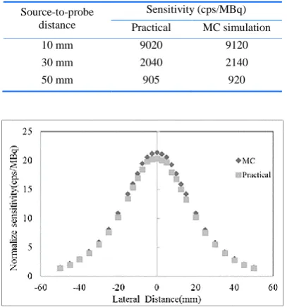
The results of the system sensitivity in the air at different distances are summarized in Table 1. The practical and MC simulation results for spatial resolution are shown in Figure 3.
After Gaussian fitting, spatial resolution at 30 mm distance with experimental measurements and MC
simulations was measured as 43mm and 44mm in terms of FWHM, respectively.
Table 1: Practical and simulated values of sensitivity for SURGEOGUIDE II probe.
Source-to-probe distance
Sensitivity (cps/MBq)
Practical MC simulation
10 mm 9020 9120
30 mm 2040 2140
50 mm 905 920
Fig 3. The practical and MC simulation results for spatial resolution.
According to the NEMA standard methods, angular resolution was reported as degrees FWHM at 30 mm source-to-probe depth using experimental and MC modeling. Angular resolution value was obtained as 70𝑜 for practical and 72𝑜 for the MC simulation tests.
Also, shielding effectiveness was measured about 98% in practice and 99% based on the MC simulation.
Optimization
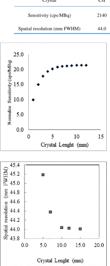
Figure 4 shows the sensitivity and spatial resolution at
30 mm distance from the probe head for different lengths of CsI scintillator crystal. Table 2 shows how the crystal type affects the sensitivity and spatial resolution of the system. BGO and NaI have the highest and the lowest sensitivity, respectively. The sensitivity and spatial resolution are in compromise relation as expected. Table 3 shows the sensitivity and spatial resolution in the air for different lengths of the collimator at distance of 30 mm.
DISCUSSION
Ir
an
J
N
u
cl
Me
d
20
1
9
,
V
ol
27
,
No
1
(
S
er
ial
N
o
52
)
h
tt
p:
//
ir
jn
m
.tu
m
s.a
c.ir
Jan
u
ar
y,
20
19
12
Table 2: Sensitivity and spatial resolution of the gamma probe for different crystal/detector materials.
Crystal CsI NaI BGO LYSO CdTe
Sensitivity (cps/MBq) 2140 2030 2250 2220 2155
Spatial resolution (mm FWHM) 44.0 42.0 46.7 46.2 44.5
Fig 4. The sensitivity (above) and spatial resolution (below) at 30 mm distance from the probe head for different lengths of CsI scintillator crystal.
Gamma probe spatial resolution is another important factor to identify lymph nodes close to each other and the nodes next to the injection site.
A gamma probe system with the optimal sensitivity and spatial resolution is an ideal system for radio-guided surgical applications. But, given interrelation of these parameters, it is difficult to design a system at optimal conditions.
Sensitivity and spatial resolution depend on parameters such as system geometry (thickness, length, and diameter of the collimator and the crystal) and the type of scintillator crystal.
Table 3: Sensitivity and spatial resolution versus length of collimator at 30 mm source-to collimator distance.
Spatial resolution (mm FWHM) Sensitivity
(cps/MBq) Collimator length
(mm)
44.0 2140
2.5
42.2 2060
3.0
40.7 1990
3.5
39.0 1920
4.0
36.7 1850
4.5
34.6 1780
5.0
In this work, we modeled the SURGEOGUIDE II gamma probe using MCNP 4C code. For validation of the MC model, some performance parameters such as sensitivity, spatial resolution, angular resolution, and shielding were measured with experimental tests and the MC simulations according to the NEMA standard. The gamma probe showed the sensitivity about 2 cps/MBq, a spatial resolution of 43 mm FWHM, and 99% shielding effectiveness. Comparison of the experimental and the simulation results shows small difference: the mean error in assessing the sensitivity parameter is 4% at 30mm distance while the spatial resolution error at 30 mm is less than 1%. The error in shielding calculations is negligible.
In all measurements, we obtained a favorable correlation between the simulation and the experimental results revealing valid simulation model when applied for optimizing the SURGEOGUIDE II system. Recently, some studies [20] were performed on a prototype of the gamma probe, SURGEOGUIDE, where a CsI(Tl) cylindrical scintillator crystal 8 mm in diameter and 10 mm in length with a pinhole tungsten collimator 3 mm long and 7.5 mm in diameter was used. Other features of the gamma probe are similar to the version used in the current work.
Ir
an
J
N
u
cl
Me
d
20
1
9
,
V
ol
27
,
No
1
(
S
er
ial
N
o
52
)
h
tt
p:
//
ir
jn
m
.tu
m
s.a
c.ir
Jan
u
ar
y,
20
19
13
In this study, to evaluate the effect of the length of the crystal, we showed that increasing the crystal length more than 5 mm has no significant impact on the sensitivity of the system. Also, it was demonstrated that the variation of the spatial resolution for the crystal lengths greater than 10 mm is not of importance. But, at crystal lengths less than 10 mm, the spatial resolution greatly increases with increasing the crystal length. Since by increasing the crystal length more than 10 mm, no significant enhancement is seen in sensitivity and spatial resolution, one can conclude that the length of 10 mm is the optimum for CsI scintillator crystal in the current gamma probe. Currently, most of the gamma probes commercially available have a CsI or NaI(Tl) scintillation crystal or a CdTe semiconductor detector [27] while some manufactures provide systems with BGO scintillator crystal. We evaluated the performance of the probe with 5 types of crystal/detector including CsI, NaI, BGO, and LYSO scintillators in addition to CdTe semiconductor. The results showed higher BGO and LYSO sensitivities, however, demonstrating worsen spatial resolution. So, CsI crystal and CdTe can be considered as the most appropriate choices for the current set-up of the gamma probe system.
Extending the collimator hole length reduces the sensitivity of the system while simultaneously improves the spatial resolution. The appropriate length of the collimator, hence, can be selected based on the special application design.
CONCLUSION
In this work, we modeled a gamma probe system, SURGEOGUIDE II, using MC simulation. This model was then validated using both practical and simulation protocols. The validated model was finally applied for optimization of the crystal length, crystal material, and collimator hole length of the probe by evaluation of the spatial resolution and the sensitivity of the system. The results showed that for the current gamma probe, a 10-mm crystal length for CsI can be an optimum choice. Also, BGO and LYSO crystals have the highest sensitivity, but as sensitivity increases, the resolution degrades, which is not optimal leading to compromised spatial resolution. The CdTe semiconductor crystal is almost similar to those of the CsI crystal, with close sensitivity and spatial resolution figures, making no difference in final results. Therefore, it can be concluded that CsI and CdTe crystals are suitable choices for application in a gamma probe system. In addition, considering the effect of the collimator hole length, this parameter can be selected according to desired application of the probe. Moreover, regarding the close agreement of the simulation results with the practical device application, the developed MC model can be applied
for evaluation and optimization of the current gamma probe systems.
Acknowledgment
The authors would like to show their debt of gratitude toward Dr. Hojjat Mahani for his invaluable help in revising the paper draft.
REFERENCES
1. Osako T, Iwase T, Kimura K, Yamashita K, Horii R,
Yanagisawa A, Akiyama F. Intraoperative molecular assay for sentinel lymph node metastases in early stage breast cancer: a comparative analysis between one-step nucleic acid amplification whole node assay and routine frozen section histology. Cancer. 2011 Oct 1;117(19):4365-74.
2. Cheng G, Kurita S, Torigian DA, Alavi A. Current status of
sentinel lymph-node biopsy in patients with breast cancer. Eur J Nucl Med Mol Imaging. 2011 Mar;38(3):562-75.
3. Gipponi M, Bassetti C, Canavese G, Catturich A, Di
Somma C, Vecchio C, Nicolò G, Schenone F, Tomei D, Cafiero F. Sentinel lymph node as a new marker for therapeutic planning in breast cancer patients. J Surg Oncol. 2004 Mar;85(3):102-11.
4. Kootstra JJ, Dijkstra PU, Rietman H, de Vries J, Baas P,
Geertzen JH, Hoekstra HJ, Hoekstra-Weebers JE. A longitudinal study of shoulder and arm morbidity in breast cancer survivors 7 years after sentinel lymph node biopsy or axillary lymph node dissection. Breast Cancer Res Treat. 2013 May;139(1):125-34.
5. Ahmed M, Purushotham AD, Douek M. Novel techniques
for sentinel lymph node biopsy in breast cancer: a systematic review. Lancet Oncol. 2014 Jul;15(8):e351-62.
6. Chen SL, Iddings DM, Scheri RP, Bilchik AJ. Lymphatic
mapping and sentinel node analysis: current concepts and applications. CA Cancer J Clin. 2006 Sep-Oct;56(5):292-309; quiz 316-7.
7. Gould EA, Winship T, Philbin PH, Kerr HH. Observations
on a "sentinel node" in cancer of the parotid. Cancer. 1960 Jan-Feb;13:77-8.
8. Tanis PJ, Nieweg OE, Valdés Olmos RA, Th Rutgers EJ,
Kroon BB. History of sentinel node and validation of the technique. Breast Cancer Res. 2001;3(2):109-12.
9. Myers WG, Vanderleeden JC. Radioiodine-125. J Nucl
Med. 1960 Jul;1:149-64.
10. Cengić T, Corluka S, Petrović T, Baranović S, Kovacić K,
Kolundzić R. Intraoperative gamma hand-held probe navigation in resection of osteoid osteoma tumor--report of two cases. Acta Clin Croat. 2013 Jun;52(2):261-5.
11. Wydra D, Matuszewski R, Romanowicz G, Bandurski T.
Evaluation of surgical gamma probes for sentinel node localization in cervical and vulvar cancer. Nucl Med Rev Cent East Eur. 2005;8(2):105-10.
12. Rogers DW. Fifty years of Monte Carlo simulations for
medical physics. Phys Med Biol. 2006 Jul 7;51(13):R287-301.
13. Buvat I, Castiglioni I. Monte Carlo simulations in SPET
and PET. Q J Nucl Med. 2002 Mar;46(1):48-61.
14. De Vries DJ, Moore SC, Zimmerman RE, Mueller SP,
Ir
an
J
N
u
cl
Me
d
20
1
9
,
V
ol
27
,
No
1
(
S
er
ial
N
o
52
)
h
tt
p:
//
ir
jn
m
.tu
m
s.a
c.ir
Jan
u
ar
y,
20
19
14
Monte Carlo simulation of photon transport in an Anger camera. IEEE Trans Med Imaging. 1990;9(4):430-8.
15. Yanch JC, Dobrzeniecki AB. Monte Carlo simulation in
SPECT:complete 3D modeling of source, collimator and tomographicdata acquisition. IEEE Trans Nucl Sci. 1993;198-203.
16. Lorincz E, Erdei G, Péczeli I, Steinbach C, Ujhelyi F, Bükki
T. Modeling and optimization of scintillator arrays for PET detectors. IEEE Trans Nucl Sci. 2010;57:48-54.
17. Sarrut D, Bardiès M, Boussion N, Freud N, Jan S, Létang
JM, Loudos G, Maigne L, Marcatili S, Mauxion T, Papadimitroulas P, Perrot Y, Pietrzyk U, Robert C, Schaart DR, Visvikis D, Buvat I. A review of the use and potential of the GATE Monte Carlo simulation code for radiation therapy and dosimetry applications. Med Phys. 2014 Jun;41(6):064301.
18. National Electrical Manufacturers Association. NEMA
Standards Publication NU 3-2004. Performance
measurements and quality control guidelines for non-imaging intra-operative gamma probes. 2004.
19. Ay MR, Zeraatkar N, Gorjizadeh N, Kaviani A, Farzaneh
Far S, Sajedi S, Arabi H, Farahani M, Teimourian B. SURGEOGUIDE: a Gamma Probe for Localization of Sentinel Lymph Nodes. Eur J Nucl Med Mol Imaging. 2013;40 (Suppl 2):S211–S212.
20. Kaviani S, Zeraatkar N, Sajedi S, Gorjizadeh N, Farahani
M, Ghafarian P, El Fakhri G, Sabet H, Ay MR. Development and characterization of a compact hand-held gamma probe system, SURGEOGUIDE, based on NEMA NU3-2004 standards. J Instrum. 2016;11(12):T12004.
21. Kroese DP, Brereton T, Taimre T, Botev ZI. Why the
Monte Carlo method is so important today. Wiley Interdiscip Rev Comput Stat. 2014;6(6):386-92.
22. Tiourina T, Arends B, Huysmans D, Rutten H, Lemaire B,
Muller S. Evaluation of surgical gamma probes for radioguided sentinel node localisation. Eur J Nucl Med. 1998 Sep;25(9):1224-31.
23. Mariani G, Vaiano A, Nibale O, Rubello D. Is the "ideal"
gamma-probe for intraoperative radioguided surgery conceivable? J Nucl Med. 2005 Mar;46(3):388-90.
24. Sarikaya I, Sarikaya A, Reba RC. Gamma probes and their
use in tumor detection in colorectal cancer. Int Semin Surg Oncol. 2008 Nov 19;5:25.
25. Bolozdynya A, Vorobiev K, Evgrafova E, Zhukov K,
Kantserov V, Sosnovtsev V, Filipov DE, Yagnyukova AK. A γ probe for radionuclide diagnostics of cancer. Instrum Exp Tech. 2015;58(1):153-7.
26. Classe JM, Fiche M, Rousseau C, Sagan C, Dravet F, Pioud
R, Lisbona A, Ferrer L, Campion L, Resche I, Curtet C. Prospective comparison of 3 gamma-probes for sentinel lymph node detection in 200 breast cancer patients. J Nucl Med. 2005 Mar;46(3):395-9.
27. Halkar RK, Aarsvold JN. Intraoperative probes. J Nucl Med