EXPRESSION OF PRO-ANGIOGENIC FACTORS, INFLAMMATORY CYTOKINE EXPRESSION, AND CIRCULATING TUMOR CELL NUMBERS
by Ken Tawara
A dissertation
submitted in partial fulfillment of the requirements for the degree of Doctor of Philosophy in Biomolecular Sciences
Boise State University
© 2017 Ken Tawara
DEFENSE COMMITTEE AND FINAL READING APPROVALS
of the dissertation submitted by
Ken Tawara
Dissertation Title: Oncostatin M Promotes Breast Cancer Metastasis: Increased Expression of Pro-Angiogenic Factors, Inflammatory Cytokine Expression, and Circulating Tumor Cell Numbers
Date of Final Oral Examination: 25 August 2017
The following individuals read and discussed the dissertation submitted by student Ken Tawara, and they evaluated his presentation and response to questions during the final oral examination. They found that the student passed the final oral examination.
Cheryl Jorcyk, Ph.D. Chair, Supervisory Committee Kenneth A. Cornell, Ph.D. Member, Supervisory Committee Daniel Fologea, Ph.D. Member, Supervisory Committee Denise G. Wingett, Ph.D. Member, Supervisory Committee
iv
I would like to thank my dissertation advisor and mentor, Dr. Cheryl Jorcyk, who has helped me grow as a scientist through my masters and doctoral studies. I am
extremely grateful for her honest feedback and critical assessment on my ideas, writing, project directions, and for always pushing me to do the best that I can do. Her
unwavering belief in my success has helped me drive myself to be much more productive as a graduate student than I could have ever imagined. I would also like to thank the Department of Biological Sciences, the Biomolecular Sciences Graduate Program (BMOL), the BMOL Program Coordinator, Beth Gee, and the Program Director, Dr. Denise Wingett, for their support of the advancement of my research. They have given me the opportunity to develop skills as a scientist within and beyond the laboratory.
I would also like to thank the many people with whom I have collaborated and worked with in the lab. In particular, it was a pleasure to work to Dr. Celeste Bolin. Her expertise in animal experimentation and writing techniques have helped me tremendously to further my skills as a scientist. I would also like to thank her for the invitation to speak at the Biology Colloquium at College of Idaho, which has given me invaluable public speaking experience. I would also like to thank Dr. Julie Oxford, Dr. Randall Ryan, Dr. Caleb Sutherland, and now Dr. Hunter Covert for working with me on multiple
v protocols with the Institutional Review Board.
I would also like to thank numerous past and present members in the lab that have helped me on my numerous projects. Specifically, I would like to thank Ryan Fox, Dollie LaJoie, Danielle Hedeen, Madhuri Nandakumar, M.S. Jake Goyden, M.S. Jordan
vi
vii
viii
ACKNOWLEDGEMENTS ... iv
ABSTRACT ... vi
LIST OF TABLES ... xvii
LIST OF FIGURES ... xviii
LIST OF ABBREVIATIONS ... xxi
CHAPTER ONE: INTRODUCTION ...1
A Brief Overview of Cancer ...1
Breast Cancer ...3
Breast Cancer Metastasis ...4
Inflammation in Breast Cancer ...6
Breast Cancer Subtypes ...7
Breast Cancer Treatment...9
Intracellular Pathways That Promote Breast Cancer Progression ...10
Oncostatin M and Breast Cancer ...11
Interleukin-6, And Interleukin-1 Beta in Breast Cancer ...13
Cancer Angiogenesis and Hypoxia Factors ...14
Circulating Tumor Cells and Their Detection ...15
Mouse Models for Breast Cancer ...17
ix
Figures...22
Figure 1.1 Chronic inflammation leads to genomic and epigenetic instability and promotes tumor formation ...23
Figure 1.2. Multi-step development of invasive ductal carcinoma (IDC). 24 Figure 1.3. Treatment strategies for breast cancer subtypes ...25
Figure 1.4. Multiple signaling pathway activation in breast cancer cells. .26 Figure 1.5. OSM induces the STAT3, PI3K, and MAPK pathways. ...27
Figure 1.6. Tumor cells promote the development of angiogenesis. ...28
References ...29
CHAPTER TWO: CO-EXPRESSION OF VEGF AND IL-6 FAMILY CYTOKINES ARE ASSOCIATED WITH DECREASED SURVIVAL IN HER2- BREAST CANCER: SUBTYPE SPECIFIC IL-6 FAMILY CYTOKINE-MEDIATED VEGF SECRETION .45 Authors ...45
Abstract ...46
Introduction ...47
Materials and Methods ...50
Oncomine analysis ...50
Tissue culture ...51
siRNA transfection...52
Immunoblot analysis ...52
VEGF ELISA ...53
RT-PCR...53
pSTAT3 ELISA ...54
x
Inflammatory cytokine and VEGF co-expression is correlated with
decreased survival of IDC patients ...54
HER2- status dictates poor survival in IDC patients with high co-expression of OSM, IL-6, OSMRβ or IL-6Rα and VEGF...55
OSM mediates VEGF induction independent of HIF1α signaling in TNBC cells ...57
OSM strongly induces STAT3 activation in ER+ and ER- HER2- breast cancer cells ...58
Activation of STAT3 signaling is required for VEGF secretion by MDA-MB-231 TNBC cells ...59
Discussion ...60
Conclusions ...64
CHAPTER TWO: FIGURES ...66
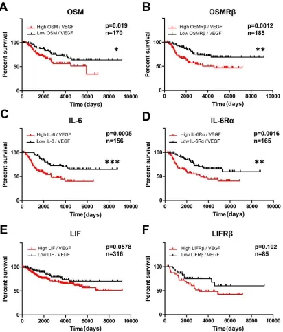
Figure 2.1. Inflammatory cytokine and VEGF co-expression are correlated with decreased survival of invasive ductal carcinoma patient. ...67
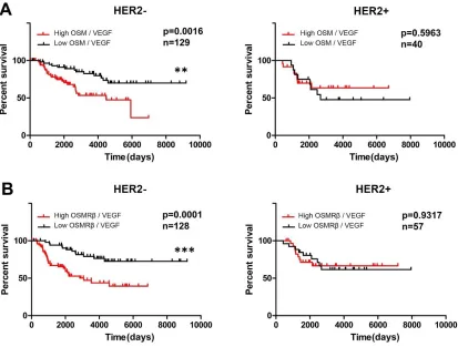
Figure 2.2 Co-expression of VEGF with OSM or OSMRβ appears to affect survival in HER2- but not HER2+ subtypes. ...69
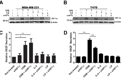
Figure 2.3. IL-6 family cytokine OSM induces VEGF secretion independent of HIF1α signaling in MDA-MB-231 cells. ...70
Figure 2.4. OSM strongly induces phosphorylation of STAT3. ...72
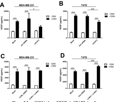
Figure 2.5. OSM induces VEGF via STAT3 signaling. ...73
Figure 2.6. Mechanistically distinct regulation of VEGF secretion in MDA-MB-231 TNBC and T47D (ER+/PR+/HER2-) cells. ...74
Supplemental Materials and Methods ...75
Endothelial Cell Tube and Branch Point Formation Assay ...75
In vivo Matrigel Plug Assay ...75
xi
Figure 2.S1. Increased co-expression of OSM or OSMRβ and VEGF
correlates with decreased survival of IDC patients...77
Figure 2.S2. Expression of cytokines or their receptors and VEGF appears to affect survival in HER2- but not HER2+ subtypes...79
Figure 2.S3. Co-expression of OSM or OSMRβ and VEGF differentially affects survival based on estrogen receptor status. ...81
Figure 2.S4. OSM induces endothelial cell tube formation and branch points in vitro. ...83
Figure 2.S5. OSM-treatment of human breast cancer cells stimulates angiogenesis in vivo. ...85
Figure 2.S6. JNK Inhibitor 2 suppresses VEGF secretion and phosphorylation of STAT3. ...87
References ...88
CHAPTER THREE: ONCOSTATIN M PROMOTES BREAST CANCER METASTASIS TO LUNG AND INCREASES CIRCULATING TUMOR CELL NUMBER ...95
Authors ...95
Abstract ...95
Introduction ...96
Materials and Methods ...99
Tissue microarrays ...99
Cell lines and culture conditions ...100
Plasmid construct design and cell transfections...100
ELISA ...100
Western blot analysis ...101
xii
Detection of circulating tumor cells (Alu qPCR) ...103
Quantitative PCR (qPCR) ...104
In-vivo magnetic resonance imaging ...104
Detection of circulating tumor cells (clonogenic assay) ...105
Epithelial-mesenchymal transition assay ...106
Cell migration assay ...106
Cell detachment assay ...106
Statistical analysis ...107
Results ...107
High OSM expression in breast ductal carcinoma in situ (DCIS) and IDC suggests autocrine signaling ...107
OSM generated from TET-inducible MDA-MB-231 (MDATO/OSM) cells increases metastases to the lung and decreases survival ...108
OSM increases metastasis to lung and circulating tumor cell numbers in an orthotopic MDA-MB-231 model of breast cancer...109
Suppression of OSM in syngeneic mouse model reduced lung metastases ...111
Suppression of OSM by shRNA increases survival from spontaneous metastasis via orthotopic injection but not via intracardiac injection in vivo ...112
CTC number and metastatic burden is reduced in OSM knockout mice compared to wild type mice ...113
OSM increases pre-intravasation metrics of metastatic capacity in 4T1.2 cells ...114
Discussion ...114
xiii
Chapter Three: Figures ...120
Figure 3.1. OSM is highly expressed in DCIS and IDC. ...121
Figure 3.2: MDATO/OSM tumors increase metastasis and decrease survival. ...123
Figure 3.3. Peri-tumoral OSM injections into mice with MDA-MB-231 D3H2LN tumors promote the development of metastases and CTCs ...126
Figure 3.4. Reduced OSM expression results in fewer spontaneous lung metastases and lower total volume of lung metastases by MRI. ...128
Figure 3.5. Reduced tumor cell OSM expression increases survival in a 4T1.2-shOSM mouse model of tumor resection. ...130
Figure 3.6. OSM promotes 4T1.2 cell detachment and migration...132
Figure 3.7. Model of OSM-mediated metastasis. ...133
CHAPTER THREE: SUPPLEMENTAL FIGURES...135
Figure 3.S1. Deterioration of body condition in MDATO/OSM tumor bearing mice treated with TET ...136
Figure 3.S2. OSM is highly expressed in orthotopic 4T1.2 primary mammary tumors in female Balb/c mice ...138
Figure 3.S3. qPCR analysis of lung metastases after intracardiac injections. ...139
Supplemental Materials and Methods ...140
Complete details for tissue microarrays. ...140
Animal Histology ...142
References ...143
CHAPTER FOUR: HIGH EXPRESSION OF OSM AND IL-6 IS ASSOCIATED WITH DECREASED BREAST CANCER SURVIVAL: SYNERGISTIC INDUCTION OF IL-6 SECRETION BY OSM AND IL-1β ...148
xiv
Introduction ...149
Materials and Methods ...153
Oncomine analysis ...153
Enzyme linked immunosorbent assay ...153
Cell lines ...154
Stable and transient transfections...155
Animal tumor xenograft model ...156
Immunoblot assay ...156
Immunoprecipitation ...157
Statistical analysis ...158
Results ...158
OSM and IL-6 expression are correlated with each other and associated with decreased invasive breast cancer survival ...158
High OSM serum levels in breast cancer patients are correlated high IL-6 levels ...159
OSM promotes IL-6 expression in a mouse model of human breast cancer ...160
OSM induces human IL-6 secretion in the absence of ER from various cancer cells in vitro ...161
STAT3 is phosphorylated by OSM signaling in both ER- and ER+ cells ...162
Invasive breast cancer patient stroma expresses high levels of OSM and IL-1 ...163
OSM and IL-1β promote increased lymph node metastases and decreased survival: a three-way correlation for OSM-IL-1β-IL-6 expression ...164
xv
ER interaction with STAT3 is suppressed by OSM and IL-1β in MCF7
cells ...167
Discussion ...168
Conclusions ...172
Declarations ...173
Chapter Four: Figures ...174
Figure 4.1. OSM and IL-6 are associated with decreased invasive breast cancer survival ...175
Figure 4. 2. OSM breast cancer patient serum levels correlate with IL-6 levels. ...176
Figure 4.3. OSM induces IL-6 in an animal model of human breast cancer. ...179
Figure 4.4. OSM induces IL-6 secretion in an ER-dependent manner ....181
Figure 4.5. OSM signals through STAT3 in ER- MDA-MB-231 cells to induce IL-6 ...183
Figure 4.6. OSM and IL-1β expression is higher in invasive breast cancer compared to normal tissue and correlates with higher lymph node metastasis, decreased survival, and IL-6 levels ...185
Figure 4.7. OSM and IL-1β activate separate signaling pathways and synergistically induce IL-6 secretion ...187
Figure 4.8. OSM and IL-1β promote IL-6 expression in a breast cancer cell-subtype specific manner...188
Chapter Four: Supplemental Figures ...189
Figure 4.S1. OSM and IL-6 serum concentration is correlated in vivo ...189
Figure 4.S2. IL-6 does not promote OSM secretion ...190
xvi
Figure 4.S5. OSM and IL-1β promotes EMT-like morphology in T47D cells. ...194 Figure 4.S6. ERK signaling is required for MCF7’s synergistic response to OSM and IL-1β ...196 Figure 4.S7. STAT3 siRNA does not affect IL-1β-induced IL-6
xvii
xviii
Figure 1.1 Chronic inflammation leads to genomic and epigenetic instability and
promotes tumor formation. ... 22
Figure 1.2. Multi-step development of invasive ductal carcinoma (IDC) ... 24
Figure 1.3. Treatment strategies for breast cancer subtypes. ... 25
Figure 1.4. Multiple signaling pathway activation in breast cancer cells. ... 26
Figure 1.5. OSM induces the STAT3, PI3K, and MAPK pathways. ... 27
Figure 1.6. Tumor cells promote the development of angiogenesis. ... 28
Figure 2.1. Increased co-expression of OSM or OSMRβ and VEGF correlates with decreased survival of IDC patients ... 66
Figure 2.2. Co-expression of VEGF with OSM or OSMRβ appears to affect survival in HER2- but not HER2+ subtypes ... 68
Figure 2.3. IL-6 family cytokine OSM induces VEGF secretion independent of HIF1α signaling in MDA-MB-231 cells ... 70
Figure 2.4. OSM strongly induces phosphorylation of STAT3 ... 71
Figure 2.5. OSM induces VEGF via STAT3 signaling ... 73
Figure 2.6. Mechanistically distinct regulation of VEGF secretion in MDA-MB-231 TNBC and T47D (ER+/PR+/HER2-) cells ... 74
Figure 2.S1. Mechanistically distinct regulation of VEGF secretion in MDA-MB-231 TNBC and T47D (ER+/PR+/HER2-) cells ... 76
Figure 2.S2. Expression of cytokines or their receptors and VEGF appears to affect survival in HER2- but not HER2+ subtypes ... 78
xix
Figure 2.S6. JNK Inhibitor 2 suppresses VEGF secretion and phosphorylation of
STAT3... 86
Figure 3.1. OSM is highly expressed in DCIS and IDC ... 120
Figure 3.2. MDATO/OSM tumors increase metastasis and decrease survival ... 122
Figure 3.3. Peri-tumoral OSM injections into mice with MDA-MB-231-D3H2LN tumors promotes the development of metastases and CTCs ... 125
Figure 3.4. Reduced OSM expression results in fewer spontaneous lung metastases and lower total volume of lung metastases by MRI ... 127
Figure 3.5. Reduced tumor cell OSM expression increases survival in a 4T1.2-shOSM mouse model of tumor resection ... 129
Figure 3.6. OSM promotes 4T1.2 cell detachment and migration ... 131
Figure 3.7. Model of OSM-mediated metastasis ... 133
Figure 3.S1. Deterioration of body condition in MDATO/OSM tumor bearing mice with TET ... 135
Figure 3.S2. OSM is highly expressed in orthotopic 4T1.2 primary mammary tumors in female Balb/c mice ... 137
Figure 3.S3. qPCR analysis of lung metastases after intracardiac injections ... 139
Figure 4.1. OSM and IL-6 are associated with decreased invasive breast cancer survival ... 174
Figure 4.2. OSM breast cancer patient serum levels correlate with IL-6 levels ... 176
Figure 4.3. OSM induces IL-6 in an animal model of human breast cancer... 178
Figure 4.4. OSM induces IL-6 secretion in an ER-dependent manner ... 180
Figure 4.5. OSM signals through STAT3 in ER- MDA-MB-231 cells to induce IL-6 ... 182
xx
Figure 4.8. OSM and IL-1β promote IL-6 expression in a breast cancer cell-subtype
specific manner ... 188
Figure 4.S1. OSM and IL-6 serum concentration is correlated in vivo ... 189
Figure 4.S2. IL-6 does not promote OSM secretion... 190
Figure 4.S3. IL-6 does not promote OSM secretion... 191
Figure 4.S4. IL-1β mRNA expression levels correlate with both OSM and IL-6 in breast cancer patients ... 193
Figure 4.S5. OSM and IL-1β promotes EMT-like morphology in T47D cells ... 194
Figure 4.S6. ERK signaling is required for MCF7’s synergistic response to OSM and IL-1β ... 195
Figure 4.S7. STAT3 siRNA does not affect IL-1β-induced IL-6 secretion ... 197
xxi CTC Circulating tumor cells DCIS Ductal carcinoma in situ ECM Extracellular matrix
EMT Epithelial to mesenchymal transition
ER Estrogen receptor
FBS Fetal bovine serum
FOV Field of view
HER2 Human epidermal growth factor receptor 2 HIF1α Hypoxia inducible factor 1 alpha
IDC Invasive ductal carcinoma
IACUC Institutional Animal Care and Use Committee IL-1β Interleukin-1 beta
IL-6 Interleukin-6
IRB Institutional review board
JAK Janus kinase
MAPK Mitogen activated protein kinase MET Mesenchymal to epithelial transition MMP Matrix metalloproteinase
NFκB Nuclear factor kappa B
xxii
PR Progesterone receptor
STAT3 Signal transducer and activator of transcription 3
TET Tetracycline
TE Exposure time
TR Repetition time
TMA Tissue microarray
TME Tumor microenvironment
CHAPTER ONE: INTRODUCTION
A Brief Overview of Cancer
Cancer describes a large group of diseases that commonly involve abnormal cell growth and invasion and the potential for metastasis to target organs. For the most part, it has been thought that cells become cancerous through a multi-step process that is
typically due to DNA mutations. Mutations in genes that regulate cellular growth, cell-cycle regulation genes, DNA repair genes, and a multitude of other genes related to cell transformation and migration are affected. However, by the turn of the century it became clear that changes in DNA sequences alone could not explain the extremely
potential role as therapeutics in certain malignancies (3). For example, HDAC inhibitors have been effective against hematological malignancies, but it is still unclear as to whether they will have positive clinical effect against solid tumors like breast cancer. Also, recent studies on ovarian cancer indicate that there may be some positive clinical potential for HDAC inhibitors in combination with chemotherapeutics (4).
While epigenetic aberrations are now known to be a major factor in the
development of various malignancies, the etiological origins of these alterations in the development of cancer are not well defined. An interesting hypothesis has been developing over the past decade that postulates that inflammation can drive epigenetic alterations. In a study on cardiovascular disease, it was shown that inflammation promotes DNA hypermethylation and is a strong risk factor for the development of cardiovascular disease (5). Additional studies have shown a link between inflammation and epigenetics in a variety of other diseases ranging from macular degeneration, depression, and systemic sclerosis to cancer (6-9). It is also well known that localized inflammation and systematic inflammation have a strong dietary component and that pathological dietary habits have been shown to be a risk factor in many types of
malignancies. Interestingly, diet, inflammation, and gut flora have been recently linked to epigenetic effects in colorectal cancer (10-12).
promote the production of breast cancer growth factors, invasion, and metastasis, as well as decreased survival. Specifically, we show that proinflammatory factors such as
oncostatin M (OSM) drive expression of other inflammatory cytokines, increase tumor angiogenesis, and the number of circulating tumor cells (CTCs) in breast cancer. Collectively, this body of research also provides a rationale for the development of anti-OSM therapeutics for the treatment of metastatic breast cancer.
Breast Cancer
During tumor progression, IDC starts as ductal hyperplasia, progresses to atypical hyperplasia with dysplasia, and then to ductal carcinoma in situ (DCIS), where the tumor cells start growing abnormally in the ductal tissue, but are not yet invasive (21) (Figure 2). Finally, when the tumor cells become malignant and acquire a more aggressive phenotype, they can penetrate through the basement membrane and into the local tumor microenvironment (TME). At this point, the tumor is designated invasive ductal
carcinoma (IDC) and can potentially metastasize to distant organs through the lymphatic or the circulatory systems.
Significant morbidity and mortality associated with breast cancer comes from its metastasis to vital organs. The primary tumor itself is not typically lethal, as the breast is not considered essential for survival. Breast cancer most commonly metastasizes to lung, brain, liver, spine, and bone, all of which can lead to functional and structural disruptions in the target organs (22-25). Mechanisms that promote breast cancer metastasis are still not well understood, but it is thought that metastasis occurs by tumor cell dissemination through the lymphatic or the blood circulatory system. In most cases, cancer cells have to go through a phenotypic change called an epithelial-mesenchymal transition (EMT). This process causes the cells to change from an adherent, non-motile cell into a non-adherent, invasive, and highly mobile cell. Recent research indicates that the higher the tumor expression level of EMT markers, the more aggressive the breast cancer cells become and the worse the overall prognosis the patient experiences (26, 27).
Breast Cancer Metastasis
multi-step process called the metastatic cascade, which involves the tumor cell’s migration from the local TME through adjacent tissue into the circulatory system. For tumor cells to be able to move through local tissue barriers, the cells must first undergo an EMT (28, 29). In the process of EMT, epithelial cells that are not motile and maintain cell-cell and cell-matrix contact become mesenchymal, motile, and lose cell-to-cell contact. These mesenchymally-transitioned cells then enter the lymphatic or blood circulatory system in a process called intravasation (29, 30). Once the cells enter the circulation, they are designated as circulating tumor cells (CTCs) and can extravasate, or exit, into a target organ. CTCs that enter the circulation typically do not survive to form metastases, and in fact only 0.1% of all CTCs survive in the circulation (31), as most CTCs undergo apoptosis or are destroyed by the immune system. The few cells that do survive, extravasate into a target organ, undergo the reverse of EMT called a
mesenchymal-epithelial transition (MET), and may either lay dormant (32) or develop into a secondary metastatic tumor (33).
A common belief is that for cancer to metastasize, the tumor has to grow to a certain size before it is able to intravasate into the circulatory system. This is true for many types of cancer; however, recent studies show that for breast cancer, cells can metastasize as early as the DCIS stage when the cells are technically not supposed to be invasive (39, 40). This may necessitate a change in the clinical paradigm for the treatment of breast cancer, where new detection modalities need to be investigated. In addition, there may be other risk factors that affect early dissemination of breast cancer cells. For example, during systemic inflammation, inflammatory factors increase the propensity of the tumor cells to become invasive (41-43).
Inflammation in Breast Cancer
Many risky behavioral factors such as smoking, high alcohol consumption, sedentary lifestyle, depression, and high body mass index can promote chronic inflammation (44-46). Furthermore, poor dietary choices in adolescence with refined sugars and grains, processed meat, and low intake of vegetables have been linked to increased serum concentrations of inflammatory proteins such as C-reactive protein and interleukin-6 (IL-6) (46). Chronic breast inflammation arises from a multitude of factors including obesity, chronic infection, and poorly designed breast implants (47-49). Inflammation appears to drive breast cancer progression due to long-term insults
remission has been associated with poor health in connection to systemic inflammation (50).
Some of the downstream mediators of inflammation are cyclooxygenase-2 (COX-2) and prostaglandin E2 (PGE(COX-2), which are both correlated with a poor breast cancer prognosis (58). Aromatase activity, which increases serum estrogen levels and promotes breast cancer cell growth, can be induced by PGE2 and other inflammatory mediators (52, 53, 59). Interestingly, the use of non-steroidal anti-inflammatory drugs (NSAIDs) to suppress COX-2 has been associated with decreased risk of breast cancer (60).
Inflammation caused by various inflammatory mediators and cytokines in the gp130 family increase tumor angiogenesis, survival, immune evasion, and metrics associated with metastasis (42, 61, 62). Interestingly, no matter what the etiological origin of IBC is, it is typically considered invariably fatal (20, 63). Together, these studies suggest that there is an intrinsic link between inflammation and breast cancer aggressiveness and possibly to breast cancer subtypes.
Breast Cancer Subtypes
and PR, they become estrogen- and progesterone-independent (also called ER- and PR-) and are unresponsive to therapies that ablate these hormones (67). When assessing ER and PR expression in tissue, a binary relationship exits at the cellular level, where each breast cancer cell either expresses or does not express ER and/or PR. On the other hand, HER2 expression is measured on a scale from low to high above the basal level of HER2 expression by normal breast epithelial cells. High HER2 expression (HER2HIGH),
generally caused by gene amplification (68), constitutively activates various signaling pathways such as the PI3K/AKT and mitogen-activated protein kinase (MAPK) pathways and leads to the promotion of tumor cell proliferation, survival, and invasion (69) (65). Furthermore, this receptor can also gain a mutation where it becomes ligand-independent and is able to self-activate. When HER2 self-activates, the overall expression level of HER2 may still be low but the effects of HER2 are dramatically increased.
While ER and HER2 are therapeutically targetable markers, Ki-67 is primarily used as a marker for cell proliferation where high levels of Ki-67 staining (Ki67HIGH) in the nucleus correlate to high levels of cellular mitosis (70). Unlike with ER, PR, and HER2 markers, Ki-67 is strictly used as a prognostic marker as it is not a very viable target for therapy due to the fact that nuclear proteins are very difficult to target with small molecule inhibitors. Currently there are no known therapies or clinical trials in progress for inhibition of Ki-67.
cancers and is also the least aggressive with a good patient prognosis (72, 73). Luminal B (ER/PR+, HER2HIGH Ki67HIGH) subtype is abundantly seen in ductal carcinoma in situ
(DCIS) and is also not very aggressive. Patients with luminal B breast cancer also have a good prognosis. The most invasive breast cancer subtypes are triple negative breast cancer (TNBC) (ER/PR- HER2LOWKi67HIGH) and Her2-type (ER/PR- HER2HIGH Ki67HIGH), which both have a poor overall prognosis (73). Contributing to the poor prognosis for patients with TNBC is the lack of targetability by therapeutic interventions, as the current anti-ER and anti-HER2 compounds are ineffective against cells lacking these targets.
Breast Cancer Treatment
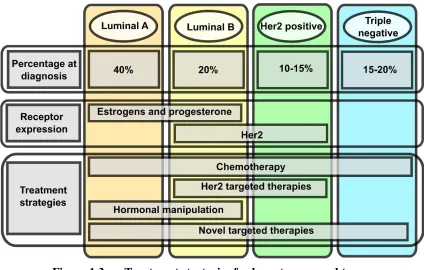
have to rely on chemotherapy and radiation alone, and if they are fortunate, in combination with new, untested drugs in clinical trials (Figure 3) (76).
Other therapies such as gefitinib, erlotinib, and cetuximab, which all target the epithelial growth factor receptor (EGFR), have been used for TNBC patients in clinical trials (77). However, these therapies were not able to statistically improve patient
outcomes when compared to standard treatment alone (77). Unlike with many other types of cancer, it is possible that TNBC is not dependent on EGFR signaling for tumor
growth, survival, and invasive capacity. The lack of treatments for distant metastases makes metastatic TNBC the least survivable subtype with a 5-year survival rate of only 26% (17). This provides a rationale for the development of better anti-cancer therapeutics that are effective against metastatic breast cancer.
Intracellular Pathways That Promote Breast Cancer Progression
Inflammation-driven breast cancer is associated with various signaling pathways that are activated by inflammatory cytokines (78, 79). While there are a multitude of intracellular signaling pathways that affect gene regulation, only a handful are well known for promoting cancer progression. In breast cancer, pathways that are involved include the signal transducer and activator of transcription 3 (STAT3),
phosphatidylinositol-3-kinase (PI3K), mitogen activated protein kinase (MAPK)
pathways, and nuclear factor kappa B (NFκB) (79-85). Specifically, STAT3 signaling has been shown to increase breast cancer invasive potential by upregulating matrix
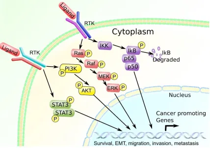
Furthermore, PI3K is associated with increased AKT activation and promotes breast cancer cell survival against chemotherapeutics (81, 87). In addition, MAPK signaling promotes similar events as STAT3 and PI3K, including cell survival (69), drug resistance (88-90), and invasion (91), as well as promoting cell proliferation. NFκB signaling, on the other hand, has been shown to be associated with the development of breast cancer stem cells, which can repopulate metastases after tumor ablation by chemotherapeutics (92). Specifically, NFκB pathway activation been shown to increase breast cancer cell proliferation and invasion by increasing cluster of differentiation 44 (CD44) expression, which promotes cell migration, survival, and increased stem cell-like characteristics (85). Based on these studies it is quite evident that there is a lot of overlap in function between the STAT3, AKT, MAPK and NFκB signaling pathways in the context of breast cancer, suggesting that there could be synergistic effects when multiple pathways are activated (Figure 4). Indeed, other studies show that these pathways can be activated at the same time, which can promote a synergistic effect that drives complex breast cancer
progression (79, 81). In summary, inflammation and inflammatory pathways related to the gp130 family of cytokines appear to be able to drive the simultaneous activation of these signaling pathways (78, 79).
Oncostatin M and Breast Cancer
Oncostatin M (OSM), a gp130 family cytokine, acquired its name due to its cytostatic effect on breast cancer cells and was originally thought to be a possible therapeutic (93, 94). While this idea was popular between 1990-2000, it was later
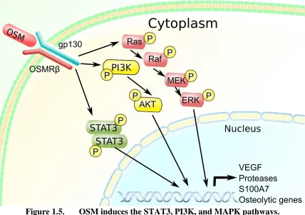
(101), and promotes metastasis (34). Furthermore, OSM signaling is associated with a poor breast cancer patient prognosis (97). OSM is secreted by a variety of cells in the tumor microenvironment such as tumor-associated neutrophils (TANs) and macrophages (TAMs), as well as adipocytes (101, 102). Interestingly, since OSM can be produced by adipose tissue, it expression levels may be increased in patients with obesity and type 2 diabetes, both of which are thought to be drivers in systemic inflammation-mediated breast cancer progression (14, 62, 100, 103). Furthermore, OSM appears to accumulate in the acidic breast cancer microenvironment, where it binds to ECM proteins and stays active (98, 104, 105). Once OSM is secreted, it can act on target cells through the OSM receptor complex (OSMR), which consists of the gp130 protein dimerized with OSMRβ (106) (Figure 5). Once OSM binds to its receptor, it can activate a multitude of intracellular
signaling cascades.
OSM is known to stimulate the activation of three signaling pathways including the STAT3 (107), PI3K/AKT (78), and MAPK pathways (78, 107) (Figure 5).
cancer cells towards a TNBC phenotype (111). Together, these studies strongly suggest that OSM is a proinflammatory factor with a multitude of effects that exacerbate breast cancer progression and metastasis. However, there are other related cytokines such as interleukin-1 beta (IL-1β) and the parental gp130 family member IL-6, which can have synergistic and overlapping functions with OSM (112).
Interleukin-6, And Interleukin-1 Beta in Breast Cancer
IL-6 is a cytokine in the gp130 family and is known to bind to a receptor composed of a dimer of IL-6Rα and a dimer of gp130 to form a tetramer receptor
IL-1β is a proinflammatory cytokine in the IL-1 family, and it binds to a receptor complex composed of IL1-R1 and IL-1RAcP (125, 126). Once the receptor complex is activated, IL-1β induces the NFB and MAPK pathways (127-129). IL-1β signaling is able to increase tumor cell growth, invasiveness, and metastasis in vivo by the stimulated secretion of proteases that degrade the ECM (125, 130-133). Anti-IL-1β therapeutics have been developed (134); however, much like with the anti-IL-6 biologics, IL-1β inhibition has not yielded positive results in patients when compared to standard
chemotherapeutics (135). These studies suggest that for breast cancer patients, inhibition of a singular cytokine may not be enough due to the overlapping functions of these cytokines. Common anti-inflammatory medications such as NSAIDs inhibit
cyclooxygenase 2 (COX2) expression, resulting in a decrease in prostaglandin production and in the reduced serum levels of a wide range of inflammatory cytokines and growth factors such as tumor necrosis factor alpha (TNF), IL-6, IL-1β, and VEGF (136-138). Additionally, studies have shown that there may be a benefit in using NSAIDS for the reduction of breast cancer risk (60). In conclusion, multiple cytokines may need to be suppressed simultaneously to obtain a positive clinical effect with reduction in metastases and increased patient survival.
Cancer Angiogenesis and Hypoxia Factors
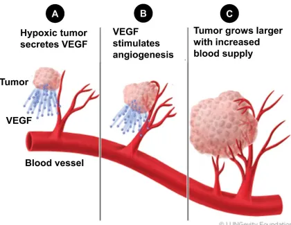
proangiogenic vascular endothelial growth factor (VEGF) (139-142) (Figure 6). Furthermore, proangiogenic transcription factors such as hypoxia-inducible factor 1 (HIF1) have also been linked to inflammation, cancer stem cells, and cancer progression (143, 144). HIF1 consists of a dimer of HIF1 and HIF1. Typically, HIF1α is regulated by rapid degradation while under normoxic conditions or in the absence of any inducing cytokines (145). Once HIF1α has been stabilized by hypoxia or by HIF1-inducing cytokines, it can translocate to the nucleus after dimerizing with the constitutively
expressed HIF1β and activate gene expression of VEGF and other proangiogenic proteins such as basic fibroblast growth factor (bFGF) (146, 147). In particular, OSM has been shown to increase HIF1α levels through STAT3 activation that leads to an increase in the production of VEGF (148-150). IL-6 has also been shown to increase VEGF secretion through the induction of STAT3 phosphorylation (116). Despite the mountain of evidence showing that VEGF promotes breast cancer progression, anti-VEGF therapies have had limited success clinically (151). Much like with the story of the failed therapies with anti-IL-6 and anti-IL-1β, it is likely that other proangiogenic factors play a role in promoting tumor angiogenesis. One of the consequences of tumor angiogenesis is that the tumor cells intravasate into the newly formed vessels and become circulating tumor cells.
Circulating Tumor Cells and Their Detection
of epithelial markers such as epithelial cell adhesion molecule (EpCAM), cytokeratin 19 (CK19), and human mammoglobin (hMAM) (155). Detection methods that are reliant on the presence of epithelial makers such as EpCAM and CK19 have run into a few major problems. For example, CTCs tend to lose their epithelial markers (such as EpCAM and CK19) when they undergo EMT and intravasate into the circulation (156, 157).
the nature of CTCs with a search for additional useful cancer cell markers that can be used in the detection of CTCs.
Mouse Models for Breast Cancer
The most common animal model for the study of tumorigenesis, metastasis, and drug development is the common mouse, mus musculus. The short life span and high metabolism of the mouse allows researchers to study the course of cancer progression in a compressed time-scale. Typical animal experiments using mus musculus for the study of cancer commonly last between 1-3 months, whereas in humans, studies take 1 to 5+ years. Broadly speaking, there are three types of mouse cancer models: the transgenic tumor-forming mouse model, the human xenograft mouse model, and the syngeneic cancer mouse model (162-164). In the transgenic animal model, the animals are genetically modified to overexpress oncogenes such as c-Myc or have loss-of-function mutations in tumor suppressor genes such as p53 (164). These animals spontaneously develop tumors and are a useful model for studying neo-tumorigenesis where normal cells transform into metastatic tumor cells in a step-wise fashion.
the same genetic background. This allows the study of tumor growth and metastatic disease in a immunocompetent model (162). An example of such a model utilizes the 4T1 mammary tumor cell line that was derived from a spontaneous mammary tumor in a BALB/c albino mouse. The 4T1.2 cell line was developed by selecting a clonal
population of 4T1 cells and possesses a metastatic capacity that roughly matches what is seen in human breast cancer (165). An advantage of the syngeneic model is that the immune system is not compromised and inflammation and immunity, so important for metastasis, can be studied (41).
These animal models collectively give researchers powerful tools by which to study breast cancer progression, yet they are not without their faults. Many therapies that appear promising in in vivo animal models of breast cancer, fail miserably in the clinic. This underlies the need for better animal models that recapitulates human breast cancer (166). Although it is unlikely that a single animal model could ever perfectly model human breast cancer, utilization of multiple animal models may help confirm positive results. In addition, results from animal studies should be corroborated with available human data to increase the likelihood of the data being clinically relevant for human breast cancer.
Human Data Analysis
downloaded from Oncomine™, and various types of gene expression and correlation analyses can be performed. Other sources of online databases include the catalog of somatic mutations in cancer (COSMIC), the human protein atlas, the cBioPortal cancer genomics database, and others (168-170). Combined with a few bioinformatics tools and some rudimentary PERL programming, these online databases can be a powerful source of data for researchers to associate in vitro or in vivo data with actual human cancer gene expression data.
Other sources of human patient data can be obtained from serum or tissues
obtained from breast cancer patients. Serum and tissue samples can generally be acquired either from commercial sources and/or from hospitals/institutes that have a repository of stored human tissue and serum samples from patients. Alternatively, a new institutional review board protocol can be drafted to recruit patients for serum or tissue biopsy collection for cancer research, if the already available samples are insufficient or inappropriate for the study design. Additionally, serum samples can also be obtained from the National Cancer Institute’s serum bank for various research studies (171). The collected samples can then be assessed by numerous assays ranging from
immunohistochemistry, immunoblot analysis, and enzyme-linked immunosorbent assays (ELISAs), to laser capture microdissection and qPCR. Overall, these sources of samples and data provide researchers a diverse tool set and help move data from tissue culture and animal models closer to clinical relevance.
Summary
metastasis, survival, and their synergistic relationship with other proinflammatory cytokines. In Chapter 2, co-expression of VEGF and gp130 family cytokines in relation to patient survival is assessed. These studies found that high expression levels of these proteins correlate with decreased breast cancer patient survival and that the level of VEGF production in response to OSM or IL-6 is breast cancer subtype-dependent. In Chapter 3, the role of OSM in the promotion of CTCs and lung metastases was
investigated in multiple in vivo mouse models of breast cancer. Specifically, high levels of OSM, through injection of recombinant OSM or induction of OSM expression in mammary tumor-bearing mice, resulted in an increase in both CTC numbers and lung metastases, as well as in decreased survival. However, OSM had no effect on the survival of mice when the earlier steps of the metastatic cascade were bypassed by injecting tumor cells directly into circulation. This finding suggests that OSM is important early in the metastatic cascade. In Chapter 4, OSM induction of IL-6 was assessed in breast cancer. It was determined that OSM-induced IL-6 production is predicated on breast cancer cells lacking ER. Specifically, ER+ T47D and MCF7 did not produce any IL-6 in response to OSM, while ER+ TNBC MDA-MD-231 cells did. Furthermore, the synergistic
Figures
Figure 1.1 Chronic inflammation leads to genomic and epigenetic instability and promotes tumor formation
A, Chronic inflammation promotes the development of downstream
pro-cancerous effects seen in B. B, Inflammation increases genomic instability resulting from DNA damage and changes the epigenetic profile of various genes. C, Genomic changes in precancerous cells can promote the secretion of inflammatory cytokines and
Figure 1.2. Multi-step development of invasive ductal carcinoma (IDC)
Figure 1.2. Multi-step development of invasive ductal carcinoma (IDC).
Figure 1.3. Treatment strategies for breast cancer subtypes.
Figure 1.3. Treatment strategies for breast cancer subtypes.
Figure 1.4. Multiple signaling pathway activation in breast cancer cells.
Figure 1.4. Multiple signaling pathway activation in breast cancer cells.
Figure 1.5. OSM induces the STAT3, PI3K, and MAPK pathways.
Figure 1.5. OSM induces the STAT3, PI3K, and MAPK pathways.
Figure 1.6. Tumor cells promote the development of angiogenesis.
Figure 1.6. Tumor cells promote the development of angiogenesis.
References
1. Jones PA, Laird PW. Cancer epigenetics comes of age. Nat Genet. 1999;21(2):163-7. doi: 10.1038/5947. PubMed PMID: 9988266.
2. Yoo CB, Jones PA. Epigenetic therapy of cancer: past, present and future. Nat Rev Drug Discov. 2006;5(1):37-50. doi: 10.1038/nrd1930. PubMed PMID:
16485345.
3. Khan O, La Thangue NB. HDAC inhibitors in cancer biology: emerging mechanisms and clinical applications. Immunol Cell Biol. 2012;90(1):85-94. Epub 2011/11/29. doi: 10.1038/icb.2011.100. PubMed PMID: 22124371.
4. Akiyama M, Sowa Y, Taniguchi T, Watanabe M, Yogosawa S, Kitawaki J, Sakai T. Three-Combined Treatment, a Novel HDAC Inhibitor OBP-801/YM753, 5-Fluorouracil and Paclitaxel, Induces G2-Phase Arrest Through the p38 Pathway in Human Ovarian Cancer Cells. Oncol Res. 2017. Epub 2017/01/23. doi:
10.3727/096504017X14850164661097. PubMed PMID: 28117030.
5. Stenvinkel P, Karimi M, Johansson S, Axelsson J, Suliman M, Lindholm B, Heimbürger O, Barany P, Alvestrand A, Nordfors L, Qureshi AR, Ekström TJ, Schalling M. Impact of inflammation on epigenetic DNA methylation - a novel risk factor for cardiovascular disease? J Intern Med. 2007;261(5):488-99. doi:
10.1111/j.1365-2796.2007.01777.x. PubMed PMID: 17444888.
6. Joyce BT, Gao T, Liu L, Zheng Y, Liu S, Zhang W, Penedo F, Dai Q, Schwartz J, Baccarelli AA, Hou L. Longitudinal Study of DNA Methylation of Inflammatory Genes and Cancer Risk. Cancer Epidemiol Biomarkers Prev.
2015;24(10):1531-8. Epub 2015/08/11. doi: 10.1158/1055-9965.EPI-15-0198. PubMed PMID: 26265203; PMCID: PMC4592468.
7. Gemenetzi M, Lotery AJ. The role of epigenetics in age-related macular degeneration. Eye (Lond). 2014;28(12):1407-17. Epub 2014/09/19. doi:
10.1038/eye.2014.225. PubMed PMID: 25233816; PMCID: PMC4268465.
8. Garfield L, Mathews HL, Witek Janusek L. Inflammatory and Epigenetic Pathways for Perinatal Depression. Biol Res Nurs. 2016;18(3):331-43. Epub 2015/11/16. doi: 10.1177/1099800415614892. PubMed PMID: 26574573.
9. Ciechomska M, van Laar JM, O'Reilly S. Emerging role of epigenetics in systemic sclerosis pathogenesis. Genes Immun. 2014;15(7):433-9. Epub 2014/07/17. doi: 10.1038/gene.2014.44. PubMed PMID: 25030429.
10. Han A, MacDonald A, Ahmed B, Whelan J, Donohoe D. Butyrate
Regulates Its Own Metabolic Fate as an HDAC inhibitor in Colorectal Cancer Cells. The FASEB Journal. 2017;31(1 Supplement):300.2-.2.
11. Dashwood RH, Myzak MC, Ho E. Dietary HDAC inhibitors: time to rethink weak ligands in cancer chemoprevention? Carcinogenesis. 2006;27(2):344-9. Epub 2005/11/02. doi: 10.1093/carcin/bgi253. PubMed PMID: 16267097; PMCID: PMC2267878.
13. Wang S, Ma N, Zhao W, Midorikawa K, Kawanishi S, Hiraku Y, Oikawa S, Zhang Z, Huang G, Murata M. Inflammation-Related DNA Damage and Cancer Stem Cell Markers in Nasopharyngeal Carcinoma. Mediators Inflamm. 2016;2016:9343460. Epub 2016/08/28. doi: 10.1155/2016/9343460. PubMed PMID: 27647953; PMCID: PMC5018333.
14. Suarez-Carmona M, Lesage J, Cataldo D, Gilles C. EMT and inflammation: inseparable actors of cancer progression. Mol Oncol. 2017. Epub 2017/06/09. doi: 10.1002/1878-0261.12095. PubMed PMID: 28599100.
15. Torre LA, Bray F, Siegel RL, Ferlay J, Lortet-Tieulent J, Jemal A. Global cancer statistics, 2012. CA: a cancer journal for clinicians. 2015;65(2):87-108. doi: 10.3322/caac.21262. PubMed PMID: 25651787.
16. Society AC. Cancer Facts and Figures 2017. In: American Cancer Society I, editor. Atlanta2017. p. 1-56.
17. Society AC. Breast Cancer Facts and Figures 2015-2016. Atlanta: American Cancer Society, Inc; 2016.
18. Harris JR, Lippman ME, Veronesi U, Willett W. Breast cancer (2). N Engl J Med. 1992;327(6):390-8. doi: 10.1056/NEJM199208063270606. PubMed PMID: 1320737.
19. Tan BY, Acs G, Apple SK, Badve S, Bleiweiss IJ, Brogi E, Calvo JP, Dabbs DJ, Ellis IO, Eusebi V, Farshid G, Fox SB, Ichihara S, Lakhani SR, Rakha EA, Reis-Filho JS, Richardson AL, Sahin A, Schmitt FC, Schnitt SJ, Siziopikou KP, Soares FA, Tse GM, Vincent-Salomon A, Tan PH. Phyllodes tumours of the breast: a consensus review. Histopathology. 2016;68(1):5-21. doi: 10.1111/his.12876. PubMed PMID: 26768026; PMCID: PMC5027876.
20. Levine PH, Veneroso C. The epidemiology of inflammatory breast cancer. Semin Oncol. 2008;35(1):11-6. doi: 10.1053/j.seminoncol.2007.11.018. PubMed PMID: 18308141.
21. Bleicher RJ. Ductal carcinoma in situ. Surg Clin North Am.
2013;93(2):393-410. Epub 2013/01/29. doi: 10.1016/j.suc.2012.12.001. PubMed PMID: 23464692.
22. DeSantis CE, Fedewa SA, Goding Sauer A, Kramer JL, Smith RA, Jemal A. Breast cancer statistics, 2015: Convergence of incidence rates between black and white women. CA: a cancer journal for clinicians. 2015. doi: 10.3322/caac.21320. PubMed PMID: 26513636.
23. Trape AP, Gonzalez-Angulo AM. Breast cancer and metastasis: on the way toward individualized therapy. Cancer genomics & proteomics. 2012;9(5):297-310. PubMed PMID: 22990109.
24. Diel IJ. Bone Metastasis in Breast Cancer. Breast care. 2012;7(2):90-1. doi: 10.1159/000338708. PubMed PMID: 22740793; PMCID: 3376358.
25. Scully OJ, Bay BH, Yip G, Yu Y. Breast cancer metastasis. Cancer genomics & proteomics. 2012;9(5):311-20. PubMed PMID: 22990110.
26. Pomp V, Leo C, Mauracher A, Korol D, Guo W, Varga Z. Differential expression of epithelial-mesenchymal transition and stem cell markers in intrinsic subtypes of breast cancer. Breast cancer research and treatment. 2015. doi:
27. Bae YK, Choi JE, Kang SH, Lee SJ. Epithelial-Mesenchymal Transition Phenotype Is Associated with Clinicopathological Factors That Indicate Aggressive Biological Behavior and Poor Clinical Outcomes in Invasive Breast Cancer. Journal of breast cancer. 2015;18(3):256-63. doi: 10.4048/jbc.2015.18.3.256. PubMed PMID: 26472976; PMCID: 4600690.
28. Liu H, Zhang X, Li J, Sun B, Qian H, Yin Z. The biological and clinical importance of epithelial-mesenchymal transition in circulating tumor cells. J Cancer Res Clin Oncol. 2015;141(2):189-201. Epub 2014/06/26. doi: 10.1007/s00432-014-1752-x. PubMed PMID: 24965746.
29. van Zijl F, Krupitza G, Mikulits W. Initial steps of metastasis: cell invasion and endothelial transmigration. Mutat Res. 2011;728(1-2):23-34. Epub 2011/05/12. doi: 10.1016/j.mrrev.2011.05.002. PubMed PMID: 21605699; PMCID: PMC4028085.
30. Carr I. Lymphatic metastasis. Cancer Metastasis Rev. 1983;2(3):307-17. PubMed PMID: 6367969.
31. Fidler IJ. Metastasis: quantitative analysis of distribution and fate of tumor emboli labeled with 125 I-5-iodo-2'-deoxyuridine. J Natl Cancer Inst. 1970;45(4):773-82. PubMed PMID: 5513503.
32. Price TT, Burness ML, Sivan A, Warner MJ, Cheng R, Lee CH, Olivere L, Comatas K, Magnani J, Kim Lyerly H, Cheng Q, McCall CM, Sipkins DA. Dormant breast cancer micrometastases reside in specific bone marrow niches that regulate their transit to and from bone. Sci Transl Med. 2016;8(340):340ra73. doi:
10.1126/scitranslmed.aad4059. PubMed PMID: 27225183.
33. Chaffer CL, Brennan JP, Slavin JL, Blick T, Thompson EW, Williams ED. Mesenchymal-to-epithelial transition facilitates bladder cancer metastasis: role of fibroblast growth factor receptor-2. Cancer Res. 2006;66(23):11271-8. doi:
10.1158/0008-5472.CAN-06-2044. PubMed PMID: 17145872.
34. Bolin C, Tawara K, Sutherland C, Redshaw J, Aranda P, Moselhy J, Anderson R, Jorcyk CL. Oncostatin m promotes mammary tumor metastasis to bone and osteolytic bone degradation. Genes & cancer. 2012;3(2):117-30. doi:
10.1177/1947601912458284. PubMed PMID: 23050044; PMCID: 3463924.
35. Bos PD, Zhang XH, Nadal C, Shu W, Gomis RR, Nguyen DX, Minn AJ, van de Vijver MJ, Gerald WL, Foekens JA, Massagué J. Genes that mediate breast cancer metastasis to the brain. Nature. 2009;459(7249):1005-9. Epub 2009/05/06. doi:
10.1038/nature08021. PubMed PMID: 19421193; PMCID: PMC2698953.
36. Weigelt B, Peterse JL, van 't Veer LJ. Breast cancer metastasis: markers and models. Nat Rev Cancer. 2005;5(8):591-602. doi: 10.1038/nrc1670. PubMed PMID: 16056258.
37. Petrut B, Trinkaus M, Simmons C, Clemons M. A primer of bone metastases management in breast cancer patients. Curr Oncol. 2008;15(Suppl 1):S50-7. PubMed PMID: 18231649; PMCID: PMC2216426.
38. Singh VA, Haseeb A, Alkubaisi AA. Incidence and outcome of bone metastatic disease at University Malaya Medical Centre. Singapore Med J.
2014;55(10):539-46. PubMed PMID: 25631896; PMCID: PMC4293965.
breast cancer. Cancer Cell. 2008;13(1):58-68. doi: 10.1016/j.ccr.2007.12.003. PubMed PMID: 18167340.
40. Hosseini H, Obradović MM, Hoffmann M, Harper KL, Sosa MS, Werner-Klein M, Nanduri LK, Werno C, Ehrl C, Maneck M, Patwary N, Haunschild G, Gužvić M, Reimelt C, Grauvogl M, Eichner N, Weber F, Hartkopf AD, Taran FA, Brucker SY, Fehm T, Rack B, Buchholz S, Spang R, Meister G, Aguirre-Ghiso JA, Klein CA. Early dissemination seeds metastasis in breast cancer. Nature. 2016. Epub 2016/12/14. doi: 10.1038/nature20785. PubMed PMID: 27974799; PMCID: PMC5390864.
41. Grivennikov SI, Greten FR, Karin M. Immunity, inflammation, and cancer. Cell. 2010;140(6):883-99. doi: 10.1016/j.cell.2010.01.025. PubMed PMID: 20303878; PMCID: PMC2866629.
42. Tawara K, Oxford JT, Jorcyk CL. Clinical significance of interleukin (IL)-6 in cancer metastasis to bone: potential of anti-IL-(IL)-6 therapies. Cancer management and research. 2011;3:177-89. doi: 10.2147/CMR.S18101. PubMed PMID: 21625400; PMCID: 3101113.
43. Crespi E, Bottai G, Santarpia L. Role of inflammation in obesity-related breast cancer. Curr Opin Pharmacol. 2016;31:114-22. Epub 2016/11/24. doi:
10.1016/j.coph.2016.11.004. PubMed PMID: 27889687.
44. Pierce BL, Neuhouser ML, Wener MH, Bernstein L, Baumgartner RN, Ballard-Barbash R, Gilliland FD, Baumgartner KB, Sorensen B, McTiernan A, Ulrich CM. Correlates of circulating C-reactive protein and serum amyloid A concentrations in breast cancer survivors. Breast Cancer Res Treat. 2009;114(1):155-67. Epub 2008/04/10. doi: 10.1007/s10549-008-9985-5. PubMed PMID: 18401703; PMCID: PMC3523176.
45. Ford DE, Erlinger TP. Depression and C-reactive protein in US adults: data from the Third National Health and Nutrition Examination Survey. Arch Intern Med. 2004;164(9):1010-4. doi: 10.1001/archinte.164.9.1010. PubMed PMID: 15136311.
46. Harris HR, Willett WC, Vaidya RL, Michels KB. An Adolescent and Early Adulthood Dietary Pattern Associated with Inflammation and the Incidence of Breast Cancer. Cancer Res. 2017;77(5):1179-87. doi: 10.1158/0008-5472.CAN-16-2273. PubMed PMID: 28249935; PMCID: PMC5335878.
47. Bhatelia K, Singh K, Singh R. TLRs: linking inflammation and breast cancer. Cellular signalling. 2014;26(11):2350-7. doi: 10.1016/j.cellsig.2014.07.035. PubMed PMID: 25093807.
48. Deng T, Lyon CJ, Bergin S, Caligiuri MA, Hsueh WA. Obesity, Inflammation, and Cancer. Annu Rev Pathol. 2016;11:421-49. doi: 10.1146/annurev-pathol-012615-044359. PubMed PMID: 27193454.
49. Gubitosi A, Docimo G, Ruggiero R, Esposito A, Esposito E, Foroni F. Breast implant (PIP), chronic inflammation and cancer: is there a connection? Case report. Annali italiani di chirurgia. 2012;83. PubMed PMID: 23075481.
50. Cole SW. Chronic inflammation and breast cancer recurrence. Journal of clinical oncology : official journal of the American Society of Clinical Oncology. 2009;27(21):3418-9. doi: 10.1200/JCO.2009.21.9782. PubMed PMID: 19470918.
52. Rose DP, Gracheck PJ, Vona-Davis L. The Interactions of Obesity, Inflammation and Insulin Resistance in Breast Cancer. Cancers. 2015;7(4):2147-68. doi: 10.3390/cancers7040883. PubMed PMID: 26516917.
53. Rose DP, Vona-Davis L. Biochemical and molecular mechanisms for the association between obesity, chronic inflammation, and breast cancer. BioFactors. 2014;40(1):1-12. doi: 10.1002/biof.1109. PubMed PMID: 23857944.
54. Suman S, Sharma PK, Rai G, Mishra S, Arora D, Gupta P, Shukla Y. Current perspectives of molecular pathways involved in chronic Inflammation-mediated breast cancer. Biochemical and biophysical research communications. 2015. doi:
10.1016/j.bbrc.2015.10.133. PubMed PMID: 26522220.
55. Brighenti E, Calabrese C, Liguori G, Giannone FA, Trere D, Montanaro L, Derenzini M. Interleukin 6 downregulates p53 expression and activity by stimulating ribosome biogenesis: a new pathway connecting inflammation to cancer. Oncogene. 2014;33(35):4396-406. doi: 10.1038/onc.2014.1. PubMed PMID: 24531714; PMCID: 4150990.
56. De Benedetti F, Alonzi T, Moretta A, Lazzaro D, Costa P, Poli V, Martini A, Ciliberto G, Fattori E. Interleukin 6 causes growth impairment in transgenic mice through a decrease in insulin-like growth factor-I. A model for stunted growth in children with chronic inflammation. The Journal of clinical investigation. 1997;99(4):643-50. doi: 10.1172/JCI119207. PubMed PMID: 9045866; PMCID: 507846.
57. Nguyen DP, Li J, Tewari AK. Inflammation and prostate cancer: the role of interleukin 6 (IL-6). BJU international. 2014;113(6):986-92. doi: 10.1111/bju.12452. PubMed PMID: 24053309.
58. Holmes MD, Chen WY, Schnitt SJ, Collins L, Colditz GA, Hankinson SE, Tamimi RM. COX-2 expression predicts worse breast cancer prognosis and does not modify the association with aspirin. Breast Cancer Res Treat. 2011;130(2):657-62. Epub 2011/07/05. doi: 10.1007/s10549-011-1651-7. PubMed PMID: 21728052; PMCID: PMC3292350.
59. Samarajeewa NU, Yang F, Docanto MM, Sakurai M, McNamara KM, Sasano H, Fox SB, Simpson ER, Brown KA. HIF-1α stimulates aromatase expression driven by prostaglandin E2 in breast adipose stroma. Breast Cancer Res. 2013;15(2):R30. Epub 2013/04/08. doi: 10.1186/bcr3410. PubMed PMID: 23566437; PMCID:
PMC3672802.
60. Takkouche B, Regueira-Méndez C, Etminan M. Breast cancer and use of nonsteroidal anti-inflammatory drugs: a meta-analysis. J Natl Cancer Inst.
2008;100(20):1439-47. Epub 2008/10/07. doi: 10.1093/jnci/djn324. PubMed PMID: 18840819.
61. Repovic P, Fears CY, Gladson CL, Benveniste EN. Oncostatin-M induction of vascular endothelial growth factor expression in astroglioma cells. Oncogene. 2003;22(50):8117-24. doi: 10.1038/sj.onc.1206922. PubMed PMID: 14603252.
62. Silver JS, Hunter CA. gp130 at the nexus of inflammation, autoimmunity, and cancer. J Leukoc Biol. 2010;88(6):1145-56. Epub 2010/07/07. doi:
63. Zhou J, Yan Y, Guo L, Ou H, Hai J, Zhang C, Wu Z, Tang L. Distinct outcomes in patients with different molecular subtypes of inflammatory breast cancer. Saudi Med J. 2014;35(11):1324-30. PubMed PMID: 25399208; PMCID: PMC4362150.
64. Kohler BA, Sherman RL, Howlader N, Jemal A, Ryerson AB, Henry KA, Boscoe FP, Cronin KA, Lake A, Noone AM, Henley SJ, Eheman CR, Anderson RN, Penberthy L. Annual Report to the Nation on the Status of Cancer, 1975-2011, Featuring Incidence of Breast Cancer Subtypes by Race/Ethnicity, Poverty, and State. J Natl Cancer Inst. 2015;107(6):djv048. Epub 2015/03/30. doi: 10.1093/jnci/djv048. PubMed PMID: 25825511; PMCID: PMC4603551.
65. Gangadhara S, Smith C, Barrett-Lee P, Hiscox S. 3D culture of Her2+ breast cancer cells promotes AKT to MAPK switching and a loss of therapeutic response. BMC Cancer. 2016;16:345. Epub 2016/06/01. doi: 10.1186/s12885-016-2377-z. PubMed PMID: 27251376; PMCID: PMC4888214.
66. Jahandoost S, Farhanghian P, Abbasi S. The Effects of Sex Protein Receptors and Sex Steroid Hormone Gene Polymorphisms on Breast Cancer Risk. J Natl Med Assoc. 2017;109(2):126-38. Epub 2017/03/01. doi: 10.1016/j.jnma.2017.02.003. PubMed PMID: 28599754.
67. Gangadhara S, Barrett-Lee P, Nicholson RI, Hiscox S. Pro-metastatic tumor-stroma interactions in breast cancer. Future Oncol. 2012;8(11):1427-42. doi: 10.2217/fon.12.134. PubMed PMID: 23148616.
68. Dybdal N, Leiberman G, Anderson S, McCune B, Bajamonde A, Cohen RL, Mass RD, Sanders C, Press MF. Determination of HER2 gene amplification by fluorescence in situ hybridization and concordance with the clinical trials
immunohistochemical assay in women with metastatic breast cancer evaluated for treatment with trastuzumab. Breast Cancer Res Treat. 2005;93(1):3-11. doi: 10.1007/s10549-004-6275-8. PubMed PMID: 16184453.
69. Menendez JA, Vellon L, Mehmi I, Teng PK, Griggs DW, Lupu R. A novel CYR61-triggered 'CYR61-alphavbeta3 integrin loop' regulates breast cancer cell survival and chemosensitivity through activation of ERK1/ERK2 MAPK signaling pathway. Oncogene. 2005;24(5):761-79. doi: 10.1038/sj.onc.1208238. PubMed PMID: 15592521.
70. Scholzen T, Gerdes J. The Ki-67 protein: from the known and the unknown. J Cell Physiol. 2000;182(3):311-22. doi:
10.1002/(SICI)1097-4652(200003)182:3<311::AID-JCP1>3.0.CO;2-9. PubMed PMID: 10653597.
71. Tamimi RM, Baer HJ, Marotti J, Galan M, Galaburda L, Fu Y, Deitz AC, Connolly JL, Schnitt SJ, Colditz GA, Collins LC. Comparison of molecular phenotypes of ductal carcinoma in situ and invasive breast cancer. Breast Cancer Res.
2008;10(4):R67. Epub 2008/08/05. doi: 10.1186/bcr2128. PubMed PMID: 18681955; PMCID: PMC2575540.
72. Sørlie T, Perou CM, Tibshirani R, Aas T, Geisler S, Johnsen H, Hastie T, Eisen MB, van de Rijn M, Jeffrey SS, Thorsen T, Quist H, Matese JC, Brown PO, Botstein D, Lønning PE, Børresen-Dale AL. Gene expression patterns of breast
73. Soliman NA, Yussif SM. Ki-67 as a prognostic marker according to breast cancer molecular subtype. Cancer Biol Med. 2016;13(4):496-504. doi:
10.20892/j.issn.2095-3941.2016.0066. PubMed PMID: 28154782; PMCID: PMC5250608.
74. Macedo LF, Sabnis G, Brodie A. Aromatase inhibitors and breast cancer. Ann N Y Acad Sci. 2009;1155:162-73. doi: 10.1111/j.1749-6632.2008.03689.x. PubMed PMID: 19250202.
75. Arkin M, Moasser MM. HER-2-directed, small-molecule antagonists. Curr Opin Investig Drugs. 2008;9(12):1264-76. PubMed PMID: 19037833; PMCID: PMC3031872.
76. Catalanotti V BV, Tariq N, Califano R. Treatment of Advanced Breast Cancer (ABC): The Expanding Landscape of Targeted Therapies. J Cancer Biol Res. 2014;2(1):1036
77. Nakai K, Hung MC, Yamaguchi H. A perspective on anti-EGFR therapies targeting triple-negative breast cancer. Am J Cancer Res. 2016;6(8):1609-23. Epub 2016/08/01. PubMed PMID: 27648353; PMCID: PMC5004067.
78. Demyanets S, Kaun C, Rychli K, Pfaffenberger S, Kastl SP, Hohensinner PJ, Rega G, Katsaros KM, Afonyushkin T, Bochkov VN, Paireder M, Huk I, Maurer G, Huber K, Wojta J. Oncostatin M-enhanced vascular endothelial growth factor expression in human vascular smooth muscle cells involves PI3K-, p38 MAPK-, Erk1/2- and
STAT1/STAT3-dependent pathways and is attenuated by interferon-gamma. Basic research in cardiology. 2011;106(2):217-31. doi: 10.1007/s00395-010-0141-0. PubMed PMID: 21174212.
79. Kim MS, Lee WS, Jeong J, Kim SJ, Jin W. Induction of metastatic potential by TrkB via activation of IL6/JAK2/STAT3 and PI3K/AKT signaling in breast cancer. Oncotarget. 2015;6(37):40158-71. doi: 10.18632/oncotarget.5522. PubMed PMID: 26515594.
80. Gariboldi MB, Ravizza R, Molteni R, Osella D, Gabano E, Monti E. Inhibition of Stat3 increases doxorubicin sensitivity in a human metastatic breast cancer cell line. Cancer letters. 2007;258(2):181-8. doi: 10.1016/j.canlet.2007.08.019. PubMed PMID: 17920763.
81. Kim JW, Kim DK, Min A, Lee KH, Nam HJ, Kim JH, Kim JS, Kim TY, Im SA, Park IA. Amphiregulin confers trastuzumab resistance via AKT and ERK activation in HER2-positive breast cancer. Journal of cancer research and clinical oncology. 2015. doi: 10.1007/s00432-015-2012-4. PubMed PMID: 26195282.
82. Sonnenblick A, Brohee S, Fumagalli D, Vincent D, Venet D, Ignatiadis M, Salgado R, Van den Eynden G, Rothe F, Desmedt C, Neven P, Loibl S, Denkert C, Joensuu H, Loi S, Sirtaine N, Kellokumpu-Lehtinen PL, Piccart M, Sotiriou C.
Constitutive phosphorylated STAT3-associated gene signature is predictive for trastuzumab resistance in primary HER2-positive breast cancer. BMC medicine. 2015;13:177. doi: 10.1186/s12916-015-0416-2. PubMed PMID: 26234940; PMCID: 4522972.
2015;16(10):24772-90. doi: 10.3390/ijms161024772. PubMed PMID: 26501276; PMCID: 4632776.
84. Zhang H, Cai K, Wang J, Wang X, Cheng K, Shi F, Jiang L, Zhang Y, Dou J. MiR-7, inhibited indirectly by lincRNA HOTAIR, directly inhibits SETDB1 and reverses the EMT of breast cancer stem cells by downregulating the STAT3 pathway. Stem cells. 2014;32(11):2858-68. doi: 10.1002/stem.1795. PubMed PMID: 25070049.
85. Smith SM, Lyu YL, Cai L. NF-κB affects proliferation and invasiveness of breast cancer cells by regulating CD44 expression. PLoS One. 2014;9(9):e106966. Epub 2014/09/03. doi: 10.1371/journal.pone.0106966. PubMed PMID: 25184276; PMCID: PMC4153718.
86. Tester AM, Ruangpanit N, Anderson RL, Thompson EW. MMP-9 secretion and MMP-2 activation distinguish invasive and metastatic sublines of a mouse mammary carcinoma system showing epithelial-mesenchymal transition traits. Clin Exp Metastasis. 2000;18(7):553-60. PubMed PMID: 11688960.
87. Chen X, Wang H, Ou-Yang XN, Xie FW, Wu JJ. Research on drug resistance mechanism of trastuzumab caused by activation of the PI3K/Akt signaling pathway. Contemporary oncology. 2013;17(4):363-9. doi: 10.5114/wo.2013.35292. PubMed PMID: 24592124; PMCID: 3934046.
88. Donovan JC, Milic A, Slingerland JM. Constitutive MEK/MAPK activation leads to p27(Kip1) deregulation and antiestrogen resistance in human breast cancer cells. The Journal of biological chemistry. 2001;276(44):40888-95. doi:
10.1074/jbc.M106448200. PubMed PMID: 11527971.
89. Eralp Y, Derin D, Ozluk Y, Yavuz E, Guney N, Saip P, Muslumanoglu M, Igci A, Kucucuk S, Dincer M, Aydiner A, Topuz E. MAPK overexpression is associated with anthracycline resistance and increased risk for recurrence in patients with triple-negative breast cancer. Annals of oncology : official journal of the European Society for Medical Oncology / ESMO. 2008;19(4):669-74. doi: 10.1093/annonc/mdm522. PubMed PMID: 18006896.
90. Normanno N, Campiglio M, Maiello MR, De Luca A, Mancino M, Gallo M, D'Alessio A, Menard S. Breast cancer cells with acquired resistance to the EGFR tyrosine kinase inhibitor gefitinib show persistent activation of MAPK signaling. Breast cancer research and treatment. 2008;112(1):25-33. doi: 10.1007/s10549-007-9830-2. PubMed PMID: 18060492.
91. Meng X, Cai C, Wu J, Cai S, Ye C, Chen H, Yang Z, Zeng H, Shen Q, Zou F. TRPM7 mediates breast cancer cell migration and invasion through the MAPK pathway. Cancer letters. 2013;333(1):96-102. doi: 10.1016/j.canlet.2013.01.031. PubMed PMID: 23353055.
92. Shostak K, Chariot A. NF-κB, stem cells and breast cancer: the links get stronger. Breast Cancer Res. 2011;13(4):214. Epub 2011/07/26. doi: 10.1186/bcr2886. PubMed PMID: 21867572; PMCID: PMC3236328.
94. Spence MJ, Vestal RE, Liu J. Oncostatin M-mediated transcriptional suppression of the c-myc gene in breast cancer cells. Cancer research. 1997;57(11):2223-8. PubMed PMID: 9187125.
95. Li C, Ahlborn TE, Tokita K, Boxer LM, Noda A, Liu J. The critical role of the PE21 element in oncostatin M-mediated transcriptional repression of the p53 tumor suppressor gene in breast cancer cells. Oncogene. 2001;20(57):8193-202. doi:
10.1038/sj.onc.1205046. PubMed PMID: 11781835.
96. Liu J, Li C, Ahlborn TE, Spence MJ, Meng L, Boxer LM. The expression of p53 tumor suppressor gene in breast cancer cells is down-regulated by cytokine oncostatin M. Cell growth & differentiation : the molecular biology journal of the American Association for Cancer Research. 1999;10(10):677-83. PubMed PMID: 10547071.
97. West NR, Murphy LC, Watson PH. Oncostatin M suppresses oestrogen receptor-alpha expression and is associated with poor outcome in human breast cancer. Endocrine-related cancer. 2012;19(2):181-95. doi: 10.1530/ERC-11-0326. PubMed PMID: 22267707.
98. Ryan RE, Martin B, Mellor L, Jacob RB, Tawara K, McDougal OM, Oxford JT, Jorcyk CL. Oncostatin M binds to extracellular matrix in a bioactive
conformation: implications for inflammation and metastasis. Cytokine. 2015;72(1):71-85. doi: 10.1016/j.cyto.2014.11.007. PubMed PMID: 25622278; PMCID: 4328881.
99. West NR, Murray JI, Watson PH. Oncostatin-M promotes phenotypic changes associated with mesenchymal and stem cell-like differentiation in breast cancer. Oncogene. 2014;33(12):1485-94. doi: 10.1038/onc.2013.105. PubMed PMID: 23584474.
100. Lapeire L, Hendrix A, Lambein K, Van Bockstal M, Braems G, Van Den Broecke R, Limame R, Mestdagh P, Vandesompele J, Vanhove C, Maynard D, Lehuédé C, Muller C, Valet P, Gespach CP, Bracke M, Cocquyt V, Denys H, De Wever O. Cancer-associated adipose tissue promotes breast cancer progression by paracrine oncostatin M and Jak/STAT3 signaling. Cancer Res. 2014;74(23):6806-19. Epub 2014/09/24. doi: 10.1158/0008-5472.CAN-14-0160. PubMed PMID: 25252914.
101. Queen MM, Ryan RE, Holzer RG, Keller-Peck CR, Jorcyk CL. Breast cancer cells stimulate neutrophils to produce oncostatin M: potential implications for tumor progression. Cancer research. 2005;65(19):8896-904. doi: 10.1158/0008-5472.CAN-05-1734. PubMed PMID: 16204061.
102. Guihard P, Boutet MA, Brounais-Le Royer B, Gamblin AL, Amiaud J, Renaud A, Berreur M, Rédini F, Heymann D, Layrolle P, Blanchard F. Oncostatin m, an inflammatory cytokine produced by macrophages, supports intramembranous bone healing in a mouse model of tibia injury. Am J Pathol. 2015;185(3):765-75. Epub 2015/01/02. doi: 10.1016/j.ajpath.2014.11.008. PubMed PMID: 25559270.
103. Sanchez-Infantes D, White UA, Elks CM, Morrison RF, Gimble JM, Considine RV, Ferrante AW, Ravussin E, Stephens JM. Oncostatin m is produced in adipose tissue and is regulated in conditions of obesity and type 2 diabetes. J Clin Endocrinol Metab. 2014;99(2):E217-25. Epub 2013/12/02. doi: 10.1210/jc.2013-3555. PubMed PMID: 24297795; PMCID: PMC3913819.