DOI: 10.2478/10004-1254-65-2014-2522 Original article
Acute toxic effects of cadmium in larvae of the green toad,
Pseudepidalea variabilis (Pallas, 1769) (Amphibia: Anura)
Mert Gürkan, Ayşe Çetin, and Sibel Hayretdağ
Department of Biology, Zoology Section, Faculty of Arts and Sciences, Çanakkale Onsekiz Mart University, Turkey
Received in April 2014 CrossChecked in April 2014
Accepted in August 2014
The environmental impact of cadmium use and its accumulation in nature have increased to alarming levels. This study aimed to morphologically and histologically investigate the acute toxic effects of cadmium on green toad, Pseudepidalea variabilis (Pallas, 1769) larvae. Embryos were obtained from specimens collected in amplexus from nature and kept under laboratory conditions until stage 26, when they were exposed to cadmium (0, 1, 5, 10, 25, and 50 µg L-1) for 96 h. The LC
10, LC50,and LC90 values of cadmium were calculated to be 26.98, 35.35, and 46.31 µg L-1, respectively. Our results showed that cadmium had a negative effect on the body size of P. variabilis larvae (over 1 µg L-1). Histological examination detected a fusion of gill lamellae, liver haemorrhage, oedema in the abdominal cavity, and deformations of pronephric tubules (over 10 µg L-1). Our findings suggest that the green toad was sensitive to the cadmium treatment, with LC50 values lower than those reported by other studies. Thus, this species could be considered a reliable indicator species of environmental stress in aquatic ecosystem.
KEY WORDS: embryotoxicity; heavy metal; histopathology; lethal concentrations
The decline in amphibian populations has reached
striking dimensions in recent years (1, 2). There are
several hypotheses about the possible reasons behind this phenomenon (3, 4). Some of these hypotheses include impact of urbanization due to rapid increase in settlements and industrialization, chemical pollution, habitat degradation/destruction, and
diseases (5-8). Among these, the destruction of
habitats and increased pollution significantly threaten the survival of amphibians, which spend their embryonic and larval stages exclusively in water and play a significant role in biomonitoring programmes (9-11). That is why amphibian larvae are among the preferred bioindicators for aquatic toxicology studies
(12-16).
Cadmium is present in the environment naturally;
however, it is not an essential element for organisms
(17). Increases in the amount of cadmium in nature
might be either due to natural factors, like the ablation
of rocks resulting from erosion and rain, or anthropogenic factors (18). In Turkey, for instance, one study (19) found the mean cadmium concentration
in surface waters to be 110 µg L-1.
The half-life of cadmium in organisms is 20 years
(20). It cannot be excreted from the body and
accumulates in the organism and throughout the food
chain. Its biomagnification poses threats for human health as well as the ecosystem as a whole. Therefore, cadmium has recently become the subject of many
investigations (16, 21-25). The toxic effects of
cadmium investigated in freshwater fish include development abnormalities detected in many teleost
species (26-28). Cadmium led to larval teratogenic
and developmental abnormalities (29), to micronucleus
induction at environmental levels of 2, 10, and 30 µg L-1, and to increased concentration-dependent
quantities of micronucleus after contamination with
cadmium exposure caused a decline in the survival
numbers of Bufo americanus (13), developmental
delays in tadpoles of Bufo raddei (31), and toxic acute effects on early-life stages in Duttaphrynus melanostictus at levels of 0.2 mg L-1 (23). On the
contrary, cadmium stimulated growth and development in amphibian larvae at concentrations from 0.25 to
5 µg L-1 (32).
Known as the green toad, Pseudepidalea variabilis
is a common amphibian species in Turkey. This
terrestrial and nocturnal species is water-dependent in the breeding season, when it is known to stay in water
for long periods of time (33). This species generally
inhabits slow-flowing and stagnant waters, seasonal ponds, and shallow pits filled with water for egg-laying (34). Its aquatic habitats are prone to increased heavy
metal concentrations due to agricultural and industrial
activities. To our knowledge, until now no related
study on the acute toxic effects of cadmium on P. variabilis larvae has been conducted.
This study aimed to determine the lethal concentrations (LC10, LC50,and LC90) of cadmium and the acute toxic effects on the tissues and organs of P. variabilis larvae after a 96-h acute exposure.
MATERIALS AND METHODS
Animals and treatment
The green toad, Pseudepidalea variabilis (Pallas, 1769), is included in the “data deficient” (DD)
category of the IUCN Red List (35). Tadpoles were obtained in the laboratory from 4 adult (2 ♂♂, 2 ♀♀)
P. variabilis caught in amplexus in March 2010, in a
seasonal pond in Çanakkale, north-western Turkey. P. variabilis specimens were transferred to the laboratory
and kept in polypropylene containers until ovulation.
After ovulation, embryos were transferred to 30×30×25 cm glass aquaria. After hatching, the larvae were fed with fish feed of plant origin (100 g/ carbohydrate: 30.16 g; sugars: 7.33 g; dietary fibre:
9.3 g; fat: 19.94 g; protein: 36.49 g; energy: 450 kcal)
and boiled lettuce leaves until stage 26 (36).
Cadmium chloride (CdCl2) was supplied from Sigma-Aldrich Chemical Company Inc. (St. Louis, MO, USA). A stock solution of 100 µg L-1 was
prepared by dissolving CdCl2 powder in distilled water. Larvae at stage 26 were exposed to concentrations of
1, 5, 10, 25, and 50 µg L-1 of cadmium. The dosing
solutions were prepared by appropriate dilutions of
the stock solution.
Twenty randomly selected P. variabilis larvae were exposed to control (water) and each cadmium
concentration, in acute (96 h) exposure experiments,
in a static bioassay test system. Aquaria were filled with 2 L of water or cadmium solutions and during
exposure, temperature, pH, and dissolved oxygen
levels were measured daily with an Elmetron CO-401 meter (analysio GmbH, Greifswald, Germany). The treatment was carried out in a 14:10-h light-dark cycle and the number of dead tadpoles was recorded and
faeces removed every 24 hours.
Morphology and histology
Ten randomly selected larvae from the control and
each treatment group were measured for total length, width, and tail length. Furthermore, wet weights of the larvae were determined (0 and 96 h). Wet weight
(precision 0.001 g) and total length (precision
0.01 mm) were measured after euthanasia with a
digital scale and digital calipers. Ten larvae survived
after the 96 h of exposure were fixed in Bouin’s
solution for histological examination after macroscopic
evaluation. Afterwards, these larvae were processed through alcohol, xylene, and paraffin series and then paraffin blocks were prepared. Serial cross sections 6-8 μm in thickness were obtained from these blocks, stained with hematoxylin and eosin (H&E), and
histopathologically examined under a light microscope.
Finally, the histological imaging of the preparations was carried out using a camera mounted on an Olympus BX51 light microscope (Japan) and analysed using the DP2-BSW software.
Statistical analysis
Finney’s Probit analysis method was used to
calculate 96-h LC10, LC50, and LC90 values of cadmium for P. variabilis larvae. Lethal concentrations were calculated at 95 % confidence intervals. Comparisons of total length, tail length, width and weight between control and treatment groups were done using one-way analysis of variance (ANOVA). Discriminant analysis was done in order to show the possible total differences
RESULTS
Acute toxicity
Mean water temperature was 21.2±2 °C, while pH and dissolved oxygen in aquaria were 6.8±0.3 and 8.2±0.4 mg L-1, respectively. No mortality was recorded in the control group and in the cadmium groups exposed to 1 and 5 µg L-1. The highest mortality
was observed at 50 µg L-1, where 10 larvae died within
the first 24 h and only one larva survived at the end of the exposure period. After 72 h, a dead larva was
found in the 10 µg L-1 group, whereas 8 larvae died in the 25 µg L-1 group. Values of 96-h LC
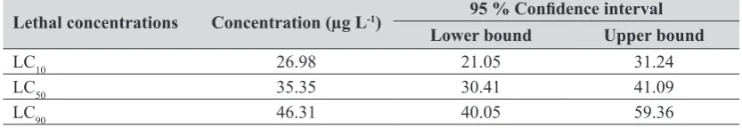
10, LC50, and LC90 are presented in Table 1.
Morphology and histology
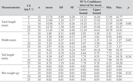
Total length was the only parameter found to be significantly different between treatments (p<0.05).
Comparisons of total length, width, tail length, and wet weight between control and treatment groups are presented in Table 2. Since only one larva survived
the 50 µg L-1 concentration to the end, this group was not included in the statistical analysis.
The canonic discriminant analysis was performed to assess differences in total body length among groups. It showed two functions, which explained 100 % of the total variance (Figure 1) and the p-value
of function 1 was significant (Table 3).
No abnormalities were found in the histological
examinations of larvae from the control group.
Likewise, no significant histological findings were
detected in the groups exposed to 1 and 5 µg L-1 of
cadmium. However, deformations and gill lamellar fusions were recorded in some larvae at 10 and
25 µg L-1 (Figure 2).
Furthermore, effects such as deformations of pronephric tubules structure were prominent and clearly observed at increasing concentrations (Figure 3).
No histopathological alterations were encountered
in the cross sections of the livers of larvae in the
control group. Nevertheless, deformation and haemorrhage in liver were observed in most of the
larvae exposed to 10 and 25 µg L-1 of cadmium. In
addition, vacuolization and increments in the distance of the intercellular area were observed in the hepatocytes of the same sections (Figure 4).
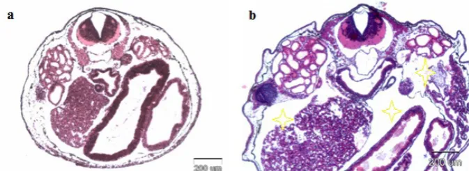
A severe visceral oedema was detected in larvae
at 25 µg L-1 in the cross-sections of the internal organs
(Figure 5). As only one larva survived at 50 µg L-1,
the histopathological findings are not presented here. The incidence of observed histopathological findings of the ten larvae is presented in Table 4.
DISCUSSION
There is a strong relationship between increasing
concentrations of cadmium and the decline in the
hatching success of frog embryos (37). Cadmium LC50
values determined in previous acute toxicity studies
on amphibian larvae vary considerably. The 96-h LC50
of cadmium for Bufo arenarum larvae was reported
in the range from 2.19 to 6.77 mg L-1 (38). The LC 50 values for cadmium in R. ridibunda larvae calculated
in two different studies were 0.45 mg L-1 (39) and
71.8 mg L-1 (40). This last value is very close to the 90-h LC50 reported in Xenopus laevis larvae - 80 to
Table 1 Lethal concentrations after cadmium exposure in P. viridis for 96 hours
Lethal concentrations Concentration (µg L-1) 95 % Confidence interval
Lower bound Upper bound
LC10 26.98 21.05 31.24
LC50 35.35 30.41 41.09
LC90 46.31 40.05 59.36
100 mg L-1 (41), but both were substantially above the LC50 calculated for X. laevis embryos - 850 μg L-1. The cadmium 96-h LC50 for stage 26 P. variabilis
larvae in our study was 35.35 μg L-1. The dissimilar lethal concentration values calculated in all of these
studies might have been due to differences in environmental conditions in habitats, such as
temperature and pH, or perhaps due to general differences in species. The developmental stage at the
moment of exposure also influences lethal concentration estimations. In a study on juvenile R. ridibunda, the LC50 calculated after a 96-h exposure to cadmium was
51.2 mg L-1 (42), a value greater than or similar to the LC50 found for the larvae of this species. It is therefore
important to analyse the water at several stages; not
only once. According to Turkish regulations, the
highest acceptable value is 100 μg L-1 (43), while this
value was set at 200 μg L-1 in the 2008 TSE limits (the
Turkish Standards Institution) (43).
This study detected serious declines in survival percentages of the larvae resulting from cadmium
exposure at between 25 and 50 μg L-1. We could say
that cadmium levels within 25-50 μg L-1 were critical for P. variabilis larvae. This is most strongly confirmed by the survival of one larva in the group exposed to 50 μg L-1 of cadmium. James and Little (13) reported
that 540 μg L-1 of chronically applied cadmium
reduced survival and extended the metamorphosis period in Bufo americanus larvae, but prematurely
induced metamorphosis at 5 and 54 μg L-1. Cadmium
exposure at 0.25-5 μg L-1 stimulated growth and metamorphosis in Rana pipiens larvae (16, 32). In
another study (15), chronic exposure to 0-855 μg L-1
of cadmium did not affect the survival rate of X. laevis
larvae. When discussing the complexity of amphibian
larval development (under extensive hormonal
regulation), it may be concluded that small
concentrations of cadmium exposure may even have a stimulating effect on this process.
Table 2 Comparison of morphological measurements of treatment groups
Measurements (µg LCd -1) n mean SD SE
95 % Confidence
interval for mean
Min. Max. p
Lower
bound Upper bound
Total length (mm)
0 10 15.76 0.89 0.28 15.12 16.40 13.95 16.77
1 10 15.08 1.18 0.37 14.23 15.92 12.72 16.99
5 10 14.88 0.75 0.26 14.25 15.51 13.95 15.88 0.00
10 10 14.78 1.30 0.45 13.69 15.86 13.18 16.47
25 10 13.45 1.38 0.48 12.29 14.61 10.70 14.91
Width (mm)
0 10 3.08 0.31 0.09 2.85 3.30 2.51 3.61
1 10 3.20 0.85 0.27 2.59 3.82 0.92 4.04
5 10 3.03 0.37 0.13 2.72 3.35 2.14 3.27 0.69
10 10 3.29 0.25 0.09 3.07 3.50 3.04 3.77
25 10 2.97 0.36 0.12 2.66 3.27 2.15 3.37
Tail length (mm)
0 10 9.62 0.78 0.24 9.07 10.18 7.90 10.38
1 10 9.19 0.78 0.24 8.64 9.75 7.72 10.11
5 10 9.39 0.87 0.35 7.75 9.53 7.54 10.78 0.34
10 10 9.43 0.87 0.30 8.70 10.15 7.90 10.70
25 10 8.78 1.19 0.42 7.79 9.78 6.25 10.20
Wet weight (g)
0 10 0.03 0.01 0.01 0.02 0.03 0.03 0.04
1 10 0.03 0.01 0.01 0.02 0.03 0.03 0.04
5 10 0.03 0.01 0.01 0.02 0.03 0.03 0.04 0.33
10 10 0.03 0.01 0.01 0.02 0.03 0.02 0.04
25 10 0.02 0.01 0.01 0.01 0.03 0.01 0.04
Figure 2 Cross sections of the gill lamellae of (a) control group, (b) 10 µg L-1 cadmium, and (c) 25 µg L-1 cadmium (lamellar
Table 3 Statistically significant values of the discriminant analysis made so as to reveal the difference among the groups following cadmium exposure in P. viridis larvae for 96 hours
Function Eigenvalue Variance (%) Total (%) CorrelationCanonical LambdaWilks Chi-square df p
1 0.648 94 94 0.627 0.583 24.556 8 0.00
2 0.041 6 100.0 0.199 0.960 1.835 3 0.60
Figure 3 Cross sections of the pronephric tubules of (a) control group, and (b) 25 µg L-1 cadmium (deformations of pronephric
tubules shown with asterisk), H&E staining
Table 4 The incidence of observed histopathological findings in the gill, kidney, and liver of larvae administered cadmium
Lesions Cadmium (µg L
-1)
0 1 5 10 25
Gills lamellar fusions 0 0 0 5 7
Kidney deformation of pronephric tubules 0 0 1 3 6
Liver deformation and haemorrhage 0 0 1 3 6
Vacuolisation 0 0 0 6 8
Visceral oedema 0 0 3 5 8
n=10. Values indicate the numbers of larvae with observed lesions in their sections
Figure 4 Cross sections of the liver of (a) control group, (b) 10 µg L-1 cadmium and (c) 25 µg L-1 cadmium (haemorrhage shown
Heavy metal exposure at sub-lethal concentrations initially affects the gills, with the potential risk of
accumulation (44). Gill damage, in particular fusion
of secondary lamellae, has been reported in Channa
punctatus exposed to 172 mg L-1 of cadmium during 10 days (45). The most important cause of gill lamellar fusion due to pollutant exposure is their function as a
barrier for preventing pollutant entry into the body (46). Likewise, fusions in gill lamellae in our study were found in larvae exposed to 10 and 25 μg L-1 of cadmium.
Cadmium causes histopathological changes in the
liver, kidney, gill, spleen, and bone marrow, as well as hypocalcemia and hypoglycemia, and inhibits the
intake of Ca2+ through gills, therefore affecting plasma ion composition and osmoregulation (27). It has also
been reported that cadmium has hepatotoxic effects
and causes hepatic dysfunctions and increases in
certain enzyme activities (14), as well as
histopathological lesions in the liver and necrosis in
hepatocytes (45, 47). Furthermore, a study on Bufo
arenarum revealed that cadmium accumulates particularly in the liver (48). Much like in other sources, our study also detected serious deformations
in the hepatic and renal tubules. The damage observed
in liver cross-sections, necrosis, lesions, and haemorrhages verify that cadmium primarily targets the liver. Effects on kidneys include epithelial cell
deformations in the pronephric tubules and enlargement of the tubular lumen. Similar results were reported for
Rana ridibunda in a study that revealed progressive nephropathy and glomerulonephropathy as a result of cadmium exposure (14).
In conclusion, even though the histopathological
findings of our study support the results of previous studies, its main contribution is that it is the first to
report results for P. variabilis, which has shown to be a very good indicator species for aquatic ecosystems.
Our LC50 values were lower than those reported by
other investigators. The mechanisms and biological route of cadmium, with special reference to amphibians, has not yet been fully clarified and further detailed acute, sublethal, and chronic toxicity studies are
necessary.
REFERENCES
1. Halliday T. Diverse phenomena influencing amphibian
population declines. In: Lannoo MJ, editor. Amphibian declines - The conservation status of United State species. Berkeley (CA): University of California Press; 2005. p. 3-6.
2. Barnum TR, Verburg P, Kilham SS, Whiles MR, Lips KR,
Colon-Gaud C, Pringle CM. Use of stable isotope ratios to characterize potential shifts in the isotopic niches of grazing insects following an amphibian decline in a Neotropical stream. J Trop Ecol 2013;29:291-9. doi: 10.1017/ S026646741300028X
3. Collins JP, Storfer A. Global amphibian declines: sorting the
hypotheses. Divers Distrib 2003;9:89-98. doi: 10.1046/j.1472-4642.2003.00012.x
4. Rohr JS, Raffel TR. Linking global climate and temperature
variability to widespread amphibian declines putatively caused by disease. Proc Natl Acad Sci USA
2010;107:8269-74. doi: 10.1073/pnas.0912883107
5. Clark EJ, Norris DO, Jones RE. Interactions of gonadal
steroids and pesticides (DDT, DEE) on gonaduct growth in
larval tiger salamanders, Ambystoma tigrinum. Gen Comp Endocrinol 1998;109:94-105.
6. Goleman W, Urguidi LJ, Anderson TA, Smith EE, Kendall
RJ, Carr JA. Environmentally relevant concentrations of ammonium perchlorate inhibit development and
metamorphosis in Xenopus laevis. Environ Toxicol Chem 2002;21:424-30. doi: 10.1002/etc.5620210227
7. Qin ZF, Zho JM, Chu SG, Xu XB. Effects of Chinese
domestic polychlorinated biphenyls (PCBs) on gonadal
differentiation in Xenopus laevis. Environ Health Perspect 2003;111:553-6. doi: 10.1289/ehp.5620
8. Bernabo I, Brunelli E, Berg C, Bonacci A, Tripepi S.
Endosulfan acute toxicity in Bufo bufo gills: ultrastructural
changes and nitric oxide synthase localization. Aquat Toxicol 2008;86:447-56. doi: 10.1016/j.aquatox.2007.12.006
Figure 5 Cross sections of the internal organs of (a) control group, and (b) 25 µg L-1 cadmium (visceral oedema shown with
9. Cooke AS. Tadpoles asindicators of harmful levels of pollution in the field. Environ Pollut 1981;25:123-33. doi: 10.1016/0143-1471(81)90012-X
10. Lajmanovich RC, Sanches-Hernandes JC, Peltzer PM,
Attademo AM, Fiorenza MC, Cabagna MC, Basso A. Levels of plasma B-esterase and Glutathione-S-transferase activities in three South American toads. J Toxicol Environ Chem
2008;90:1145-61. doi: 10.1080/02772240801923107
11. Zhelev ZM, Popgeorgiev GS, Angelov MV. Investigating the
changes in the morphological content of the blood of
Pelophylax ridibundus (Amphibia: Ranidae) as a result of
the anthropogenic pollution and its use as an environmental
bioindicator. Acta Zool Bulg 2013;65:187-96.
12. Flament S, Kuntz S, Chesnel A, Grillier-Vuissoz I, Tankozic
C, Penrad-Mobayed M, Auque G, Shirali P, Schroeder H,
Chardard D. Effects of cadmium on gonadogenesis and metamorphosis in Pleurodeles waltl (urodele amphibian). Aquat Toxicol 2003;64:143-53. doi: 10.1016/S0166-445X(03)00042-0
13. James SM, Little EE. The effects of chronic cadmium
exposure on American toad (Bufo americanus) tadpoles. Environ Toxicol Chem 2003;22:377-80. doi: 10.1002/ etc.5620220219
14. Loumbourdis NS. Hepatotoxic and nephrotoxic effects of
cadmium in the frog Rana ridibunda. Arch Toxicol 2005;79:434-40. doi: 10.1007/s00204-005-0652-x
15. Sharma B, Patino R. Exposure of Xenopus laevis tadpoles to
cadmium reveals concentration-dependent bimodal effects on growth and monotonic effects on development and thyroid gland activity. Toxicol Sci 2008;105:51-8. doi: 10.1093/
toxsci/kfn119
16. Gross JA, Johnson PTJ, Prahl LK, Karasov WH. Critical
period of sensitivity for effects of cadmium on frog growth
and development. Environ Toxicol Chem 2009;28:1227-32. doi: 10.1897/08-205.1
17. Jolibois LS, Shi LS, George W, Henson WJ, Anderson MB.
Cadmium accumulation and effects on progesterone release
by cultured human trophoblast cells. Reprod Toxicol 1999;13:215-21. doi: 10.1016/S0890-6238(99)00009-X
18. McCarthy JF, Shugart LR, editors. Biomarkers of
Environmental Contamination. Boca Raton (FL): Lewis Publishers; 1990.
19. Satarug S, Baker JR, Urbenjapol S, Haswell-Elkins M, Reilly
PEB, Williams DJ, Moore MR. A global perspective on
cadmium pollution and toxicity in non-occupationally exposed population. Toxicol Lett 2003;137:65-83. doi:
10.1016/S0378-4274(02)00381-8
20. Altındağ A, Yiğit S. Assessment of heavy metal concentrations
in the food web of lake Beyşehir, Turkey. Chemosphere 2005;60:552-6. doi: 10.1016/j.chemosphere.2005.01.009
21. Baker JR, Satarug S, Urbenjapol S, Edwards RJ, Williams
DJ, Moore MR, Reilly PEB. Potential for early involvement of CYP isoforms in aspects of human cadmium toxicity. Toxicol Lett 2003;137: 85-93. doi: 10.1016/S0378-4274(02)00382-X
22. Jarub L, Akesson A. Current status of cadmium as an
environmental health problem. Toxicol Appl Pharmacol 2009;238: 201-8. doi: :10.1016/j.taap.2009.04.020
23. Ranatunge RAAR, Wijesinghe MR, Ratnasooriya WD,
Dharmarathne HASG, Wijesekera RD. Cadmium-induced
toxicity on larvae of the common asian toad Duttaphrynus melanostictus (Schneider 1799): Evidence from emperial
trials. Bull Environ Contam Toxicol 2012;89:143-6. doi:
10.1007/s00128-012-0635-6
24. Weltjze L, Simpson P, Gross M, Crane M, Wheeler JR.
Comparative acute and chronic sensitivity of fish and
amphibians: A critical review of data. Environ Toxicol Chem
2013;32:984-94. doi: 10.1002/etc.2149
25. Gay F, Laforgia V, Caputo I, Esposito C, Lepretti M, Capaldo
A. Chronic exposure to cadmium disrupts the adrenal gland
activity of the newt Triturus carnifex (Amphibia, Urodela).
BioMed Res Int 2013;1-6. doi: 10.1155/2013/424358
26. Spry DJ, Wiener JG. Metal bioavailability and toxicity to
fish in low-alkalinity lakes; A critical review. Environ Pollut
1991;71:243-304. doi: 10.1016/0269-7491(91)90034-T 27. Ricard AC, Daniel C, Anderson P, Hontela A. Effects of
subchronic exposure to cadmium chloride on endocrine and
metabolic functions in rainbow trout Oncorhynchus mykiss.
Arch Environ Contam Toxicol 1998;34:377-81. doi: 10.1007/ s002449900333
28. Vetillard A, Bailhache T. Cadmium: An endocrine disrupter
that affects gene expression in the liver and brain of juvenile rainbow trouts. Biol Reprod 2005;72:119-26. PMID:
15317685
29. Calevro F, Campani S, Ragghianti M, Bucci S, Mancino G.
Tests of toxicity and teratogenicity in biphasic vertebrates
treated with heavy metals (Cr3+, Al3+, Cd2+). Chemosphere
1998;37:3011-7. doi: 10.1016/S0045-6535(98)00342-7
30. Mouchet F, Baudrimont M, Gonzalez P, Cuenot Y,
Bourdineaud JP, Boudou A, Gauthier L. Genotoxic and stress
inductive potential of cadmium in Xenopus laevis larvae.
Aquat Toxicol 2006;78:157-66. doi: 10.1016/j. aquatox.2006.02.029
31. Zhang Y, Huang D, Zhao D, Long J, Song G, Li A. Long-term
toxicity effects of cadmium and lead on Bufo raddei tadpoles.
Bull Environ Contam Toxicol 2007;79:178-83. doi: 10.1007/
s00128-007-9152-4
32. Gross JA, Chen TH, Karasov WH. Lethal and sublethal
effects of chronic cadmium exposure on northern leopard frog (Rana pipiens) tadpoles. Environ Toxicol Chem 2007;26:1192-7. doi: 10.1897/06-479R.1
33. Başoğlu M, Özeti N, Yılmaz I. Türkiye Amfibileri [The
Amphibians of Turkey, in Turkish]. Izmir: Ege Üniversitesi Fen Fakültesi; 1992.
34. Kinzelbach R, Kasparek M. Amphibia and Reptilia. Zool
Middle East 1992;3:27.
35. International Union for Conservation of Nature (IUCN). The
IUCN Red List of Threatened Species. [Displayed 25 August 2014]. Available at http://www.iucnredlist.org/
36. Gosner KL. A simplified table for staging anuran embryos
and larvae with notes on identification. Herpetologica
1960;16:183-90.
37. Haywood LK, Alexander GJ, Byrne MJ, Cukrowska E.
Xenopus laevis embryos and tadpoles as models for testing pollution by zinc, copper, lead and cadmium. Afr Zool
2004;39:163-74.
38. Ferrari L, Saliban A, Muino CV. Selective protection of
temperature against cadmium acute toxicity of Bufo arenarum tadpoles. Bull Environ Contam Toxicol 1993;50:212-8. doi: 10.1007/BF00191724
39. Grillitch B, Chovanec A. Heavy metals and pesticides in
anuran spawn and tadpoles, water, and sediment. Toxicol
40. Loumbourdis NS, Kyriakopoulou-Sklavounou P, Zachariadis G. Effects of cadmium exposure on bioaccumulation and
larval growth in the frog Rana ridibunda. Environ Pollut
1999;104:429-33. doi: 10.1016/S0269-7491(98)00172-9
41. Woodal C, Maclean N, Crossley F. Responses of the trout
fry (Salmo gairdneri) and Xenopus laevis tadpoles to
cadmium and zinc. Comp Biochem Physiol C 1988;89:93-9. doi: 10.1016/0742-8413(88)90151-X
42. Selvi M, Gül A, Yılmaz M. Investigation of acute toxicity of
cadmium chloride (CdCl2·H2O) metal salt and behavioral
changes it causes on water frog (Rana ridibunda Pallas,
1771). Chemosphere 2003;52:259-63. doi:
10.1016/S0045-6535(03)00262-5
43. Kayhan FE. Su ürünlerinde kadmiyum biyobirikimi ve
toksisitesi [Bioaccumulaton and toxicity of cadmium in the water products, in Turkish]. J Fish Aquat Sci 2006;23:215-20.
44. Flos R, Tort L, Balasch J. Effects of zinc sulphate on
haematological parameters in the dogfish Scyliorhinus
canicula and influences of MS222. Mar Environ Res
1987;21:289-98. doi: 10.1016/0141-1136(87)90051-1
45. Babu RA. Effects of cadmium on digestive organs of teleost
fish Ophiocephalus (Channa). Universal J Environ Res
Technol 2013;3:173-80.
46. Mallat J. Fish gill structural changes induced by toxicants
and other irritants: A statistical review. Can J Fish Aquat Sci
1985;42:630-48. doi: 10.1139/f85-083
47. Mitsumori K, Shibutani M, Sato S, Onodera H, Nakagava J,
Hayashi Y, Ando M. Relationship between the development
of hepato-renal toxicity and cadmium accumulation in rats given minimum to large amounts of cadmium chloride in the long-term: Preliminary study. Arch Toxicol 1998;72:545-52. doi: 10.1007/s002040050541
48. Pérez-Coll CS, Herkovits J, Salibian A. [Effects of cadmium
Sažetak
Akutni toksični učinci kadmija na ličinke zelene žabe, Pseudepidalea variabilis (Pallas, 1769) (Amphibia: Anura)
Štetni utjecaji kadmija na okoliš i njegovo nakupljanje u prirodi posljednjih su godina poprimili zabrinjavajuće razmjere. U okviru ove studije istražili smo akutne toksične učinke kadmija na morfologiju i histologiju ličinaka zelene žabe, Pseudepidalea variabilis (Pallas, 1769). Embriji žabe dobiveni su od jedinki koje su u fazi ampleksusa prikupljene u prirodi. U laboratorijskim uvjetima embriji su bili držani do razvojnog stadija 26, kada su izloženi kadmiju u koncentracijama od 0, 1, 5, 10, 25 i 50 µg L-1 tijekom 96 sati. U pokusu smo odredili sljedeće letalne koncentracije kadmija: LC10 26,98 µg L-1,LC
50 35,35 µg L-1 iLC90 46,31 µg L-1. Izloženost kadmiju u koncentracijama većim od 1 µg L-1 negativno je utjecala na veličinu tijela ličinaka. Histološke analize upućuju na sljepljivanje škržnih listića, krvarenja u jetrima, pojavu edema u trbušnoj šupljini i deformacije pronefričkih kanalića (pri koncentracijama većim od 10 µg L-1). Dobiveni rezultati pokazali su da je zelena žaba vrlo osjetljiva na kadmij, na što upućuje vrijednost LC50 koja je u našem pokusu bila niža od vrijednosti zabilježenih u drugim istraživanjima. Prema tome, ta se vrsta može smatrati pouzdanim pokazateljem okolišnog stresa u slatkovodnim ekosustavima.
KLJUČNE RIJEČI: embriotoksičnost; histopatologija; letalne koncentracije; teški metal
CORRESPONDING AUTHOR:
Sibel Hayretdağ Terzioğlu Campus, 17100 Çanakkale, Turkey