The impact of concentration and administration time on
the radiomodulating properties of undecylprodigiosin
in vitro
Sandra Petrović
1, Vesna Vasić
1, Tatjana Mitrović
3, Saša Lazović
2, and Andreja Leskovac
1Vinča Institute of Nuclear Sciences1, Institute of Physics Belgrade2, University of Belgrade, Institute for Development
of Water Resources “Jaroslav Černi”3, Belgrade, Serbia
[Received in October 2016; Similarity Check in October 2016; Accepted in January 2017]
Undecylprodigiosin pigment (UPP) is reported to display cytotoxic activity towards various types of tumours. Nevertheless,
its efficacy in modifying the cellular response to ionising radiation is still unknown. In this study, the radiomodulating
effects of UPP were investigated. The effects of UPP were assessed in vitro by treating cultures of human peripheral blood with UPP and ionising radiation using two treatment regimens, the UPP pre-irradiation treatment and UPP post-irradiation treatment. The activity of UPP was investigated evaluating its effects on the radiation-induced micronuclei formation, cell proliferation, and induction of apoptosis. The redox modulating effects of UPP were examined measuring the catalase activity and the level of malondialdehyde, as a measure of oxidative stress. The results showed that UPP effects on cellular response to ionising radiation depend on its concentration and the timing of its administration. At low concentration, the
UPP displayed radioprotective effects in γ-irradiated human lymphocytes while at higher concentrations, it acted as a radiosensitiser enhancing either mitotic catastrophe or apoptosis depending on the treatment regimen. The UPP modified redox processes in cells, particularly when it was employed prior to γ-irradiation. Our data highlight the importance of
further research of the potential of UPP to sensitize tumour cells to radiation therapy by inhibiting pathways that lead to treatment resistance.
KEY WORDS: apoptosis; mitotic catastrophe; oxidative stress; radiosensitisation; undecylprodigiosin
The main therapeutic modalities to treat cancer are surgery, radiotherapy, and chemotherapy. Radiotherapy is one of the most effective forms of cancer treatment. However, ionising radiation (IR) used for the elimination of malignant tumours induces persistent DNA double-strand breaks (DSBs) leading to the permanent cell growth arrest, apoptosis, necrosis, or mitotic catastrophe (1). The major
difficulties encountered during the treatment of cancer are
tumour resistance to therapy and therapy-associated normal tissue toxicity (2). Alternative approaches to enhancing the
efficacy of radiotherapy and minimising its harmful side
effects focus on a combined treatment with anticancer drug and radiation (2). The potential radiotherapeutic agents are commonly categorised as (i) radioprotectors, which reduce normal tissue toxicity, thus, minimising the side effects of radiotherapy, and (ii) radiosensitisers, which enhance the radiosensitivity of tumour cells, thereby minimising the radiation dosage and damage to surrounding normal tissues (3, 4). Radiosensitisation of tumour cells by different agents is mainly achieved by enhancement of radiation-induced DNA damage in tumour cells compared to surrounding tissues, inhibition of DNA synthesis and cell growth, and
enhancement of radiation-induced apoptosis (2, 5). Another approach is to modulate reduction/oxidation reactions within tumour cells (4).
In the last few decades, a wide variety of naturally occurring compounds have been tested in order to identify effective radiosensitisers that could enhance the sensitivity of cancer to the effects of IR and improve the cancer patients’ survival rate (6-8). Plant-derived anticancer drugs
continue to play a significant role in the development of
new therapeutics aimed at modifying the radiobiological response of cells. Plant compounds such as etoposide, pactitaxel, and Vinca alkaloids have been recognised as radiosensitisers with low toxicity and high effectiveness whose synergism with radiation therapy increases the likelihood of cancer treatments (9). However, besides plants, microorganisms represent a profuse source of diverse bioactive metabolites with anticancer and possible radiomodulating activities.
Prodigiosins (PGs), produced as secondary metabolites by many terrestrial and marine bacterial strains such as the species of Serratia (10) and Streptomyces (11, 12), are a family of natural red pigments that are reported to possess antimicrobial, immunosuppressive, and cytotoxic activities (13-15). It has been reported that PGs mainly target cancer cells (hematopoietic, gastrointestinal, breast, and lung
Correspondence to: Dr Andreja Leskovac, Vinča Institute of Nuclear Sciences, University of Belgrade, M. Petrovića Alasa 12-14, 11001
Numerous studies indicated that PGs displayed selective cytotoxicity against cancer cells, anti-metastatic activity, and p53-independent proapoptotic effects, which points to their potency as anticancer agents (16, 19).
Recent studies put a PG family member, undecylprodigiosin pigment (UPP), in the focus of anticancer investigation based on its proapoptotic potency demonstrated in various types of tumours (20-23). It has been suggested that UPP inhibits cell proliferation by inducing G2/M phase arrest and apoptosis of cancer cells. The tumoricidal action of UPP alone and UPP conjugated to gold nanoparticles was observed in melanoma, lung, breast, and colon cancer cells, enabling the platform for
further development of efficient anticancer drugs suitable
for clinical application (22). On the other side, it has been shown that UPP provides protection against oxidative stress by delaying the lipid peroxidation process (24) and displaying the antioxidant properties and mitochondrial
function improvement (25). Similar findings were obtained
in our previous investigation showing that treatment of human lymphocytes with low UPP concentrations (0.1 µg mL-1) significantly reduced DNA damages and
maintained the redox homeostasis of cells, indicating its cytoprotective nature. At moderate concentrations (1 µg mL-1), UPP displayed effects similar to that of the
untreated control, while the highest applied concentration (5 µg mL-1) was genotoxic (unpublished data). The
protective effects of UPP against UV irradiation were described previously (26).
As far as we know, the effect of UPP as a modifier of
cellular response to ionising radiation is still unknown. Therefore, in this study, we investigated potential radioprotective/radiosensitising effects of UPP in
γ-irradiated human peripheral blood cells using different
combined treatment regimens with UPP and IR. The influence of UPP on radiation-induced apoptosis, micronuclei (MNi) induction, and oxidative stress alterations was investigated.
MATERIALS AND METHODS
Undecylprodigiosin pigment (UPP) preparation
Undecylprodigiosin (UP) pigment was synthesised in
a shake flask culture of Streptomyces sp. JS520 using mannitol-soy-yeast medium supplemented with methyl-oleate (0.2 %, v/v) for six days at 30 °C and recovered and
purified by column chromatography. The quality of the purified UP was analysed by liquid chromatography coupled
to mass spectroscopy (LC-MS) as described previously (24).
Blood sample was obtained from a healthy, non-smoking, 30-year-old male volunteer donor in accordance with current Health and Ethical Regulations in Serbia (27).
Aliquots of heparinised whole blood (0.5 mL) were first
added to culture tubes containing 4.5 mL of RPMI-1640 medium supplemented with 15 % calf serum (Invitrogen-Gibco, Paisley, UK) and were then treated with an increasing concentration of UPP (final concentrations 0.1 µg mL-1, 1 µg mL-1, and 5 µg mL-1).
The UPP effects were studied employing the experimental set up used in the study of Shukla et al. (28). For each UPP concentration, two treatment regimens were applied: pre-treatment - UPP was added to cell cultures and
then exposed to γ-irradiation, and post-treatment - cell cultures were treated with γ-irradiation and then exposed to UPP treatment. γ-irradiated cultures not treated with UPP
served as control.
For pre-treatment, UPP was added to cell cultures and incubated at 37 °C for one hour. Cultures were subsequently
exposed to 2 Gy of 60Co γ-radiation, at a dose rate of
0.45 Gy min-1. After irradiation, UPP was washed off by
centrifugation and cultures were reconstituted by adding 4.5 mL of RPMI-1640 medium supplemented with 15 % calf serum and 2 % phytohemagglutinin (Invitrogen-Gibco, Paisley, UK). They were then incubated at 37 °C for 72 h. For post-treatment, cell cultures were exposed to
γ-irradiation as described above. Immediately after
irradiation, phytohemagglutinin and UPP were added to the cultures, which were then incubated for 72 h.
The adequate number of blood cultures was established to enable examinations of the MNi frequency,
cytokinesis-block proliferation index (CBPI), catalase activity (CAT), and malondialdehyde (MDA) level.
For the apoptosis (AP) assay, blood aliquots of 0.5 mL
were incubated in a RPMI-1640 medium (Invitrogen-Gibco, Paisley, UK) supplemented with 15 % calf serum (Invitrogen-Gibco, Paisley, UK) for 24 h.The treatments
with UPP and γ-irradiation were performed as described
above.
For each analysis performed, three independent experiments were carried out. The obtained data were pooled and results were expressed as the mean and standard deviation (SD) of the mean.
Micronucleus assay
Radiosensitivity was assessed using the cytokinesis block method of Fenech (29).For MN preparation, Cytochalasin B (Sigma-Aldrich, St. Louis, MO, USA) at a
final concentration of 4 µg mL-1 was added to each culture
44 h after incubation in order to inhibit cytokinesis. Lymphocyte cultures were incubated for further 28 h. Cells were collected by centrifugation and treated with a hypotonic solution (0.56 % KCl+0.90 % NaCl, mixed in
fixative, and dropped onto clean slides. Slides were air-dried
and stained in alkaline Giemsa. For each sample, at least 1000 binucleated (BN) cells were scored and MNi were recorded using an Optech microscope (Munich, Germany)
with 400x or 1000x magnification.
Cytokinesis-block proliferation index
The ability of cells to proliferate in vitro was evaluated by counting the number of cells with one to four nuclei on the same slides. The results of these analyses are presented as a cytokinesis-block proliferation index. CBPI was calculated according to the method of Surrales at al. (30) as follows: CBPI =[MI + 2MII + 3(MIII +MIV)]/N, where MI-MIV represents the number of cells with one to four nuclei, respectively, and N is the number of cells scored.
Apoptosis of leukocytes
After 24 h of incubation, cells were gently washed with
physiological saline (0.9 % NaCl) at 37 °C, and fixed in methanol/acetic acid (3:1). Afterwards, the pellet was fixed
in 96 % ethanol and stored at +4 °C. Samples were incubated at room temperature for 10-15 min in incubation phosphate buffer. Propidium iodide (PI, Sigma-Aldrich) and Ribonuclease A (RNase A, Sigma-Aldrich) were added
5 min prior to the flow cytometry analysis.
Apoptosis was assessed by flow cytometric (Partec, Germany) identification of cells displaying
apoptosis-associated DNA condensation. DNA content was assessed
by measuring the UV fluorescence of propidium iodide
stained DNA. Apoptotic population was calculated using the Flow Max software (Partec, Germany).
Blood culture preparation
After 72 h of incubation, blood cultures were separated on a Lymphoprep (Lymphocyte separation medium, PAA Laboratories GmbH, Pasching, Austria); lymphocytes were collected by centrifugation, washed in a physiological saline, and frozen at -20 ºC for the TBA assay, while erythrocytes were haemolysed in ice cold deionised water
and frozen at -20 ºC for subsequent analyses of the catalase
activity.
Assay of the catalase activity
The catalase activity was measured using the method
of Aebi (31) with minor modifications by following the
catalytic reduction of hydrogen peroxide. The decomposition of the substrate H2O2 was measured using a Perkin Elmer
Lambda 25 Spectrophotometer (Perkin Elmer Instruments, Norwalk, CT, USA) at 240 nm. The activity was expressed
as K – rate constant of the first-order reaction per minute
per mg of Hb. Haemoglobin concentration was determined using the Drabkin’s method.
A thawed lymphocyte suspension was treated with thiobarbituric acid, and used to determine malondialdehyde levels, spectrophotometrically at 532 nm (32). Values were expressed as nmol TBA-reactive substance (MDA
equivalent)/mg protein, using a standard curve of
1,1,3,3-tetramethoxypropane. Protein concentration was determined according to the method of Lowry et al. (33).
Statistics
Statistical analysis was carried out using the Statistica 8 and OriginPro 8.5.1 software packages for Microsoft Windows. Statistical analysis was done using the Mann-Whitney U test. P values of less than 0.05 were considered
to be significant.
RESULTS
The radiomodulating effects of UPP were assessed in vitro by treating human peripheral blood cultures with UPP and IR using two treatment regimens: UPP was added to
the cultures one hour prior to γ-irradiation as pre-treatment and immediately after γ-irradiation as post-treatment. The
activity of UPP was investigated evaluating its effects on the radiation-induced MNi formation, cell proliferation, and induction of apoptosis. The redox modulating effects of UPP were examined measuring the catalase activity and the level of malondialdehyde, the lipid peroxidation product, as markers of oxidative stress.
Combined treatment with IR and UPP – the UPP pre-treatment regimen
As shown in Table 1, the UPP pre-treatment, at low
concentration, induced a significant reduction of the MNi frequency (by 13 %) compared to control (irradiated sample
not treated with UPP) (p<0.05). At higher applied UPP concentrations, no considerable differences in the MNi
frequency compared to control were observed. The
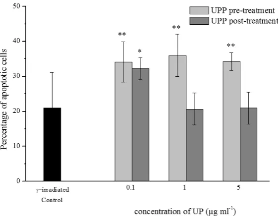
proliferation potential of cells was almost unchanged up to the highest applied concentration when significant suppression of cell proliferation was found (p<0.05). As shown in Figure 1, the percentage of apoptotic cells was
significantly higher (by approximately 50 %) than that in
control (p<0.01), irrespective of the UPP concentration. Considering the redox modulating activities of UPP, in cell cultures that were pre-treated with the lowest UPP
concentration, a significant decrease in the MDA level (by 32 %) was observed (p<0.05), while the subsequent increase
in UPP concentrations enhanced MDA level (p<0.05), potentiating hence the effects of IR (Figure 2a). As shown in Figure 2b, the catalase activity slightly increased in a
dose-dependent manner reaching significant enhancement
γ-irradiation (UPP pre-irradiation treatment and UPP post-irradiation treatment)
Frequency of micronuclei
(MN) Proliferation potential (CBPI)
Unirradiated Control 10.56±1.22 1.82±0.02
γ-irradiated Control 215.87±8.46 1.51±0.04
UPP concentration
UPP
pre-irradiation treatment
0.1 µg ml-1 189.04±9.41* 1.53±0.01
1 µg ml-1 203.33±4.55 1.48±0.02
5 µg ml-1 215.43±8.38 1.40±0.03*
UPP
post-irradiation treatment
0.1 µg ml-1 228.30±9.30 1.48±0.01
1 µg ml-1 231.27±6.81* 1.41±0.02*
5 µg ml-1 247.52±4.30* 1.38±0.02*
*Comparison with γ-irradiated control, p<0.05
Figure 1 Apoptosis of leukocytes in a combined treatment with IR and UPP (mean± SD); Comparison with γ-irradiated control, *p<0.05, **p<0.01
post-treatment regimen
The UPP post-treatment resulted in significant enhancement of radiation-induced MN (by 15 %) and reduction of cell proliferation in a concentration dependent manner (p<0.05) (Table 1).
The percentage of apoptotic cells was significantly
enhanced (by approximately 50 %) compared to control only at the lowest applied UPP concentration (p<0.05), while a further increase in UPP concentrations decreased apoptosis to control level (Figure 1).
As shown in Figure 2a, the UPP post-treatment caused
no significant changes in the MDA level except for the
highest UPP concentration, which enhanced radiation-induced lipid peroxidation by 35 % (p<0.05).
The UPP post-treatment caused mild, insignificant enhancement of the catalase activity (Figure 2b). Schematic representation of the treatment of human peripheral blood
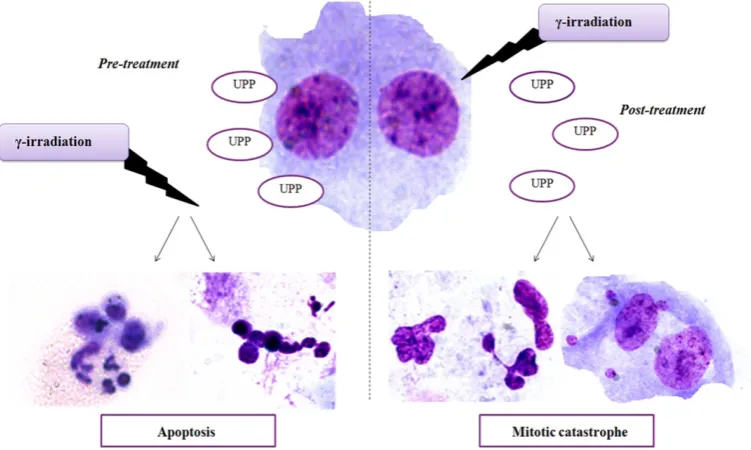
lymphocytes with UPP and γ-irradiation is given in Figure 3.
DISCUSSION
Different classes of natural compounds have been investigated for their potencies to augment the therapeutic index of radiotherapy (1). Although PGs have been found
to display anticancer activities, their efficacy in modifying
cellular response to IR has not been investigated so far. In
this study, we examined the influence of UPP on different
end-points of radiation damage. Results of the study clearly indicate that UPP radiomodulating effects depend on its concentration and treatment regimens.
DNA damage is caused by free radicals derived from the ionisation or excitation of the water component of cells (34). Reactive oxygen species (ROS) generated upon irradiation induce DNA strand breaks and structural and functional alterations of biomolecules (35). Some ROS i.e. HO2- and H2O2 can pass through cell membranes, which
results in important changes in cellular functions (36). Acting on cells before radiation exposure, UPP
displayed significant proapoptotic effects enhancing the
radiation-induced apoptosis independently of the concentration applied. This result is in accordance with the previously reported propensity of PGs to induce apoptosis. It has been shown that prodigiosins modulate the expression of Bcl-2 family proteins, IAP proteins, and death ligand/ receptors involving both mitochondrial and death-receptor
apoptotic pathways (19). The influence of UPP on
radiation-induced MNi formation appeared to be concentration dependent. Specifically, the low UPP concentration
attenuated the radiation-induced MNi frequency while the subsequent increase of UPP concentrations kept the level
of MNi similar to that of control. On the other hand, the UPP post-treatment induced enhanced radiation-induced MNi and suppressed cell proliferation in a concentration dependant manner. A dose-dependent increase in the
frequency of MNi in the lymphocytes treated with UPP was
also observed in our previous study (unpublished data). These results suggest that UPP added to cell cultures after irradiation displayed cytostatic effects and promoted mitotic catastrophe, a type of cell death that results from abnormal mitosis and leads to the formation of cells with multiple MNi (37). In this treatment regimen, the observed decrease in radiation-induced apoptosis is compensated by an
should not be applied. When applied at pre-irradiation stage, UPP, at low concentration, displayed radioprotective effects in gamma-irradiated human lymphocytes reducing MNi formation and acting as an antioxidant, thus exhibiting potential for further research.
Acknowledgments
This work has been supported by the Ministry of Education, Science and Technological Development of the Republic of Serbia (Project no. 172023). The authors are
thankful to Dr Branka Vasiljević, Dr Jasmina Nikodinović and Dr Lidija Šenerović from the Institute of Molecular
Genetics and Genetic Engineering, Belgrade, Serbia, for providing UPP for the research.
REFERENCES
1. Labay E, Efimova EV, Quarshie BK, Golden DW, Weichselbaum RR, Kron SJ. Ionizing radiation-induced foci persistence screen to discover enhancers of accelerated senescence. Int J High Throughput Screen 2011:2:1-13. doi: 10.2147/IJHTS.S17076
2. Garg AK, Buchholz TA, Aggarwal BB. Chemosensitization and radiosensitization of tumors by plant polyphenols. Antioxid Redox Signal 2005;7:1630-47. doi: 10.1089/ ars.2005.7.1630
3. Hazra B, Ghosh S, Kumar A, Pandey BN. The prospective role of plant products in radiotherapy of cancer: a current overview. Front Pharmacol 2012;2:94. doi: 10.3389/ fphar.2011.00094
4. Rosenberg A, Knox S. Radiation sensitization with redox modulators: a promising approach. Int J Radiat Oncol Biol Phys 2006;64:343-54. doi: 10.1016/j.ijrobp.2005.10.013 5. Papazisis KT, Zambouli D, Kimoundri OT, Papadakis ES,
Vala V, Geromichalos GD, Voyatzi S, Markala D, Destouni E, Boutis L, Kortsaris AH. Protein tyrosine kinase inhibitor, genistein, enhances apoptosis and cell cycle arrest in K562 cells treated with gamma-irradiation. Cancer Lett 2000;160:107-13. doi: 10.1016/S0304-3835(00)00569-3 6. Begg AC, Stewart FA, Vens C. Strategies to improve
radiotherapy with targeted drugs. Nat Rev Cancer 2011;11:239-53. doi: 10.1038/nrc3007
7. Joksic G, Leskovac A, Petrovic S. Modulation of radiation-induced damage by Serbian natural plant products: implications for radioprotection. In: Arora R, editor. Herbal radiomodulators: applications in medicine, homeland defence and space. Oxfordshire: CAB International; 2008. p. 67-82. 8. Moding EJ, Kastan MB, Kirsch DG. Strategies for optimizing the response of cancer and normal tissues to radiation. Nat Rev Drug Discov 2013;12:526-42. doi: 10.1038/nrd4003 9. Burris HA, 3rd, Hurtig J. Radiation recall with anticancer
agents. Oncologist 2010;15:1227-37. doi: 10.1634/ theoncologist.2009-0090
10. Thomson NR, Crow MA, McGowan SJ, Cox A, Salmond GP. Biosynthesis of carbapenem antibiotic and prodigiosin pigment in Serratia is under quorum sensing control. Mol
Microbiol 2000;36:539-56. doi: 10.1046/j.1365-2958. 2000.01872.x
of mitotic catastrophe. Mitotic catastrophe represents the main form of cell death induced by IR (38). Thus, in a post-treatment regimen, UPP contributed to the inappropriate entry of cells into mitosis possibly via suppression of multiple central checkpoint proteins or disturbance of the mitotic spindle formation.
The effect of UPP on radiation-induced lipid peroxidation is mainly observed in the UPP pre-treatment; the experimental data showed that high concentration of UPP displayed pro-oxidant effects, while at low concentration UPP possessed antioxidant properties and acted as a scavenger of highly reactive free radicals generated upon irradiation. The attenuation of radiation-induced injury was also seen through the reduction of IR-induced MNi as described above. However, UPP at the same concentration proved to be ineffective as antioxidant if administered at the post-irradiation stage. Augmented levels of intrinsic oxidative stress have been found in a variety of tumours, possibly due to more active metabolism, mitochondrial
mutation, cytokines, and inflammation (39). Therefore,
modulating the redox status of cancer cells might be critical for their survival (40). In that way, UPP as the IR-induced oxidative stress enhancer may sensitise cancer cells to undergo apoptotic death, which was the scenario observed in cell cultures pre-treated with UPP. It can be assumed that
UPP promotes the fixation of IR-induced free radicals and
prevents the repair of cellular radiation damage. In addition, the highest UPP concentration added to cells prior to gamma
irradiation induced a significant enhancement of the catalase
activity. Knowing that many human cancers including melanoma, neuroblastoma, colon carcinoma, and ovarian carcinoma, constitutively generate a high amount of H2O2 (41), it can be assumed that UPP may induce cell cycle arrest and apoptosis of these cancer cells through the scavenging of H2O2 by overexpressed catalase. It is worth noting that in a post-treatment regimen, the UPP redox modulating activities were attenuated. Radiation-induced lipid peroxidation was potentiated by UPP only at the highest employed concentration.
CONCLUSION
Taken together, the results of this study suggest that UPP effects on cellular response to IR vary from mild radioprotection to radiosensitisation depending on its concentration and the timing of its administration, i.e. pre- or post-irradiation stage. At higher concentrations, the UPP’s genotoxic and pro-oxidant behaviour augmented radiation-induced injury and it acted as a radiosensitiser enhancing either mitotic catastrophe or apoptosis depending
on the treatment regimen. These findings highlight the
importance of further research of UPP effects in cancer cells in terms of its utilisation for sensitising tumour cells to
Pigmented antibiotic production by Streptomyces coelicolor A3(2): kinetics and the influence of nutrients. J Gen Microbiol
1990;136:2291-6. doi: 10.1099/00221287-136-11-2291 12. Kawasaki T, Sakurai F, Hayakawa Y. A prodigiosin from the
roseophilin producer Streptomyces griseoviridis. J Nat Prod 2008;71:1265-7. doi: 10.1021/np7007494
13. Nakamura A, Magae J, Tsuji RF, Yamasaki M, Nagai K.
Suppression of cytotoxic T cell induction in vivo by
prodigiosin 25-C. Transplantation 1989;47:1013-6. doi: 10.1097/00007890-198906000-00019
14. Songia S, Mortellaro A, Taverna S, Fornasiero C, Scheiber EA, Erba E, Colotta F, Mantovani A, Isetta AM, Golay J. Characterization of the new immunosuppressive drug undecylprodigiosin in human lymphocytes: retinoblastoma protein, cyclin-dependent kinase-2, and cyclin-dependent kinase-4 as molecular targets. J Immunol 1997;158:3987-95. PMID: 9103470
15. Tsuji RF, Magae J, Yamashita M, Nagai K, Yamasaki M. Immunomodulating properties of prodigiosin 25-C, an antibiotic which preferentially suppresses induction of cytotoxic T cells. J Antibiot (Tokyo) 1992;45:1295-302. PMID: 1399851
16. Pandey R, Chander R, Sainis KB. Prodigiosins as anti cancer agents: living upto their name. Curr Pharm Design 2009;15:732-41. doi: 10.2174/138161209787582192 17. Yamamoto C, Takemoto H, Kuno K, Yamamoto D, Nakai K,
Baden T, Kamata K, Hirata H, Watanabe T, Inoue K. Cycloprodigiosin hydrochloride, a H+/Cl- symporter, induces apoptosis in human colon cancer cell lines in vitro. Oncol Rep 2001;8:821-4. PMID: 11410791
18. Yamamoto D, Kiyozuka Y, Uemura Y, Yamamoto C, Takemoto H, Hirata H, Tanaka K, Hioki K, Tsubura A. Cycloprodigiosin hydrochloride, a H+/Cl- symporter, induces apoptosis in human breast cancer cell lines. J Cancer Res Clin Oncol 2000;126:191-7. doi: 10.1007/s004320050032 19. Chang CC, Chen WC, Ho TF, Wu HS, Wei YH. Development
of natural anti-tumor drugs by microorganisms. J Biosci Bioeng 2011;111:501-11. doi: 10.1016/j.jbiosc.2010.12.026 20. Ho TF, Ma CJ, Lu CH, Tsai YT, Wei YH, Chang JS, Lai JK, Cheuh PJ, Yeh CT, Tang PC, Tsai Chang J, Ko JL, Liu FS, Yen HE, Chang CC. Undecylprodigiosin selectively induces apoptosis in human breast carcinoma cells independent of p53. Toxicol Appl Pharmacol 2007;225:318-28. doi: 10.1016/j.taap.2007.08.007
21. Liu P, Wang YY, Qi X, Gu Q, Geng M, Li J. Undecylprodigiosin induced apoptosis in P388 cancer cells is associated with its binding to ribosome. PLoS One 2013;8:e65381. doi: 10.1371/ journal.pone.0065381
22. Nikodinovic-Runic J, Mojic M, Kang Y, Maksimovic-Ivanic D, Mijatovic S, Vasiljevic B, Stamenkovic VR, Senerovic L. Undecylprodigiosin conjugated monodisperse gold
nanoparticles efficiently cause apoptosis in colon cancer cells in vitro. J Mater Chem B. 2014;2:3271-81. doi: 10.1039/ C4TB00300D
23. Stankovic N, Senerovic L, Ilic-Tomic T, Vasiljevic B, Nikodinovic-Runic J. Properties and applications of undecylprodigiosin and other bacterial prodigiosins. Appl Microbiol Biotechnol 2014;98:3841-58. doi: 10.1007/ s00253-014-5590-1
24. Stankovic N, Radulovic V, Petkovic M, Vuckovic I, Jadranin
M, Vasiljevic B, Nikodinovic-Runic J. Streptomyces sp.
undecylprodigiosin with antibacterial, antioxidative, and UV-protective properties. Appl Microbiol Biotechnol 2012;96:1217-31. doi: 10.1007/s00253-012-4237-3 25. Leiros M, Alonso E, Sanchez JA, Rateb ME, Ebel R, Houssen
WE, Jaspars M, Alfonso A, Botana LM. Mitigation of ROS insults by Streptomyces secondary metabolites in primary cortical neurons. ACS Chem Neurosci 2014;5:71-80. doi: 10.1021/cn4001878
26. Boric M, Danevcic T, Stopar D. Prodigiosin from Vibrio sp. DSM 14379; a new UV-protective pigment. Microb Ecol 2011;62:528-36. doi: 10.1007/s00248-011-9857-0
27. Law on health care.Official Gazette of the Republic of Serbia.
Parliament of the Republic of Serbia. 2005. 107:112-161. 28. Shukla S, Anjaria K, Bhat N, Shirsath K, Sreedevi B. Effect
of caffeine on radiation induced micronuclei in human lymphocytes. Radiat Protect Environ 2010;33:195-8 [displayed 11 January 2017]. Available at http://www.rpe. org.in/article.asp?issn=0972-0464;year=2010;volume=33;i ssue=4;spage=195;epage=198;aulast=Shukla
29. Fenech M. The cytokinesis-block micronucleus technique:
a detailed description of the method and its application to genotoxicity studies in human populations. Mutat Res 1993;285:35-44. doi: 10.1016/0027-5107(93)90049-L 30. Surralles J, Xamena N, Creus A, Marcos R. The suitability
of the micronucleus assay in human lymphocytes as a new biomarker of excision repair. Mutat Res 1995;342:43-59. doi: 10.1016/0165-1218(95)90089-6
31. Aebi H. Catalase. In: Bergmeyer HU, editor. Methods of enzymatic analysis. Weinheim: Verlag Chemie; 1974. p. 673-6. 32. Aruoma OI, Halliwell B, Laughton MJ, Quinlan GJ,
Gutteridge JM. The mechanism of initiation of lipid
peroxidation. Evidence against a requirement for an
iron(II)-iron(III) complex. Biochem J 1989;258:617-20. doi: 10.1042/ bj2580617
33. Lowry OH, Rosebrough NJ, Farr AL, Randall RJ. Protein measurement with the Folin phenol reagent. J Biol Chem 1951;193:265-75. PMID: 14907713
34. Baskar R, Lee KA, Yeo R, Yeoh KW. Cancer and radiation therapy: current advances and future directions. Int J Med Sci 2012;9:193-9. doi: 10.7150/ijms.3635
35. Halliwell B, Gutteridge J. Free Radicals in Biology and Medicine. 4 ed. Oxford: Oxford University Press; 2007. 36. Azzam EI, Jay-Gerin JP, Pain D. Ionizing radiation-induced
metabolic oxidative stress and prolonged cell injury. Cancer Lett 2012;327:48-60. doi: 10.1016/j.canlet.2011.12.012 37. Roninson IB, Broude EV, Chang B-D. If not apoptosis, then
what? Treatment-induced senescence and mitotic catastrophe in tumor cells. Drug Resist Updat 2001;4:303-13. doi: 10.1054/drup.2001.0213
38. Vakifahmetoglu H, Olsson M, Zhivotovsky B. Death through a tragedy: mitotic catastrophe. Cell Death Differ 2008;15:1153-62. doi: 10.1038/cdd.2008.47
39. Gupte A, Mumper RJ. Elevated copper and oxidative stress in cancer cells as a target for cancer treatment. Cancer Treat Rev 2009;35:32-46. doi: 10.1016/j.ctrv.2008.07.004 40. Dröge W. Free radicals in the physiological control of cell
function. Physiol Rev 2002;82:47-95. doi: 10.1152/ physrev.00018.2001
Undecilprodigiozin pigment (UPP) pokazuje citotoksičnu aktivnost kod različitih tipova tumora. Međutim, njegova učinkovitost u modulaciji staničnog odgovora na ionizirajuće zračenje i dalje je nepoznata. U ovoj studiji ispitani su radiomodulirajući učinci UPP-a in vitro tretiranjem kultura ljudske periferne krvi s UPP-om i ionizirajućim zračenjem. Pri ispitivanju su se koristile dvije vrste tretmana: tretman s UPP-om prije ozračivanja (predtretman) i tretman s UPP-om poslije ozračivanja (posttretman). Djelotvornost UPP-a ispitivana je procjenom njegovih učinaka na zračenjem inducirano formiranje mikronukleusa, staničnu proliferaciju i apoptozu. Redoks-modulirajući učinci UPP-a ispitivani su mjerenjem aktivnosti katalaze i razine malondialdehida kao parametara oksidacijskoga stresa. Rezultati pokazuju da učinci UPP-a na stanični odgovor na ionizirajuće zračenje ovise o njegovoj koncentraciji i vrsti tretmana. Pri niskim koncentracijama UPP pokazuje radioprotekcijski učinak u ozračenim humanim limfocitima, a pri visokim koncentracijama djeluje kao radiosenzibilizator inducirajući ili mitotsku katastrofu ili apoptozu, ovisno o vrsti tretmana. UPP modificira redoks procese u stanici, osobito ako se primjenjuje prije zračenja. Naši rezultati upućuju na značaj daljnjeg ispitivanja UPP-a u cilju njegove primjene za senzibilizaciju tumorskih stanica u terapiji zračenjem inhibicijom puteva koji vode rezistenciji na
tretman.