Sandra Donkervoort, MS, CGC
Diana Bharucha-Goebel, MD
Pomi Yun, MPH Ying Hu, MS Payam Mohassel, MD Ahmet Hoke, MD, PhD Wadih M. Zein, MD Daniel Ezzo, BS
Andrea M. Atherton, MS, CGC
Ann C. Modrcin, MD Majed Dasouki, MD A. Reghan Foley, MD Carsten G. Bönnemann,
MD
Correspondence to Dr. Bönnemann: [email protected]
Supplemental data at Neurology.org/ng
HSP and deafness
Neurocristopathy caused by a novel mosaic
SOX10
mutation
ABSTRACT
Objective:To identify the underlying genetic cause in 2 sisters affected with progressive lower extremity spasticity, neuropathy, and early-onset deafness.
Methods: Whole-exome sequencing was performed, and segregation testing of variants was investigated using targeted Sanger sequencing. An inherited paternal mosaic mutation was fur-ther evaluated through quantitative analysis of the ratio of mutant vs wild-type allele in genomic DNA from various tissues, including blood, dermal fibroblasts, and saliva.
Results:A novel heterozygous nonsense mutation (c.1140C.A; p.Y380X) inSOX10was identified in the affected sisters. Paternal mosaicism was suspected based on a small chromatogram peak, which was less than the heterozygous peak of the mutated allele. Consistent with mosaicism, the mosaic paternal samples had notable variability in the ratio of mutant vs wild-type allele in various tissues (compared with the fully heterozygous daughter), with the highest paternal mutant levels in saliva (32.7%) and lowest in dermal fibroblasts (13.9%). Targeted clinical re-examination of the father revealed a sensorimotor neuropathy that was previously clinically unrecognized.
Conclusions: These findings expand the phenotypic spectrum ofSOX10-related neurocristop-athy. Mutations inSOX10should be considered in patients presenting with a complicated form of hereditary spastic paraplegia that includes neuropathy and deafness. Diagnostic workup may be complicated, as SOX10 mutations can present in a mosaic state, with a mild clinical manifestation.Neurol Genet2017;3:e151; doi: 10.1212/NXG.0000000000000151
GLOSSARY
CV5conduction velocity;HSP5hereditary spastic paraplegia;NCC5neural crest cell;NMD5nonsense-mediated decay; RT5real time;SRY5sex determining region Y;WS45Waardenburg syndrome type 4;WT5wild type.
SOX10 (SRY [sex determining region Y]-box 10) is a SRY-related transcription factor that plays
a critical role in the early development of the pluripotent neural crest lineage and is necessary for
cell fate determination and cell lineage development.
1–3These cell types include neurons and glia
of the peripheral nervous system, Schwann cells, enteric neurons, facial skeleton and connective
tissues, and melanocytes of the skin and of the inner ear.
4–6A defect in the neural crest cell
(NCC) lineage can cause a clinical and genetic heterogeneous group of NCC disorders known as
neurocristopathies.
7–9Historically,
SOX10
mutations were known to cause a relatively restricted
auditory-pigmentary phenotype known as Waardenburg syndrome type 4C (WS4), manifesting as
Waardenburg syndrome with Hirschsprung disease (OMIM 613266).
10,11It was not until
more recently that a distinct neurologic phenotype resulting from
SOX10
mutations was
recognized, which includes peripheral demyelinating neuropathy, central dysmyelinating
leukodystrophy, Waardenburg syndrome and Hirschsprung disease (PCWH, OMIM
From the Neuromuscular and Neurogenetic Disorders of Childhood Section (S.D., D.B.-G., P.Y., Y.H., P.M., D.E., A.R.F., C.G.B.), and National Eye Institute (W.M.Z.), National Institutes of Health, Bethesda, MD; Children’s National Medical Center (D.B.-G.), Washington, DC; Department of Neurology (A.H.), The Johns Hopkins University School of Medicine, Baltimore, MD; Children’s Mercy Hospital (A.M.A., A.C.M.), Kansas City, MO; and Department of Neurology (M.D.), University of Kansas Medical Center, Kansas City, KS.
Funding information and disclosures are provided at the end of the article. Go to Neurology.org/ng for full disclosure forms. The Article Processing Charge was funded by the authors.
609136).
9Here, we present 2 sisters with
a unique presentation of early-onset bilateral
sensorineural hearing loss, progressive distal
lower extremity spasticity, hypomyelinating
sensorimotor neuropathy, mild pigmentary
abnormalities, and normal cognition. A
novel truncating mutation in
SOX10
was
identified in each sister through exome
sequencing and was found to be inherited
from a mosaic father. Somatic mosaicism
was confirmed through quantitative analysis
of the relative ratio of the mutant allele in
blood (23.3%), dermal fibroblasts (13.9%),
and saliva (32.7%).
METHODS Standard protocol approvals, registrations, and patient consents.The study was approved by the Institu-tional Review Board of the NaInstitu-tional Institute of Neurological Disorders and Stroke, NIH (Protocol 12-N-0095). Written informed consent and appropriate assent were obtained by a qualified investigator. Medical history was obtained, and clinical evaluations were performed as part of the standard neurologic and ophthalmologic evaluations. Genomic DNA was obtained from blood, saliva, and dermal fibroblasts based on standard proce-dures. Muscle and nerve biopsies were obtained as part of the
regular clinical diagnostic workup and were evaluated by standard light and electron microscopy protocols.
Mutation detection.Whole-exome sequencing on blood sam-ples obtained from patient 1 (P1), patient 2 (P2), 1 unaffected brother, and the parents was performed at the NIH Intramural Sequencing Center using the Illumina (San Diego, CA) TruSeq Exome Enrichment Kit, and Illumina HiSeq 2500 sequencing in-struments. Variants were analyzed using VarSifter12and searched
for in the National Heart, Lung, and Blood Institute Exome Sequencing Project database (evs.gs.washington.edu/EVS/) and Exome Aggregation Consortium database (exac.broadinstitute. org). PCR amplification of exon 4 ofSOX10in the complete family was followed by Sanger sequencing on an ABI 313031 capillary sequencer, in forward and reverse directions. Results were then confirmed in an outside Clinical Laboratory Improvement Amendment–certified laboratory.
Quantification of mutant vs normal allele.Genomic DNA was extracted from blood, dermal fibroblasts, and saliva using Custom TaqMan SNP Genotyping Assays (Life Technologies, Grand Island, NY), consisting of mutation-specific primers, and fluorescent-labeled allele discrimination probes were de-signed for the mutation using a custom design tool provided by Applied Biosystems (Grand Island, NY [table e-1 at Neurology. org/ng]). Twenty-five nanograms of genomic DNA was used with total volume of 5mL for each reaction. The master mix was ordered from the manufacturer (4371353; Life Technologies, Grand Island, NY). The fluorescent readings were recorded after 40 amplification cycles. The amplification was performed using the QuantStudio 6 Flex real-time (RT) PCR system (Applied Biosystems), and signals were recorded by QuantStudio Real-time PCR software v1.1. Reactions were run in triplicates.
COL6A1was used as an endogenous quantity control for the samples. Quantitative analysis of the percentage of mosaicism was performed as previously reported.13Mutant allele quantities were
calculated by using the DD cycle threshold (DDCt) method: DDCt5DCt (mosaic)2DCt (het). (DCt was defined as the difference between the mean Ct values of mutant and wild type [WT].)
RESULTS Case report.Patient 1 (P1) is a 19-year-old woman with a history of lower extremity spasticity, neuropathy, and bilateral early-onset deafness who presented for diagnostic evaluation. First concerns arose prenatally with reduced fetal movements. The hearing loss was first recognized at 2 months of age, and brain MRI revealed the absence of the semi-circular canals bilaterally. She had early gross motor delays and began walking with an unsteady gait at 3 years of age. By 7 years of age, she had developed a prominent waddling-type gait, bilateral foot drop, and had frequent falls. By 9 years of age, she could only ambulate short distances independently, and by 13 years of age, she was unable to walk independently without assistance. Progressive lower extremity spas-ticity has been noted since 16 years of age. P1 reportedly had evidence of some early fine motor difficulties. She has no history of seizures or learning difficulties. She has a history of urinary urgency and constipation (with no history of Hirschsprung disease).
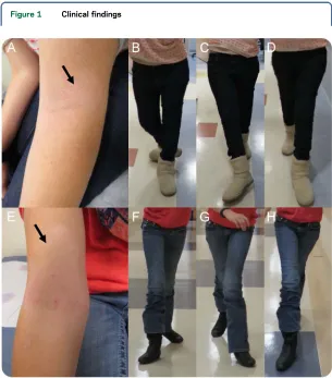
Figure 1 Clinical findings
Neurologic examination at 19 years of age revealed mild dysmorphic facial features, including a high-arched palate, a small-sized mouth, and thin, up-turned nostrils. There was evidence of a kyphotic posture. Skin examination was notable for areas of hy-popigmentation on the face and the flexor surfaces of the elbows (figure 1A). There was a head tremor at rest. Extraocular movements were full. Occasional fine nystagmus was noted during funduscopic exam-ination only. Facial strength was normal. Upper extremity strength was approximately 5/5 (the Med-ical Research Council grade) except for the thumb abduction which was 41/5, and lower extremity strength was in the 52/5 range. Sensation was reduced to pinprick and vibration in a length-dependent fashion. Proprioception was normal.
Reflexes were absent at the biceps and Achilles ten-dons bilaterally, 11at the brachioradialis, and 31at the patellae bilaterally. Babinski response was equiv-ocal. There was prominent lower extremity spasticity with evidence of a spastic catch bilaterally. Ambula-tion was dependent on full truncal support. She had a scissoring gait with circumduction of the legs and bilateral foot drop (figure 1, B–D). Rapid alternating movements were slow with reduced amplitude and evidence of mirror movements. There was dysmetria on finger-to-nose testing which was increased with eyes closed (fixed target).
Nerve conduction studies (19 years of age) were consistent with a demyelinating sensory and motor neuropathy, evidenced by diffuse, symmetric reduc-tion in nerve conducreduc-tion velocities (CVs) (peroneal
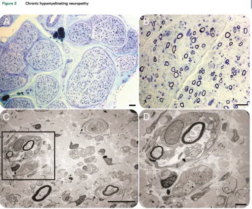
Figure 2 Chronic hypomyelinating neuropathy
motor: CV 11.7 m/s with amplitude 1.5 mV; tibial motor: CV 19.4 m/s with amplitude 2.3 mV) and prolonged distal latencies. A sural nerve biopsy per-formed at 8 years of age was consistent with a chronic predominantly hypomyelinating neuropathy (figure 2), and muscle biopsy of the quadriceps performed at the same time was suggestive of neurogenic changes with evidence of fiber-type grouping. X-ray of the spine showed a 14-degree curvature of the thoracic and lum-bar spine from T11 to L3.
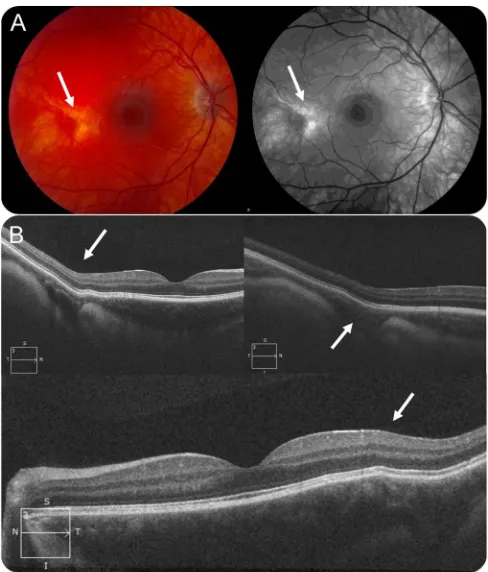
Fundoscopic examination at age 19 years showed a well-developed foveal structure but with evidence of bilateral patches of thinning and anomalous pig-mentation (mostly depigmented) temporal to the central macula (figure 3A). Optical coherence tomog-raphy indicated bowing out of the retina/choroid underlying the patchy areas of anomalous pigmenta-tion (figure 3B), suggestive of a developmental anom-aly of embryogenesis likely affecting the sclera in those regions.
Patient 2 (P2) is a 15-year-old girl and the youn-ger sister of P1. Family history is shown in figure 4. She presented similarly to her sister with decreased
fetal movements in utero as well as breech position at delivery. She was diagnosed with profound senso-rineural hearing loss at 12 months of age, and brain MRI performed in infancy reportedly revealed the absence of 1 semicircular canal. P2 also has a history of delayed gross motor development (walking inde-pendently at 2½ years of age) and mild early fine motor difficulties. She was noted to have a wide-based gait at 2½ years of age, developed spasticity over time, and by 9 years of age was using a wheel-chair for longer distances. At 15 years of age, she can ambulate with minimal assistance (figure 1, F–H). She had been diagnosed with a progressive demye-linating sensorimotor neuropathy. On examination, upper extremity strength was approximately 5/5 except for finger extension and thumb abduction which were 41/5. Lower extremity strength was subgravity in hip flexion, hip abduction, hip adduc-tion, dorsiflexion, eversion, plantarflexion, and inversion. Sensation was reduced to pinprick and vibration in a length-dependent fashion. Proprio-ception was normal. Reflexes were absent at the biceps and Achilles tendons bilaterally, 11 at the brachioradialis, and 41 at the patellae bilaterally with cross-adductor spread. There was a spastic catch in the bilateral lower extremities. Rapid alter-nating movements were slow with reduced ampli-tude. There was evidence of dysmetria with eyes closed only. Skin examination was notable for mul-tiple areas of hypopigmentation (figure 1E). Detailed clinical information for both sisters is sum-marized in table 1.
P1 and P2’s mother and father did not report any symptoms. On further questioning, however, the 39-year-old father (P3) reported a history of frequent ankle sprains, apparent high-arched feet, and mild hammertoe deformities, suggestive of a chronic neu-ropathy of likely genetic etiology. On neurologic examination, he was found to have a symmetric, length-dependent sensory greater than motor neurop-athy with reduction of pain and vibration sensation in the distal lower extremities. He had reduced reflexes throughout, including in the upper extremities (table 1). Proprioception was normal. His ophthal-mologic examination was unremarkable except for mild myopia. Nerve conduction studies showed absent sural sensory nerve action potential, suggest-ing the possibility of an existsuggest-ing neuropathy given the absent response at his age. He was found to have baseline sinus bradycardia and orthostasis (with an increase in the heart rate by more than 20 beats when transitioning from supine to standing), which may be suggestive of a mild autonomic neuropathy as well. Other quantitative autonomic testing was unremarkable. Detailed clinical information can be found in appendix e-1.
Figure 3 Ophthalmologic findings
Identification of aSOX10mutation.Exome sequencing identified a heterozygous mutation in SOX10: (c.1140C.A [p.Y380X] [NM_006941.3]) in both sisters. This nonsense mutation was not present in the control database of the National Heart, Lung, and Blood Institute Exome Sequencing Project and Exome Aggregation Consortium. The mutation was confirmed through Sanger sequencing, and familial segregation studies were negative in the 3 asymptom-atic brothers of P1 and P2 and their mother. Paternal mosaicism was suspected because a small peak of the mutated allele was observed in the father’s chromato-gram (figure 4).
Mutant vs normal allele quantification.To confirm and determine the degree of paternal mosaicism, the rela-tive ratio of the mutant allele in the father was ana-lyzed in different somatic tissues, with 3 pairs of RT PCR probes specific to eitherSOX10mutant or WT allele. Genomic DNA from saliva showed the highest proportion of approximately 32.7% of the mutant allele, while the mutant ratio was 23.3% in peripheral blood and 13.9% in dermal fibroblasts.
DISCUSSION Here, we present 2 sisters with
a unique phenotype of complicated hereditary spastic paraplegia (HSP) manifesting as progressive lower extremity spasticity, demyelinating neuropathy, and early-onset sensorineural hearing loss with normal cognition, resulting from a novel mutation inSOX10.
Paternal mosaicism was confirmed through quanti-tative RT-PCR analysis of mutant allele vs WT allele in blood, dermal fibroblasts, and saliva in the affected patients compared with the mosaic and control par-ent. Although no symptoms were volunteered by the parent with the mosaicism, on careful clinical evalu-ation, he was found to have a mild sensory neurop-athy and mild autonomic dysfunction.
Mosaicism is a result of a postzygotic mitotic muta-tional event with the mutation confined to somatic cells (somatic mosaicism), germline cells (gonadal mosaicism), or both, depending on the developmental stage and lineage at which the event occurred. The clinical manifestations of somatic mosaicism are highly variable and depend on the type of mutation, the tissue distribution, and the relative mutation load. Recent ad-vances in genetic technologies have shown that paren-tal mosaicism underlying the transmission of mutations from parents without a clinical phenotype to children with a simplex genetic disorder is more common than previously appreciated.14 Mosaicism can thus complicate the diagnostic workup and the goal of providing accurate genetic counseling.
The known neurologic manifestations of SOX10-related neurocristopathy include peripheral demyelinating neuropathy, central dysmyelinating leukodystrophy, Waardenburg syndrome, and Hirschsprung disease (PCWH). Spasticity is rare, having been reported in 4 different patients and at
Figure 4 Pedigree and molecular characterization of the family
Family history showing the mosaic father (P3), his unaffected spouse (M), the 2 affected daughters (P1 and P2), and 3 unaf-fected sons (B1, B2, and B3). Genomic DNA sequence chromatograms show the somatic c.1140C.A; p.Y380XSOX10
times associated with additional PCWH manifesta-tions including seizures and cognitive impairment, features which were notably absent in our pa-tients.9,15,16 Moreover, our patients did not have distinct symptoms of Waardenburg syndrome or Hirschsprung disease. Both sisters have a history of constipation, and after deep phenotyping, cuta-neous and macular hypopigmentation were identi-fied in both, compatible with a “forme fruste”of Waardenburg syndrome.
TheSOX10gene consists of 5 exons and contains a high mobility group, a DNA-binding domain and a C-terminal transactivation domain.17 Truncating SOX10mutations can lead to PCWH or WS4, and recent work has suggested that the 2 distinct pheno-types may be explained by 2 different molecular pathogenic mechanisms.9 Truncating mutations in any exons, except the last one, lead to mutant mRNA which is recognized and degraded through nonsense-mediated decay (NMD). This results in
haploinsufficiency, which causes the restricted and classic WS4 phenotype of Waardenburg syndrome and Hirschsprung disease, without peripheral or CNS involvement. By contrast, mutations in the last exon escape NMD and generate a stable truncated SOX10 mutant protein with increased DNA-binding affinity, which acts in a dominant-negative manner, and cause the severe PCWH neurocristop-athy with neurologic manifestations. More proximal mutations within exon 5 (Q234X, Q250X, and S251X) exhibit a stronger dominant-negative effect and lead to more severe congenital-onset symptoms and possible neonatal death.18–20The novel mutation (Y380X) identified in our family is very close to the C-terminal end of SOX10, with only 2 mutations reported that are more distal9: the X467K mutation, which was identified in a patient with severe motor and cognitive delays, and the 1400del12 mutation, which was found in a patient with complete defi-ciency of brain myelination and seizures who never
Table 1 Clinical findings
Patient/sex P1/F P2/F P3/M
SOX10mutation c.1140C.A; p.Y380X c.1140C.A; p.Y380X c.1140C.A; p.Y380X
Mutation status Heterozygous Heterozygous Mosaic
Age at examination 19 y 15 y 39 y
Fetal movements/birth Reduced/uncomplicated Reduced/C-section (breech) NA
Gross motor development Sat 10 mo; walked 2½ y; unsteady gait 7 y; lost independent ambulation 13 y
Sat 10 mo; walked 2½ y; decreased endurance 10 y
NL
Speech delay 1 2 2
EMG/NCS (age study) Severe demyelinating sensorimotor
polyneuropathy (CV range 10–31 m/s);
needle EMG with mixed myopathic and neurogenic MUPs (19 y)
Moderate demyelinating sensorimotor polyneuropathy (CV
range 18–27 m/s); needle EMG with
myopathic recruitment/MUPs (11 y)
Mild distal sensory neuropathy (CV
range 38–61 m/s); needle EMG NL
(39 y)
Brain MRI (age of study) Absence of semicircular canals; normal brain (2 y); images not available for rereview
Absence of 1 semicircular canal; normal brain (10 mo); images not available for rereview
NL (39 y)
Urinary incontinences Daily urinary urgency and leakage 2 2
Constipation/Hirschsprung disease 1/2 1/2 2/2
Hearing loss/age at diagnosis/type Severe (bilateral)/2 mo/Mondini malformation and absence of the semicircular canals
Severe (bilateral)/newborn screen/ Mondini malformation and absence of 1 semicircular canal
2/2/2
Cochlear implants/ (age) 1(26 mo) 1(12 mo) 2/NA
Nystagmus Horizontal bilateral nystagmus noted at 1 mo, resolved at 3 y
Noted at 5 wk, resolved at 3 y 2
Bright blue irides/iris heterochromia 2/2 2/2 2/2
Hypopigmentation (eyes) Pigmentary change temporal to macula
Pigmentary change temporal to
macula 2
Hypopigmentation (skin) Cubital fossae, cheeks Cheeks, neck, upper chest, cubital fossae, back
2
Hyperpigmentation (skin) Right lower leg (4 cm light brown patch)
2 2
White forelock 2 2 2
reached independent ambulation.16,21It is of interest that in our family, the Y380X mutation is not asso-ciated with cognitive impairment or seizures, indicat-ing that there may be other protective factors at play. SOX10is involved in the early development of the NCCs and is involved in Schwann cells and oligoden-drocytes later in development as well as into adult-hood.17 Sural nerve pathology in patient 1 showed findings consistent with a chronic hypomyelinating neuropathy with reduction of myelinated fibers and defective myelination of axons. Because of the presence of cochlear implants, both sisters were unable to undergo brain MRI to evaluate for possible central demyelination. The progressive weakness and spastic-ity seen in these 2 patients may potentially suggest a role ofSOX10that is not only restricted to periods of neural development but is also involved in the pro-duction and maintenance of myelin and axonal health. An understanding of the underlying pathogenic disease mechanism is essential for developing targeted therapeutic interventions. Clinical and genetic data evaluated in this family show that a reduction in the relative dose of the mutant allele, as seen in the mosaic parent, ameliorates the clinical severity, indi-cating that as a therapeutic approach in PCWH, knockdown of the mutant allele expression could be achieved through antisense-mediated or RNAi-based therapy. Careful titration is essential, as haploinsuffi-ciency leads to the classic WS4 phenotype. The WS4 presentation may be more developmental in origin; therefore, the presence of haploinsufficiency postna-tally (as induced via allele-specific knockdown) may have less of a clinical effect. Moreover, this approach may potentially only alleviate the progressive neuro-muscular symptoms, as the classic WS4 symptoms are present at birth.
Based on this family history of 2 affected sisters born to apparently unaffected parents, autosomal recessive inheritance was initially suspected, and the family was counseled accordingly. A confirmed genetic diagnosis allowed for accurate genetic coun-seling as the recurrence risk in future pregnancies of the patients is 50%, which is higher than that previ-ously anticipated. Genetic counseling for the mosaic parent (the father) was more complicated, as recur-rence risk depends on the ratio of mutated germline progenitor cells, which cannot be predicted from analysis of somatic tissues. In addition to his 2 affected daughters, the mosaic parent at present has 3 unaffected children. In reviewing families with parental mosaicism for dominant mutations causing osteogenesis imperfecta and retinoblastoma, the recurrence rate was estimated to be 27% and 10%, respectively.22,23Caution is necessary when counsel-ing for somatic mosaicism, as an individual’s recur-rence risk may be as high as 50%.
We have presented 2 sisters with a unique phe-notype of early-onset bilateral sensorineural hear-ing loss, progressive distal lower extremity spasticity, demyelinating sensorimotor neuropa-thy, mild pigmentary abnormalities, and normal cognition. Our report expands the phenotypic spectrum ofSOX10-related neurocristopathy. Mu-tations inSOX10should be considered in patients presenting with a phenotype of complicated HSP with hypomyelinating neuropathy and deafness, while SOX10 somatic mosaicism may manifest with only a mild sensory neuropathy.
AUTHOR CONTRIBUTIONS
S. Donkervoort: study concept and design, analysis, interpretation of genetic and clinical data, and drafting manuscript. D. Bharucha-Goebel and P. Yun: acquisition and interpretation of clinical data and revision of manuscript. Y. Hu: analysis and interpretation of genetic data. P. Mo-hassel, A. Hoke, W.M. Zein, AM. Atherton, AC Modrcin, and M Da-souki: acquisition and interpretation of clinical and biopsy data. AR Foley and C.G. Bönnemann: supervising the acquisition and interpreta-tion of clinical and genetic data and critical revision of the manuscript for important intellectual content. All authors critically reviewed and approved the final manuscript.
ACKNOWLEDGMENT
The authors thank the family for their participation in the study. They thank the NIH Intramural Sequencing Center and Dr. Hakonarson and Dr. Guo from The Center of Applied Genomics, Children’s Hospital of Philadelphia, for performing the exome sequencing. They thank Dr. Anne Rutkowski and CureCMD for their help with patient recruitment.
STUDY FUNDING
Work in C.G. Bönnemann’s laboratory is supported by intramural funds by the National Institute for Neurological Disorders and Stroke/NIH. Exome sequencing was funded through the Clinical Center Genomics Opportunity (CCGO), which is sponsored by the National Human Genome Research Institute (NHGRI), the NIH Deputy Director for Intramural Research, and the NIH Clinical Center.
DISCLOSURE
Dr. Bharucha-Goebel, Mohassel, Hoke, Zein, Modrcin, Dasouki, Foley, Bönnemann, Ms. Donkervoort, Yun, Hu, and Mr. Ezzo report no dis-closures. Andrea M. Atherton is now a full-time employee of Shire Phar-maceuticals. Go to Neurology.org/ng for full disclosure forms.
Received September 23, 2016. Accepted in final form March 22, 2017.
REFERENCES
1. Kim J, Lo L, Dormand E, Anderson DJ. SOX10 main-tains multipotency and inhibits neuronal differentiation of neural crest stem cells. Neuron 2003;38:17–31. 2. Southard-Smith EM, Kos L, Pavan WJ.Sox10mutation
disrupts neural crest development inDomHirschsprung mouse model. Nat Genet 1998;18:60–64.
3. Kapur RP. Early death of neural crest cells is responsible for total enteric aganglionosis in Sox10(Dom)/Sox10(Dom) mouse embryos. Pediatr Dev Pathol 1999;2:559–569. 4. Le Douarin NM, Kalcheim C. The Neural Crest, 2nd ed.
Cambridge, United Kingdom: Cambridge University Press; 1999.
6. Bondurand N, Dastot-Le Moal F, Stanchina L, et al. Dele-tions at the SOX10 gene locus cause Waardenburg syndrome types 2 and 4. Am J Hum Genet 2007;81:1169–1185. 7. Bolande RP. The neurocristopathies: a unifying concept of
disease arising in neural crest maldevelopment. Hum Pathol 1974;5:409–429.
8. Read AP, Newton VE. Waardenburg syndrome. J Med Genet 1997;34:656–665.
9. Inoue K, Khajavi M, Ohyama T, et al. Molecular mechanism for distinct neurological phenotypes conveyed by allelic trun-cating mutations. Nat Genet 2004;36:361–369.
10. Omenn GS, McKusick VA. The association of Waardenburg syndrome and Hirschsprung megacolon. Am J Med Genet 1979;3:217–223.
11. Pingault V, Bondurand N, Kuhlbrodt K, et al. SOX10 mutations in patients with Waardenburg-Hirschsprung disease. Nat Genet 1998;18:171–173.
12. Teer JK, Green ED, Mullikin JC, Biesecker LG. VarSifter: Visualizing and analyzing exome-scale sequence variation data on a desktop computer. Bioinformatics 2012;28:599–600. 13. Hsu AP, Sowerwine KJ, Lawrence MG, et al. Intermediate
phenotypes in patients with autosomal dominant hyper-IgE syndrome caused by somatic mosaicism. J Allergy Clin Immunol 2013;131:1586–1593.
14. Campbell IM, Yuan B, Robberecht C, et al. Parental somatic mosaicism is underrecognized and influences recurrence risk of genomic disorders. Am J Hum Genet 2014;95:173–182.
15. Pingault V, Pierre-Louis L, Chaoui A, et al. Phenotypic similarities and differences in patients with a p.Met112Ile mutation in SOX10. Am J Med Genet Part A 2014;164A: 2344–2350.
16. Inoue K, Tanabe Y, Lupski JR. Myelin deficiencies in both the central and the peripheral nervous systems associated with a SOX10 mutation. Ann Neurol 1999;46:313–318. 17. Kuhlbrodt K, Herbarth B, Sock E, Hermans-Borgmeyer I, Wegner M. Sox10, a novel transcriptional modulator in glial cells. J Neurosci 1998;18:237–250.
18. Inoue K, Shilo K, Boerkoel CF, et al. Congenital hypo-myelinating neuropathy, central dysmyelination, and Waardenburg-Hirschsprung disease: phenotypes linked by SOX10 mutation. Ann Neurol 2002;52:836–842. 19. Pingault V, Girard M, Bondurand N, et al. SOX10
muta-tions in chronic intestinal pseudo-obstruction suggest a complex physiopathological mechanism. Hum Genet 2002;111:198–206.
20. Touraine RL, Attie-Bitach T, Manceau E, et al. Neuro-logical phenotype in Waardenburg syndrome type 4 cor-relates with novel SOX10 truncating mutations and expression in developing brain. Am J Hum Genet 2000; 66:1496–1503.
21. Sham MH, Lui VC, Chen BL, Fu M, Tam PK. Novel mutations of SOX10 suggest a dominant negative role in Waardenburg-Shah syndrome. J Med Genet 2001;38: E30.
22. Sippel KC, Fraioli RE, Smith GD, et al. Frequency of somatic and germ-line mosaicism in retinoblastoma: im-plications for genetic counseling. Am J Hum Genet 1998; 62:610–619.
DOI 10.1212/NXG.0000000000000151
2017;3;
Neurol Genet
Sandra Donkervoort, Diana Bharucha-Goebel, Pomi Yun, et al.
HSP and deafness: Neurocristopathy caused by a novel mosaic SOX10 mutation
This information is current as of May 15, 2017
Services
Updated Information &
http://ng.neurology.org/content/3/3/e151.full.html including high resolution figures, can be found at:
Supplementary Material
http://ng.neurology.org/content/suppl/2017/05/15/3.3.e151.DC1 Supplementary material can be found at:
References
http://ng.neurology.org/content/3/3/e151.full.html##ref-list-1 This article cites 22 articles, 2 of which you can access for free at:
Subspecialty Collections
http://ng.neurology.org//cgi/collection/spastic_paraplegia Spastic paraplegia
http://ng.neurology.org//cgi/collection/all_genetics All Genetics
following collection(s):
This article, along with others on similar topics, appears in the
Permissions & Licensing
http://ng.neurology.org/misc/about.xhtml#permissions its entirety can be found online at:
Information about reproducing this article in parts (figures,tables) or in
Reprints
http://ng.neurology.org/misc/addir.xhtml#reprintsus Information about ordering reprints can be found online:
reserved. Online ISSN: 2376-7839.
Published by Wolters Kluwer Health, Inc. on behalf of the American Academy of Neurology. All rights an open-access, online-only, continuous publication journal. Copyright Copyright © 2017 The Author(s).
is an official journal of the American Academy of Neurology. Published since April 2015, it is