Prostatic Acid Phosphatase: A Potpourri*
CHARLES W. MONCURE, M.D.
Assistant Professor of Pathology, Medical College of Virginia,
Health Sciences Division of Virginia Commonwealth University,
Richmond, Virginia
In 1938, Gutman et al. (6) described increased levels of acid phosphatase in metastatic carcinoma of the prostate. Since then, when most of us hear the term "acid phosphatase" we immediately think of prostatic cancer, and justifiably so. The link, how-ever, between the test and the diagnosis is not as clear and simple as many of us would like to believe. I would first like to discuss this enzyme, re-viewing some of its chemistry, its clinical value and the pitfalls and problems associated with the routine tests. Then I would like to tell about some work in which we have been involved that we hope will increase the diagnostic value of this enzyme and perhaps point the way to advances in clinical enzy-mology in other areas.
Chemical Assays. First let us remember that the term "acid phosphatase" simply means an enzyme which, at pH below 7 .0, hydrolyzes phosphate esters liberating the phosphate ion and replacing it with an -OH group. Prostatic acid phosphatase is only one of many enzymes in this category. The organic portion of the phosphate ester involved is not speci-fically defined by the term acid phosphatase, and this brings up our first problem. We do not know the natural physiological substrate of prostatic acid phosphatase. Indeed we know very little about the biological function of this enzyme. Consequently, we must measure its activity using synthetic phosphate esters which are, as far as we know, completely unrelated to prostatic physiology and reproduction. Each new substrate gives rise to a new assay, a new set of units, new degrees of specificity and sensitivity and new problems of interpretation.
As previously mentioned, there are many forms
*
Presented by Dr. Moncure at the 26th Annual Stone-burner Lecture Series, February 22, 1973, at the Medical College of Virginia, Richmond.MCV QUARTERLY 9(3): 235-239, 1973
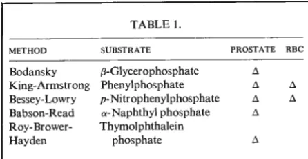
of this enzyine which are not derived from the prostate. Indeed, it is present to some degree in virtually all tissues as a lysosomal enzyme. In blood chemistry, however, the most common source of non-prostatic acid phosphatase is the red blood cell. For this reason several clinical assays have been de-veloped which readily differentiate between prostatic and erythrocytic acid phosphatase. Table 1 lists the methods and substrates commonly employed in the clinical laboratory. The Bodansky ( 4) and Babson-Read (2) as well as the newer thymolphthalein phosphate method described by Roy, Brower, and Hayden ( 14) will not detect red cell acid phospha-tase. The King-Armstrong (7) and Bessey-Lowry
( 3) methods will detect the red cell enzyme, how-ever, and employ an inhibitor, usuall:r L( +) tartrate, to differentiate prostatic and erythrocytic acid phos-phatase. Table 2 lists inhibitors which can be used for this purpose. Although selective substrates and inhibitors can make this distinction, it should be emphasized that they do not provide a truly specific test for prostatic acid phosphatase. Indeed, ~-glycero-phosphate and a-naphthyl ~-glycero-phosphate are histochemi-cal substrates (7) commonly employed for staining
acid phosphatase in a variety of tissues.
For tissue analysis and for occasional patho-logical sera, inhibitors and substrate selectivity do not clearly distinguish between prostatic and rion-prostatic acid phosphatase. Table 3 lists some of the nonprostatic diseases which have been reported to cause elevations in serum acid phosphatase and while they rarely present a problem of differential diagnosis in carcinoma of the prostate, we should not ignore their existence.
A far more common problem is the interpreta-tion of serum acid phosphatase levels where the en-zyme is of prostatic origin and where we are con-cerned with the diagnosis and staging of prostatic
TABLE 1.
METHOD SUBSTRATE PROSTATE RBC
Bodansky 13-Glycerophosphate ii
King-Armstrong Phenyl phosphate ii ii
Bessey-Lowry p-Nitrophenylphosphate ii ii
Babson-Read a-Naphthyl phosphate ii
Roy-Brower- Thymolphthalein
Hayden phosphate ii
cancer. Most prostatic carcinomas produce this en-zyme, and low serum levels in the face of apparent widespread diseases are usually the result of heat denaturation at neutral or alkaline pH as this enzyme rapidly loses its activity if the serum sample is not acidified and refrigerated.
An elevated serum prostatic acid phosphatase in general suggests prostatic cancer which has extended beyond the prostate; however, prostatic surgery, diagnostic palpation, and prostatic infarction may cause transient enzyme elevations. Careful attention to the time when the sample is drawn and to speci-men handling will greatly improve the reliability of routine serum assays.
Immunology. In order to provide a highly spe-cific means of diagnosing prostatic cancer, we began investigating the antigenicity of prostatic tissue and secretions first iIT dogs ( 8) and later in man ( 10, 11). Using antisera raised in rabbits immunized with dog prostatic fluid or human sperm-free ejaculate we could demonstrate an immunological organ speci-ficity of prostatic acid phosphatase. This specispeci-ficity had also been observed by Shulman and his co-workers ( 15) and has been confirmed by several other investigators ( 12, 13). Our methods of demon-strating antiprostatic acid phosphatase involved re-acting the antisera with prostatic homogenate in an Ouchterlony agar gel immunodiffusion system. After precipitin lines formed, the gel was washed and stained histochemically for acid phosphatase (14).
TABLE 2.
INHIBITOR PROSTATE RBC
Ethyl alcohol 40% . . . ( +) ( - ) L (+)tartaric acid 0.02 M. . . ( +) ( - ) Formaldehyde 2% . . . ( - ) ( +)
Cupricion0.001 M... (-) (+)
TABLE 3.
NON·PROSTATIC CAUSES OF ELEVATED ACID PHOSPHATASE
Gaucher's disease Osteitis deformans Thrombocythemia Lipid histocytosis Hemorrhagic shock Lymphoblastic leukemia Nonprostatic carcinoma
Extensive absorption of the antisera with non-prostatic tissues confirmed the tissue specificity of both dog and human prostatic acid phosphatase. Antihuman prostatic acid phosphatase was then raised against a partially purified enzyme prepara-tion. Human sperm-free ejaculate was passed through an ion-exchange column packed with DEAE Sephadex ( A-50) equilibrated with 0.1 M Tris-HCI pH 8.0. Acid phosphatase was trapped in the column while most other seminal proteins passed through freely. The enzyme was then eluted with a sodium chloride molarity gradient, concentrated by ultra-filtration and used with complete Freund's adjuvant to immunize New Zealand White rabbits (15). This antiserum was also checked by Ouchterlony im-munodiffusion together with a diazo coupling stain for acid phosphatase as described previously ( 15). The serum was tested against prostate, liver, spleen, kidney, pancreas, salivary gland, gastric mucosa; large and small bowel, breast, lung, muscle, lymph node, heart and freshly prepared packed platelets. The only positive reaction was with prostatic acid phosphatase. Antiserum of this quality can be used to specifically identify and quantitate the prostatic enzyme.
The sensitivity of gel diffusion tests is too low to detect normal or moderately elevated serum acid phosphatase; however, these simple, inexpensive tests can be used to identify prostatic cancer since the tissue itself is usually quite rich in the enzyme. Clinical Study. A logical application of such a test would seem to be the identification of prostatic carcinoma in bone marrow as the tumor charac-teristically spreads to bone artd the presence of bony metastases is a critical factor in staging the disease.
•
•
0
Fig. 1-Serial dilution of prostatic acid phosphatase showing reduction of precipitive ring with decreasing enzyme concentration.
s
A
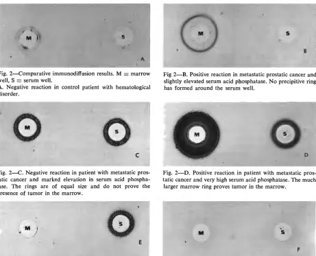
Fig. 2-Comparative immunodiffusion results. M
=
marrow well, S=
serum well.A. Negative reaction in control patient with hematological disorder.
0
c
Fig. 2-C. Negative reaction in patient with metastatic pros-tatic cancer and marked elevation in serum acid phospha-tase. The rings are of equal size and do not prove the presence of tumor in the marrow.
1,.
M
)
.
..
.,,,E
Fig. 2-E. Negative reaction in the patient with benign pros-tatic hyperplasia and elevated serum acid phosphatase fol-lowing transurethral resection. No precipitive ring is formed
around the marrow well.
s
Fig 2-B. Positive reaction in metastatic prostatic cancer and
slightly elevated serum acid phosphatase. No precipitive ring has formed around the serum well.
0
0Fig. 2-D. Positive reaction in patient with metastatic pros-tatic cancer and very high serum acid phosphatase. The much
larger marrow ring proves tumor in the marrow.
M
F
Fig. 2-F. Negative reaction in patient with Gaucher's
at a concentration of 1: 100. This technique was based on the method developed for the quantitation of serum immunoglobulins ( 16) . Low levels of
en-zyme produced small red precipitin rings when stained for acid phosphatase. As the enzyme concen-tration was increased the ring diameter increased
(fig. 1). Bone marrow aspirates and serum samples were obtained in the course of routine hematological evaluation of 61 patients with prostatic cancer and 32 patients with benign prostatic disease, nonpros-tatic neoplasms, or hematological disorders, includ-ing one patient with Gaucher's disease. Marrow smears were stained with Wright-Giemsa and
screened for tumor cells. Paired smears stained for acid phosphatase were also used to screen for pros-tatic cancer cells. Comparative radial immunodif-fusion was carried out on quantitative gel plates with two sample wells. Serum was placed in one well and marrow in the other and diffusion carried out at room temperature for 24 hours. The plates were then washed and stained. If the precipitin ring around the marrow well was larger than that of the serum, the test was considered positive. If no ring occurred around the marrow well or if serum and marrow produced rings of equal diameter, the test was con-sidered negative for bony metastases (fig. 2A-F).
Results of Bony Marrow Study. Malignant cells were seen in three of the control group and Gauch-er's cells in one. Only the GauchGauch-er's cells were stained by the phosphatase reaction. None of the 32 controls was positive by immunodiffusion. One pa-tient with benign prostatic hyperplasia who was studied immediately after cystoscopy showed detect-able serum prostatic acid phosphatase but no detect-able enzyme in the marrow, thus documenting a serum elevation due to prostatic manipulation. Among the 61 patients with prostatic cancer, 12 had malignant cells in their marrow and gave a positive immunodiffusion test. Four other cases of prostatic carcinoma were positive by immunodiffusion; how-ever, no tumor cells were found in the marrow aspirates. All four of these patients had bony lesions by x-ray.
These data suggest that a comparative im-munoassay for prostatic acid phosphatase may pro-vide a simple, inexpensive yet specific means of establishing that a patient has carcinoma of the prostate in an advanced stage. In two cases, we have obtained positive immunodiffusion tests on men with carcinoma of undetermined primary. Both of these were subsequently proven to have prostatic cancer.
While we have only applied this technique clinically on bone marrow aspirates, autopsy studies indicate that it is equally successful in identifying prostatic cancer in lymph nodes and other nonprostatic organs.
It is our belief that the techniques applied here to prostatic cancer could be extended to other neo-plasms which continue to produce some tissue spe-cific substance. Ultimately, a large battery of antisera
might be developed which could be employed for identifying neoplasms and for immunochemical stag-ing of these diseases. The antigenic specificity of
enzymes and other cellular products provides a new parameter for clinical laboratory testing which should lead to improved diagnostic procedures in both neo-plastic and non-neoplastic diseases.
Author's note: The immunological studies de-scribed in the above article were accomplished in a cooperative study carried out by Drs. Charles L. Johnston, Jr., M. J. V. Smith, Warren W. Koontz, Jr. and the author.
REFERENCES
I. ABLIN, R. J. Tissue and species: Specific antigens of
normal human prostatic tissue. /. Immuno/. 104: 1329,
1971.
2. BABSON, A. L. AND READ, P. A. A new assay for
pros-tatic acid phosphatase in serum. Am. /. Clin. Patho/.
32:6, 1959.
3. BESSEY, 0. A., LOWRY, 0. H. AND BROCK, M. J. A
method for the rapid determination of alkaline
phos-phatase with five cubic mm of serum. /. Biol. Chem.
164:321, 1946.
4. BoDANSKY, A. Phosphatase studies: II. Determination of serum phosphatase. Factors influencing the accuracy
of the determination./. Biol. Chem. 101:93, 1933.
5. BRUSTONE, M. S. Histochemical comparison of
naphthol-As-phosphates for the demonstration of phosphatases.
/. Natl. Cancer Inst. 20:601, 1958.
6. GUTMAN, A. B. AND GUTMAN, E. B. An Acid
phospha-tase occurring in the serum of patients with metastasiz-ing carcinoma of the prostrate gland. /. Clin. Invest. 17: 473, 1938.
7. KING, E. J. AND ARMSTRONG, A. R. Convenient method
for determining serum and bile phosphatase activity.
Can. Med. Assoc. I. 31:376, 1934.
8. MANCINI, G., CARBONARA, A. 0. AND HEREMANS, J. F.
Immunochemical quantitation of antigens by single
ra-dial immunodiffusion. Immunochemistry 2:235, 1965.
9. MONCURE, C. W., PROUT, G. R., JR. AND BLAYLOCK, W.
K. The immunological detection of prostatic acid
10. MONCURE, C. W., PROUT, G. R., JR. AND BLAYLOCK, W. K. Prostatic acid phosphatase antisera. Invest. Urol.
5: 3 31, 1968.
11. MONCURE,
c.
w. AND PROUT, G. R., JR. Antigenicity of human prostatic acid phosphatase. Cancer 25:463, 1970.12. PFEIFFER, L., ABLIN, R. J., GANDER, M. J. AND SOANES, W. A. Antibodies to human prostatic acid phosphatase.
Fertil. Steril. 21 :344, 1970.
13. PROUT, G. R., JR. AND MONCURE, C. w. Prostatic car-cinoma-1968. In: Sixth National Cancer Conference
Proceedings. Philadelphia, J. B. Lippincott Co. p. 195,
1968.
14. ROY, A. v., BROWER, M. E. AND HAYDEN, J.E. Sodium thymolphthalein phosphate: A new acid phosphatase substrate with greater specificity for the prostatic en-zyme in serum. Clin. Chem. 17:1093-102, 1971. 15. SHULMAN, S., MAMROD, L., GANDER, M. J. AND SOANES,
W. A. The detection of prostatic acid phosphatase by antibody reactions in gel diffusion. J. lmmunol. 93 :474, 1964.
16. THOMPSON, S. W. Selected histochemical and histo-pathological methods. Springfield, Illinois, Charles C.