Official Journal of Iranian Neuroscience Society
In Collaboration with: Iran University of Medical Sciences Tehran University of Medical Sciences Shahid Beheshti University of Medical Sciences
Chairman and Editor-in-Chief Mohammad Taghi Joghataei, PhD
Cellular and Molecular Department, Iran University of Medical Sciences, Tehran, Iran
Associate Editor Hamed Ekhtiari, MD Translational Neuroscience Program Institute for Cognitive Sciences Studies, Iran
Editorial Board
Shahin Akhondzadeh, PharmD, PhD Tehran University of Medical Sciences, Iran
Abolhassan Ahmadiani, PharmD, PhD Shahid Beheshti University of Medical Sciences, Iran
Hasan Ashayeri, MD Iran University of Medical Sciences, Iran
Mazyar Azar, MD Iran University of Medical Sciences, Iran Tourandokht Baluchnejadmojarad, PhD
Iran University of Medical Sciences, Iran Mohammad Reza Daliri, PhD Iran University of Science and Technology, Iran
Sanju George, MD
Birmangham & Solihull Mental Health NHS, UK Ali Gorji, MD, PhD
Munster University, Germany Abbas Haghparast, PhD
Shahid Beheshti University of Medical Sciences, Iran Gholamreza Hasanzadeh, PhD Tehran University of Medical Sciences, Iran
Richard Hawkes, PhD Calgary University, Canada Mitsuhiro Kavata, MD, PhD Perfetural Medical University, Japan
Alireza Khoshnevisan, MD Tehran University of Medical Sciences, Iran
Mahmoud Kiaei, PhD
University of Arkansas for Medical Sciences, USA Mehdi Mehdizadeh, PhD Iran University of Medical Sciences, Iran
Masoud Mehrpour, MD Iran University of Medical Sciences, Iran Hamidreza Mostafawi Abdolmaleki, MD
Harvard University, USA Fereshteh Motamedi, PhD
Shahid Beheshti University of Medical Sciences, Iran Arezo Nahavandi, MD, PhD Iran University of Medical Sciences, Iran
Klaus Obermayer, PhD Technische Universität Berlin, Germany
Amir H. Rezvani, PhD Duke University, USA Ali Rashidy-Pour, PhD Semnan University of Medical Sciences, Iran
Mehrdad Roghani, PhD Shahed University, Iran Jacqueline Sagen, PhD University of Miami, USA
Ali Shahbazi, PhD Iran University of Medical Sciences, Iran
Esmaeil Shahsavand Ananloo MD, PhD Tehran University of Medical Sciences, Iran
Masoud Shekarabi, PhD University of Montreal, Canada
Amir M. Sodagar, PhD University of Michigan, USA Hamid Soltanian Zadeh, PhD
Tehran University, Iran Mark J. West, PhD Aarhus University, Denmark
Ali Yoonessi, MD, PhD Tehran University of Medical Sciences, Iran Mohammad Reza Zarrindast, PharmD, PhD
Tehran University of Medical Sciences, Iran Executive Board
Sobhan Rezaee, MA Allame Tabatabaei University, Iran
Ali Shahbazi, PhD Iran University of Medical Sciences, Iran
Proof Reading Editors Nima Khlighinejad, MD
Shiva Alinaqian, MA Mohammad Torabi Nami, MD, PhD
Elham Golpoosh, MA Graphic & Layout Designer
Haleh Ghorbani Tehrani Cover Designer Bahram Teymoory Dereshky
Mohsen Farhadi
Editorial Office
Neuroscience Section, School of Medicine, Hemmat Campus, Iran University of Medical Sciences,
Shahid Hemmat EXP. Tehran, P.O. Box: 15144-6183, Islamic Republic of Iran Tel:+98-21- 86704576/Fax:+98-21-88622709
Website: bcn.iums.ac.ir Email: [email protected]
Disclaimer: While the information in this journal are believed to be true and accurate at the date of its publica-tion, neither the authors, the editors, nor the publisher can
accept any legal responsibility for any errors or omissions that maybe made. The publisher makes no warranty, ex-press or implied, with respect to material contained herein.
Copyright © 2013 by Iran University of Medical Sciences Published in Iran University of Medical Sciences in Cooperation with Mehromah-e-No Publication Institute
Tehran University of Medical Sciences Iranian
Neuroscience Society Shahid Beheshti University of Medical Sciences Autumn, November 2013, Volume 4, Number 4
Behavioral Neuroscience
Section Editor
Abbas Haghparast, PhD
Shahid Beheshti University of Medical Sciences, Iran
Deputy Section Editor
Ali Shahbazi, PhD Iran University of Medical Sciences, Iran
Associate Section Editors
Abdolrahman Sarihi, PhD Hamadan University of Medical Sciences, Iran
Abedin Vakili, PhD Semnan University of Medical Sciences, Iran
Ali Shamsizadeh, PhD Rafsanjan University of Medical Sciences, Iran
Alireza Sarkaki, PhD
Jundishapur University of Medical Sciences, Iran Esmail Riahi, PhD
Pasteur Institute, Iran Firouz Ghaderi Pakdel, PhD Urmia University of Medical Sciences, Iran
Ghassem Attarzadeh-Yazdi, PhD Hormozgan University of Medical Sciences, Iran
Mahdi Goudarzvand, PhD Alborz University of Medical Sciences, Iran
Mir-Shahram Safari, PhD RIKEN Brain Science Institute, Japan Mohammad Mohammad-Zadeh, PhD Sabzevar University of Medical Sciences, Iran
Clinical Neuroscience
Section Editor
Hamed Ekhtiari, MD Institute for Cognitive Sciences Studies, Iran
Deputy Section Editor
Mohammad Nami Torabi, MD, PhD Shiraz University of Medical Sciences, Iran
Associate Section Editors
Shahid Bashir, PhD Harvard University, USA
Arian Behzadi, MD McGill University, Canada Behzad Elahi, MD, PhD University of Toronto, Canada
Sanju George, PhD Mental Health NHS Foundation Trust, UK
Nima Khalighinejad, MD University College London, UK Behzad Mansouri, MD, PhD University of Manitoba, Canada Mandana Modirrousta, MD, PhD
University of Manitoba, Canada Cellular and Molecular Neuroscience
Section Editor
Masoud Shekarabi, PhD University of Montreal, Canada
Deputy Section
Hossein Aleyassin PhD Burke Medical Research Institute, USA
Mahmoud Kiaei PhD University of Arkansas, USA
Associate Section Editors
Arezoo Campbell PhD Western University, USA Mehdi Dotoudchi PhD,
Biotech Company, USA Effat Emamian MD Biotech Company, USA
Amir Rezvani PhD Duke University, USA Ali Salahpour PhD University of Toronto, Canada
Ahmad Salehi MD, PhD, Stanford University, USA Saba Valadghan MD, PhD
University of Ohio, USA Computational Neuroscience
Section Editor
Mohammad R. Daliri, PhD Iran University of Science and Technology, Iran
Deputy Section Editor
Vahab Nekoukar Islamic Azad University of Karaj, Iran
Associate Section Editors
Mahan Azadpour Syracuse University, USA
Mehdi Azarnoosh Islamic Azad University of Mashhad, Iran
Abbas Erfanian
Iran University of Science and Technology, Iran Reza Kobravi
Islamic Azad University of Mashhad, Iran Reza Lashgari
State University of New York, USA Amin Mahnam University of Isfahan, Iran
Hamid Reza Marateb University of Isfahan, Iran
Mohsen Omrani Queen's University, Canada
Hojjat Sabzpoushan
Iran University of Science and Technology, Iran Seyed Ehsan Tahami
Islamic Azad University of Mashhad, Iran Cognitive Neuroscience
Section Editor
Ali Yoonessi, MD, PhD Tehran University of Medical Sciences, Iran
Associate Section Editors
Ali Asadollahi, PhD Stanford University, US Reza Ebrahimpour, PhD Shahid Rajaee Teacher Training University, Iran
Arash Fazl, MD, PhD Icahn School of Medicine, US
Reza Lashgari, PhD State University of New York, US Arash Yazdanbakhsh, MD, PhD
EDITORIAL
RESEARCH PAPERS Brain Extracellular Space:
Geometry, Matrix and Physiological Importance
4 Kamali-Zare, P., Nicholson, Ch.
Antiepileptic and Antioxidant Effect of Hydroalcoholic Extract of Ferula Assa Foetida Gum on Pentylentetrazoleinduced Kindling in Male Mice
Kiasalari, Z., Khalili,M., Roghani, M., Heidari, H., Azizi, Y. 21
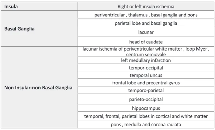
Stroke Modifies Drug Consumption in Opium
Addicts: Role of the Insula Yousefzadeh-fard, Y., Gharedaghi, M.H., Esmaeili, S., Pourbakhtyaran,E., Salehi Sadaghiani, M. Ghorbani, A., MoSahraian, M.A.
29
Effects of Systemic Administration of Oxytocin on Contextual Fear Extinction in a Rat Model of Post-Traumatic Stress Disorder
Eskandarian, SH., Vafaei, A.A., Hassan Vaezi, G., Taherian, F., Kashefi, A., Rashidy-Pour, A.
37
Use of Colchicine in Cortical Area 1 of the Hippocampus Impairs Transmission of Non-Motivational Information by the Pyramidal Cells
Riahi, N., Karami, M., Porkhodadad, S. 45
Ecstasy-Induced Caspase Expression Alters
Following Ginger Treatment Soleimani Asl, S., Pourheydar, B., Dabaghian, F., Nezha-di, A., Roointan, A., Mehdizadeh, M. 51
Intracerebroventricular Injection of Lipopolysaccharide Increases Gene Expression of Connexin32 Gap Junction in Rat Hippocampus
Abbasian, M., Sayyah, M., Babapour, V., Mahdian, R. 56
Does the Ability to Make a New Business Need More Risky Choices during Decisions? Evidences for the Neurocognitive Basis of Entrepreneurship
Nejati, V., Shahidi, Sh. 9
Evaluation of the Functional Recovery in Sciatic Nerve Injury following the Co-transplantation of Schwann and Bone Marrow Stromal Stem Cells in Rat
Zarbakhsh, S., Moradi, F., Joghataei, M.T., Bahktiari, M., Mansouri, K., Abedinzadeh, M.
13
The Effect of Food Deprivation on Nociception in Formalin Test and Plasma Levels of Noradrenaline and Corticosterone in Rats
Gheibi, N., Saroukhani, M.R., Azhdari-Zarmehri, H. 63
What is ECS and why is it
important?
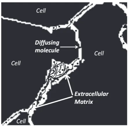
rain tissue is essentially composed of two regions: cellular elements (neurons and glial cells), and the gap between the elements, which is known as the extra-cellular space (ECS; Figure 1) (Sykova & Nicholson, 2008). The ECS resembles the water phase of a foam and remains a highly connected domain even though it is convoluted in shape and may form dead-space microdomains (e.g. local expansions or voids) (Hrabetova, Hrabe, & Nicholson, 2003). The width of the ECS is about 20-60 nm (Thorne & Nicholson, 2006), nevertheless in totality it occupies approximately 20% of the entire tissue volume (Sykova & Nicholson, 2008) The surprisingly large relative volume of the ECS makes it an important area for neuroscience research.
Extracellular space is the immediate external environ-ment of brain cells. This proximity to the cell membrane makes the structure and content of the ECS important for cellular homeostasis and function. The ECS contains a fluid similar in composition to that found in the brain ventricles that maintains an ionic balance for Ca2+, Na+, K+ and Cl- across the cell membrane. Such an ionic bal-ance establishes the cellular resting potential and permits neuronal action potentials and synaptic transmission. The ECS also provides a communication channel
be-tween cells through which chemical signals travel; this is known as volume transmission (Agnati, Fuxe, Nich-olson, & Sykova, 2000). Clinically, the ECS is an im-portant route for the delivery of drugs after they have entered the brain (Wolak & Thorne, 2013).
Editorial:
Brain Extracellular Space:
Geometry, Matrix and Physiological Importance
B
Figure 1. Schematic of brain cells and ECS. The ECS may have local expansions.
Figure 2. Schematic of the extracellular matrix as a mesh-work of long-chain molecules distributed in ECS.
Besides an ionic fluid, the ECS accommodates an extra-cellular matrix formed from a meshwork of long-chain polymeric molecules and proteins (Figure 2). These in-clude chondroitin sulfate, heparin sulfate and tenasin that often branch off from a hyaluronic acid backbone (Zim-mermann & Dours-Zim(Zim-mermann, 2008).
Molecular Diffusion in ECS
Diffusion is the dominant mechanism for transport of substances in ECS and determines both the local and global distribution of many molecules.
Among other factors, such as the local sources or sinks of molecules, diffusion depends on the space or volume fraction accessible to the molecules and the geometry of their path through the ECS. Volume fraction of ECS, α, is defined as the dimensionless parameter:
α
=VECS VTissue (1)where VECS and VTissue are the volumes of ECS and the whole tissue respectively.
Molecular diffusion in ECS is similar to that in a po-rous medium and surprisingly, using only the classical theoretical framework for diffusion, we are able to char-acterize molecular diffusion in the brain (Nicholson & Phillips, 1981; Nicholson, 2001). This enables us to use a single diffusion coefficient (D*) to capture all the ef-fects of the environment. Therefore D* is called the ‘ef-fective diffusion coefficient’.
The magnitude of D* reflects the hindrance imposed by the geometry of the path, therefore D* < D, where D is the free diffusion coefficient. The dimensionless parameter tortuosity, λ, may be used to characterize the hindrance to diffusion where:
λ
=
D
D
* (2)In addition to being affected by the geometry, the dif-fusing molecule may also interact with the matrix; this too can be incorporated into the tortuosity (Nicholson, Kamali-Zare, & Tao, 2011).
A measurement of tortuosity may be thought of as re-vealing properties of ECS itself (Nicholson & Sykova, 1998). When molecules are released from a source and make multiple random walks that are reflected from the multiple boundaries of the ECS, they effectively explore their microenvironment. If their collective behavior can be visualized, the local structure will ‘appear’. The sim-ulation shown in Figure 3 exemplifies this concept by tracking a number of molecules released from a point source in the ECS at the center of an initially unseen structure. Then, following the molecules in time, the shape of the structure emerges. The structure is revealed to be an ensemble of cubes where one in every eight is missing, providing a local expansion of ECS that consti-tutes a dead-space (Figure 3, last panel).
The ECS has been most studied in neocortex, however there have been measurements in corpus callosum, hip-pocampus, cerebellum, caudate nucleus and spinal cord.
In the cerebellar molecular layer and in regions contain-ing major fiber bundles, diffusion is anisotropic, becontain-ing different in different axes (Rice, Okada, & Nicholson, 1993).
Figure 3. Projection from top of a simulation with a popu-lation of molecules released from a point source located at the center of a structure. As molecules diffuse, they make random walks and thereby reveal the structure of the local environment. The MCell program was used for simulation and DReAMM for visualization (see Modeling section).
How to Characterize ECS
Experiments provide values for volume fraction, tortu-osity and some other parameters. Complementing exper-iments, modeling tests hypotheses about the factors that determine these parameter values. In addition, modeling establishes a solid theoretical framework for molecular diffusion in ECS. Modeling may provide a simpler al-ternative to experiments and sometimes may be the only way to proceed.
Experiments
& Tao, 1993). The RTI and IOI methods were introduced by the Nicholson laboratory and provide real-time data in small brain regions.
Modeling
Here we summarize one modeling approach based on Monte Carlo simulation using the program MCell, (Stiles & Bartol, 2001; Nicholson, Kamali-Zare, & Tao, 2011). MCell was developed at the University of Pittsburgh, Supercomputing Center and Salk Institute for Biological Studies, as a modeling tool for realistic simulation of the behavior of molecules in the complex 3D microenviron-ments found in biological tissue. The main advantage of using MCell in ECS studies is that it can represent both the ramified geometry and the molecular interactions with the extracellular matrix.
The Monte Carlo method mimics actual diffusion. A large population of ‘molecules’ is released from an ap-propriate source and the molecules execute random walks in a specified geometry. They may interact with other molecules or sites through suitable kinetic reac-tions. After a certain time the distribution of the mol-ecules is analyzed. In our applications the main output of an MCell simulation is D* (which is easily converted to the tortuosity). For a population of molecules released from the origin in a 3D medium, D* is calculated using the classical equation:
D* = <r 2>/6t (3)
where r is the distance of each molecule from the source and <r 2> represents the mean square distance of all mol-ecules at time t after the molmol-ecules have been released.
Key Facts about ECS Derived from Experiments and Modeling
The basic quantitative parameters of ECS structure are volume fraction, α, and tortuosity, λ. Using the RTI method with TMA+ as a small probe molecule it has been established that α = 0.2 (this implies that the ECS occupies 20% of the brain) and λ = 1.6 (this implies that D* 0.4 D). This value of tortuosity is valid for mol-ecules that are much smaller than the width of the ECS and do not interact with the extracellular matrix. If the molecules are much larger (Thorne & Nicholson, 2006) or reversibly bind to the matrix (Hrabetova, Masri, Tao, Xiao, & Nicholson, 2009), λ may be greater than 1.6. Figure 4 summarizes the two key players in all ECS nar-ratives: ‘Geometry’ and ‘matrix’ and emphasizes that a study of diffusion is a key to understanding ECS struc-ture and content.
Geometry of the Extracellular Space
It appears that λ = 1.6 represents a fundamental constant of the ECS and it is pertinent to ask where the value comes from. Assuming that no matrix interaction is involved it is reasonable to look to ECS geometry for the answer.
To address the role of structure, the Nicholson labora-tory developed several models to explore idealized ECS geometry. The basic element of these structures is a sin-gle cube with a size similar to that of a brain cell body. In MCell simulations where many such cubes are packed together in a 3D geometry and separated from each other by a uniform ECS with a realistic width to ensure α = 0.2, it is found that λ = 1.18 (Tao & Nicholson, 2004). Clearly, this value is much smaller than the experimen-tally measured value of λ = 1.6. To try to increase this low tortuosity, a number of local voids, or dead-spaces, were introduced in the cube-ensembles (Figure 5). This strategy was able to increase λ to about 1.6 because when molecules enter these local voids, they are tran-siently held up in the region and their diffusion time is prolonged (Tao, Tao, & Nicholson, 2005). Dead-spaces in real biological tissue may be formed by local expan-sions of the ECS (voids), membrane invaginations or by glial cells wrapping around neurons (Hrabetova, Hrabe, & Nicholson, 2003; Hrabetova & Nicholson, 2004).
Extracellular Matrix
In addition to the effect of the complex geometry of ECS, molecular diffusion is affected by the extracellular matrix. The matrix may react with suitable molecules through electrostatic or steric (actual binding and un-binding) interactions with the chondroitin sulfate (Hra-betova, Masri, Tao, Xiao, & Nicholson, 2009) or hepa-ran sulfate (Thorne, Lakkaraju, Rodriguez-Boulan, & Nicholson, 2008) components of the matrix. Our current modeling studies aim to combine geometry and matrix to study how the two components interact to affect mo-lecular diffusion (Figure 5C and 5D; Nicholson, Kamali-Zare, Tao, 2011).
ECS Applications
Research involving ECS ranges from basic to applied. Basic research aims to answer fundamental questions about how simple mechanisms lead to complex func-tions. For example, it is extending the concept of the ‘microdomain’ from being only the small region around single channels to being a larger domain spanning the spaces surrounding groups of cells. This links molecular and cellular level events to networks of cells with subtle interactions. This perspective may help our understand-ing of complex diseases, such as cancer, (Vargova et al., 2003) and brings ECS research into the realm of transla-tional research where the knowledge of basic science is applied to find innovative ways to treat diseases.
Another translational research area where ECS studies are essential is drug delivery (Wolak & Thorne, 2013). Drugs may be introduced to the brain by infusion into ventricular or spinal cavities; this offers good distribu-tion to the targets near the cavity, but poor penetradistribu-tion to more distant regions. Drugs may also enter the brain via the blood supply (following oral, intramuscular or in-travenous administration) but they have to pass through blood-brain-barrier, which often restricts drug candi-dates to small lipophilic compounds. This method also lacks targeting to specific brain regions.
Finally, drugs may be introduced via direct injection through a cannula into brain or spinal cord. This method is called convection enhanced delivery (CED) and al-lows focal application to the target but it is invasive and has the potential for damage (Morrison, Laske, Bobo, Oldfield, & Dedrick, 1994). In all these methods of de-livery the final common path for the drug to arrive at its destination is usually diffusion through the ECS.
Conclusions
The ECS is a vital but neglected component of the cell microenvironment. The properties of the ECS affect diffusion and local concentrations of many molecules within this narrow but complex space. It is important for creating the conditions that permit neuronal electri-cal and chemielectri-cal activity and extracellular signaling via volume transmission.
The geometry of extracellular space and interaction with matrix combine to modify the free diffusion of mol-ecules in the brain. This gives ECS the potential to regu-late diffusion of each molecule individually and dispatch them to specific targets.
Research on the ECS has a wide range of applications from addressing fundamental questions to finding inno-vative ways to treat diseases and deliver drugs.
Padideh Kamali-Zare* and Charles Nicholson
Deptartment of Physiology and Neuroscience, New York University, Medical School NY 10016, New York
E-mail: [email protected]
References
Agnati, L. F., Fuxe, K., Nicholson, C., Sykova, E., 2000. Volume Transmission Revisited. Progress in Brain Research, vol. 125. Amsterdam: Elsevier.
Fenstermacher, J., & Kaye, T. (1988). Drug diffusion within the brain. Annals of the New York Academy of Sciences, 531, 29-39.
Hrabetova, S., Hrabe, J. & Nicholson, C. (2003). Dead-space microdomains hinder extracellular diffusion in rat neocortex during ischemia. Journal of Neuroscience 23(23): 8351-8359.
Hrabetova, S., Masri, D., Tao, L., Xiao, F., & Nicholson, C. (2009). Calcium diffusion enhanced after cleavage of nega-tively charged components of brain extracellular matrix by chondroitinase ABC. Journal of Physiology, 587(16), 4029-4049.
Hrabetova, S., & Nicholson, C. (2004). Contribution of dead-space microdomains to tortuosity of brain extracellular space. Neurochemistry International 45(4): 467-477.
Morrison, P. F., Laske, D. W., Bobo, H., Oldfield, E. H., Dedrick, R. L., 1994. High-flow microinfusion: tissue penetration and pharmacodynamics. American Journal of Physiology 266, R292-R305.
Nicholson, C. (2001). Diffusion and related transport mecha-nisms in brain tissue. Reports on Progress in Physics, 64(7), 815-884.
Nicholson, C., Kamali-Zare, P., & Tao, L. (2011). Brain extracel-lular space as a diffusion barrier. Computing and Visualiza-tion in Science 14(7): 309-325.
Nicholson, C., & Phillips, J. M. (1981). Ion diffusion modified by tortuosity and volume fraction in the extracellular micro-environment of the rat cerebellum. Journal of Physiology 321: 225-257.
Nicholson, C., & Sykova, E. (1998). Extracellular space struc-ture revealed by diffusion analysis. Trends in Neuroscience, 21(5), 207-215.
Nicholson, C., & Tao, L. (1993). Hindered diffusion of high molecular weight compounds in brain extracellular microen-vironment measured with integrative optical imaging. Bio-physical Journal, 65(6), 2277-2290.
Rice, M. E., Okada, Y. C., & Nicholson, C. (1993). Anisotropic and heterogeneous diffusion in the turtle cerebellum: impli-cations for volume transmission. Journal of Neurophysiol-ogy, 70(5): 2035-2044.
Stiles, J., & Bartol, T. (2001). Monte Carlo methods for simulat-ing realistic synaptic microphysiology ussimulat-ing MCell. Compu-tational Neuroscience: Realistic Modeling for Experimental-ists, ed. De Schutter E. CRC Press, Boca Raton: 87-127.
Sykova, E., & Nicholson, C. (2008). Diffusion in brain extracel-lular space. Physiological Reviews, 88(4), 1277-1340.
Tao, L., & Nicholson, C. (2004). Maximum geometrical hin-drance to diffusion in brain extracellular space surrounding uniformly spaced convex cells. Journal of Theoretical Biol-ogy, 229(1), 59-68.
Tao, A., Tao, L., & Nicholson, C. (2005). Cell cavities increase tortuosity in brain extracellular space. Journal of Theoretical Biology, 234(4), 525-536.
Thorne, R. G., Lakkaraju, A., Rodriguez-Boulan, E., & Nichol-son, C. (2008). In vivo diffusion of lactoferrin in brain extra-cellular space is regulated by interactions with heparan sul-fate. Proceedings of the National Academy of Sciences of the United States of America, 105(24), 8416-8421.
Thorne, R. G., & Nicholson, C. (2006). In vivo diffusion analysis with quantum dots and dextrans predicts the width of brain extracellular space. Proceedings of the National Academy of Sciences of the United States of America 103(14): 5567-5572.
Vargova, L., Homola, A. Zamecnik, J., Tichy, M., Benes, V., & Sykova, E. (2003). Diffusion parameters of the extracellular space in human gliomas. Glia 42(1): 77-88.
Wolak, D. J., & Thorne, R. G. (2013). Diffusion of macromol-ecules in the brain: implications for drug delivery. Molecular Pharmaceutics. doi: 10.1021/mp300495e.
ear Editor, As we know, individual differ-ences play an important role in entrepre-neurship ability (Zhao & Seibert, 2006). Some studies have found significant cor-relations between individuals' personality and entrepreneurship (White, et. al., 2006, 2007; Furn-ham and Nederstrom, 2010). Personality characteristics such as tolerance for risk, preference for autonomy, and innovativeness are important factors in the selection of entrepreneurs (White, et. al., 2007). Recently the study of personality as a concrete psychological concept has concentrated on brain cognitive functions (Michalski & Shackelford, 2010; Prabhakaran et. al., 2011). Hence, it has been argued that understanding entrepreneurial be-haviors require understanding entrepreneurial thinking. The identification of entrepreneurship behavior requires the codification of neural base of entrepreneurial thought at a deeper level (Prabhakaran et. al., 2011). Some stud-ies have shown that attitudes toward risk, entrepreneurial ability, preferences for autonomy, and locus of control are important in determining who starts and operates businesses (Caliendo, Fossen, & Kritikos, 2010; Zhao & Seibert, 2006).
A whole new field of neuroeconomics has made im-portant progress in studying economic behavior at neural level (Glimcher et. al., 2009). This field has attempted to link aspects of economics such as finance (Knutson and Bossaerts, 2007), marketing (Plassmann et. al., 2007) and entrepreneurship (Krueger, 2007) with neuroscien-tific research methodology. Studies have identified neu-ral mechanisms underlying the representations of value, reward, and risk, which are important factors affecting economic behavior (Platt and Huettel, 2008; Rangel et al., 2008; Schultz, 2006).
Risk-taking in some studies has been considered as a deficit in the cognitive system so that it has been linked with violence related to addiction, drug and alcohol use, and sexual risk-taking (Blum & Nelson-Mmari, 2004; Williams, Holmbeck, & Greenley, 2002; Fridberg, et. al., 2010). However, some researchers have shown that
Letter to Editor:
Does the Ability to Make a New Business Need More Risky
Choices during Decisions? Evidences for the Neurocognitive
Basis of Entrepreneurship
risk-taking behavior may serve some positive functions in adulthood (Dworkin, 2005; Hendry &Kloep, 2003).
Reyna and Farley (2006) suggest that evaluating a par-ticular situation in an obsessive manner, may increase the risk of making errors on the part of the evaluator. In other words, if the individual spends too much time and energy evaluating the various possible costs and benefits when making a decision, he or she may increase the risk of making mistakes. Hence, intelligent risk taking may actually be a positive action, adapted by entrepreneurs as a skill. The skills which may be required by entrepre-neurs include the ability to react well in highly unpre-dictable, uncertain, and rapidly changing environments (Picot et al. 2005). For an entrepreneur to be successful, it may be necessary to divert from certain well-learned or routine actions or protocols and show risk taking be-havior (Baron 2004). For example, in a hypothetical job opportunity, an entrepreneur may be more successful if he or she uses intuitive skills more than simply analyzing the situation cognitively (Mitchell, et. al., 2007). Based on our review, there is no evidence from risk taking of entrepreneurs and similarly risk taking is considered as a negative cognitive function. In the present study, a sim-ple neurocognitive task was used to measure risk taking behavior in entrepreneurs.
Participants were told that at some point each bal-loon would explode and that this explosion could occur as early as the first pump all the way up to the point at which the balloon had expanded to fill the entire computer screen. The BART was designed to provide a context in which actual risky behavior could be examined (Vigil-Colet, 2007; Ravenzwaaij, Dutilh & Wagenmakers, 2011; Khodadadi, Dezfouli, Fakhari & Ekhtiari, 2010).
The result of various studies indicated that the BART may be a useful tool in the assessment of risk taking. In fact, there is a high correlation between Zukerman’s sensation seeking test and risk taking test in real life situ-ations (Lejuez et. al., 2003).
Results are shown in table 1. As can be seen, the num-ber of pumps exerted by participants on the exploded balloons is significantly higher in the entrepreneurs group. On the other hand, the non-entrepreneurs showed a higher tendency for saving money. Number of pumps exerted on the whole balloon was marginally significant
so that entrepreneurs have higher grade in this variable. Other dependent variables, including maximum and minimum number of pumps were not significantly dif-ferent between the two groups.
To our best knowledge, present study is the first study that used BART in entrepreneurs. The results of the cur-rent study indicate that the number of pumps exerted on the exploded balloons was higher in the entrepreneur group and the number of attempts to save money was higher in the non-entrepreneurs group. Hence, it may be concluded that the level of risk taking tendency is higher in entrepreneurs. Furthermore, non-entrepreneurs inclined to save more money (i.e. take less risks) as com-pared to entrepreneurs who were likely to collect money by taking more risks.
It is interesting to note that some researchers have tried to explain entrepreneurial behavior by pointing out to certain personality correlates. For example, Zabel et al. (2008) argued that sensation seeking, defined as the tendency to experience new situations, may be a salient among postgraduate students in Shahid Beheshti and
Tehran Universities.
The Balloon Analogue Risk Taking Test (BART) was used for evaluation of risk taking in the present experi-ment. This test was first used by Lejuez et al. (2003) in the University of Maryland to show "actual risk taking behavior in real circumstances".
In this task, participants engage in a computer simu-lation where a balloon is pumped in order to collect money. Each click on the pump inflates the balloon and
makes it look bigger. With each pump, 50 tomans (mon-etary unit in Iran) is collected by the participant and the collected amount is shown on the screen. If the balloon explodes, all the money is lost, and the next uninflated balloon appears on the screen. At any point during the trial, the participant has the choice to stop pumping the balloon and click the collect money button. Clicking this button would transfer all the money to a permanent box. After each balloon explosion or money collection, the trial ends and a new trial begins with a new balloon ap-pearing on the screen. A total of 30 balloons were in-flated (Figure1).
part of the personality profile of entrepreneurs. Sensa-tion seeking has also been correlated with risky decision making.
Entrepreneurs some times have to make decisions un-der extreme uncertainty and ambiguity and this char-acteristic may partly explain entrepreneurial success (McVea, 2009). A new theory of entrepreneurship, states that entrepreneurs are less likely to avert risks and are in-clined to face up to or indeed welcome various situations which may elicit risky behaviors (Newmann, 2007). This is in contrast to regular workers who may avert risky situations in favor of the status quo (e.g. assurance that wages are maintained). This theory is supported us-ing an experimental paradigm in the present study.
Mullins and Forlani (2000) studied the possible risky situations in entrepreneurial ventures and found that en-trepreneurs tend to choose various ventures and projects on the basis of the amount of risk involved in these ven-tures and projects rather than relying on purely logical situational and/or perceptual analyses. In other words, the greater risk potentials of a situation, the more likely that the person would select that particular situation.
Some researchers have suggested that intuition may play an important part in entrepreneurial thinking (Di-jksterhuis, Bos, van der Leij, & van Baaren, 2009). It is argued that in rational thinking we may lose a huge amount of information which is available in the form of intuitive and subconscious (i.e. impressions and hunches which are gained through experience) thinking. An ex-ample is creative thinking during sleep when rational thinking is "switched off", and the unconscious
informa-tion may have greater freedom and may be used for cre-ative thinking (Chavez-Eakle & Sanchez, 2011). Future research on entrepreneurship should address the possible role played by intuitive thinking in decision making of entrepreneurs. Indeed, Khatri and Ng (2000) have al-ready shown that intuition may play a role in strategic decision-making. Furthermore, Levander and Raccuia (2001) have shown that in entrepreneurial personality, rationality may have a lower priority than instinct in shaping entrepreneurs' behaviors.
In the current study, risk taking tendency was found to be higher in entrepreneurs than non-entrepreneurs and thus risk taking behavior may be a key factor in the screening and selection of entrepreneurs. One limitation of the present study is lack of an objective test for evalu-ation of entrepreneurship that should be considered in future studies along with a larger sample size.
Acknowledgments
The authors wish to thank all the participants for their assistance in collecting the data. This work was support-ed by Raftar Cognitive Neuroscience Research Center (www.ourbrain.net).
Vahid Nejati, PhD Shahriar Shahidi, PhD
Shahid Beheshti University, Tehran, Iran, Tel-fax: 982122431569,
E-mail: [email protected]
Test Variables Entrepreneurs Mean (SD) Non EntrepreneursMean (SD) T- Ratio P- Value
Number of pumping on the exploded balloons 10.76 (5.83) 7.15 (3.28) 3.36 0.024
Number of pumping on the whole balloons 32.84 (11.42) 25.44 (11.09) 1.99 0.054
Number of decisions to save money 20.11 (5.01) 23.20 (2.87) -2.33 0.025
Maximum number of pumps exerted 71.29 (29.87) 56.80 (22.83) 1.67 n.s.
Minimum number of pumps exerted 4.35 (4.03) 2.45 (3.36) 1.566 n.s.
References
Baron, R. A. (2004). The cognitive perspective: a valuable tool for answering entrepreneurship’s basic “why” questions. Journal of Business Venturing 19:221–239.
Blum, R., & Nelson-Mmari, K. (2004). The health of young people in a global context. Journal of Adolescent Health, 35: 402–418.
Caliendo, M., Fossen, F. M., & Kritikos, A. S. (2009). Risk at-titudes of nascent entrepreneurs – New evidence from an experimentally-validated survey. Small Business Economics, 32(2), 153–167.
Chavez-Eakle, R. A., Sanchez, F. R. C. (2011). Beyond incuba-tion: Creative breakthroughs associated with sleep . Sleep Medicine, 12(4):313-314.
Dijksterhuis, A., Bos, M. W., van der Leij, A., & van Baaren, R. B. (2009). Predicting soccer matches after unconscious and conscious thought as a function of expertise. Psychological Science, 20, 1381–1387.
Dworkin,J.(2005).Risktakingasdevelopmentallyappropriate-experimentationforcollegestudents. Journal of Adolescent Research, 20: 219–241.
Furnham, A., Nederstrom, M. (2010). Ability, demographic and personality predictors of creativity.Personality and Indi-vidual Differences 48: 957–961.
Glimcher, P. W., Camerer, C. F., Fehr, E., Poldrack, R. A. (2009). Introduction: a brief history of neuroeconomcis. In: Glim-cher, P.W., Camerer, C.F., Fehr, E., Poldrack, R.A. (Eds.), Neuroeconomics. Decision Making and the Brain. Elsevier, London.
Jones Christensen, L., Parsons, H. and Fairbourne, J. (2010) Building entrepreneurship in subsistence markets: Microf-ranchising as an employment incubator. Journal of Business Research, 63, 595-601.
Khodadadi A, Dezfouli A, Fakhari P, Ekhtiari H. (2010) Effects of Methadone Maintenance Treatment on Decision-Making Processes in Heroin-Abusers: A Cognitive Modeling Analy-sis. Behavioural an Clinical Neuroscience, 1 (3), 44-49
Knutson, B., Bossaerts, P. (2007). Neural antecedents of finan-cial decisions. Journal of Neuroscience 27: 8174–8177.
Krueger, N. (2007). What lies beneath: The experiential essence of entrepreneurial thinking, Entrepreneurship Theory & Practice, 31(1), 123-138.
Lejuez, C. W., Aklin, W. M., Zvolensky, M. J., Pedulla, C. M. (2003). Evaluation of the Balloon Analogue Risk Task (BART) as a predictor of adolescent real-world risk-taking behav-iours. Journal of Adolescence, 26(4): 475-479.
McVea, J. F. (2009). A field study of entrepreneurial decision-making and moral imagination. Journal of Business Ventur-ing 24: 491–504.
Michalski, R. L., Shackelford, T. K. (2010). Evolutionary person-ality psychology: Reconciling human nature and individual differences. Personality and Individual Differences, 48(5): 509-516.
Mitchell, R. K., Busenitz, L., Bird, B., Gaglio, C., McMullen, J., & Morse, E. (2007). The central question in entrepreneurial cog-nition research, Entrepreneurship Theory & Practice, 31(1), 1-27.
Newman, A. F. (2007). Risk-bearing and entrepreneurship. Journal of Economic Theory 137: 11 – 26.
Picot, A., Reichwald, R., Wigand, R. T. (2008). Information, Or-ganization and Management. Springer, Berlin.
Plassmann, H., O’Doherty, J., Rangel, A. (2007). Orbitofrontal cortex encodes willingness to pay in everyday economic transactions. Journal of Neuroscience 27: 9984–9988.
Platt, M.L., Huettel, S.A. (2008). Risky business: the neuroeco-nomics of decision making under uncertainty. Nature Neu-roscience 11: 398–403.
Prabhakaran, R., Kraemer, D. J. M., & Sharon L. (2011). Ap-proach, avoidance, and inhibition: Personality traits predict cognitive control abilities. Personality and Individual Differ-ences, Corrected Proof.
Rangel, A., Camerer, C., Montague, P. R. (2008). A framework for studying the neurobiology of value-based decision mak-ing. Nature Review Neuroscience 9: 545–556.
Ravenzwaaij, D. V., Dutilh, G., & Wagenmakers, E. (2011). Cog-nitive model decomposition of the BART: Assessment and application Original Research Article Journal of Mathemati-cal Psychology, 94-105
Schultz, W. (2006). Behavioral theories and the neurophysiol-ogy of reward. Annual Review Psychololneurophysiol-ogy 57: 87–115.
Vigil-Colet, A. (2007). Impulsivity and decision making in the balloon analogue risk-taking task. Personality and Individu-al Differences, 43(1): 37-45.
White, R. E., Thornhill, S., & Hampson, E. (2006). Entrepre-neurs and evolutionary biology: The relationship between testosterone and new venture creation. Organizational Be-havior and Human Decision Processes, 100: 21–34.
White, R. E., Thornhill, S., & Hampson, E. (2007). A biosocial model of entrepreneurship: The combined effects of nurture and nature. Journal of Organizational Behavior, 28: 451–466.
Williams, P., Holmbeck, G., & Greenley, R. (2002). Adolescent health psychology. Journal of Consulting and Clinical Psy-chology, 70: 828–842.
Zabel, K. L., Christopher, A. N., Marek, P., Wieth, M. B., Carl-son, J. J. (2009). Mediational effects of sensation seeking on the age and financial risk-taking relationship. Personality and Individual Differences, 47(8): 917-921.
Evaluation of the Functional Recovery in Sciatic Nerve Injury
following the Co-transplantation of Schwann and Bone
Marrow Stromal Stem Cells in Rat
Sam Zarbakhsh1, Fatemeh Moradi2,3, Mohammad Taghi Joghataei2, 3, Mehrdad Bahktiari*2, 3, Korosh Mansouri4, Mahmood Abedinzadeh5
1. Department of Anatomy, Faculty of Medicine, Semnan University of Medical Sciences, Semnan, Iran. 2. Department of Anatomy, Faculty of Medicine, Tehran University of Medical Sciences, Tehran, Iran.
3. Cellular and Molecular Research Center, Faculty of Medicine, Tehran University of Medical Sciences, Tehran, Iran. 4. Department of Physical Medicine, and Rehabilitation, Tehran University of Medical Sciences, Tehran, Iran. 5. Department of Physiology, Faculty of Para medicine, Gilan University of Medical Sciences, Gilan, Iran.
* Corresponding Author:
Mehrdad Bahktiari, MD.
Cellular and Molecular Research Center, Faculty of Medicine, Tehran University of Medical Sciences, Tehran, Iran. Tel: +98-21-86704569
E-mail: [email protected]
Introduction: Transplantation of bone marrow stromal cells (BMSCs) or Schwann cells (SCs) can increase axonal regeneration in peripheral nerve injuries. Based on our previous investigations, the goal of the present work was to examine the individual and synergistic effects of the two different cell types in sciatic nerve injury . We pursued to evaluate the effects of BMSCs and SCs co-transplantation on the functional recovery after sciatic nerve injury in rat.
Methods: In this experimental research, adult male Wistar rats (n=32, 250-300g) were used, BMSCs and SCs were cultured, and the SCs were confirmed with anti S100 antibody. Rats were randomly divided into 4 groups (n=8 in each group): 1- control group: silicon tube filled with fibrin gel without cells; 2- BMSCs group: silicon tube filled with fibrin gel seeded with BMSCs; 3- SCs group: silicon tube filled with fibrin gel seeded with SCs and 4- co-transplantation group: silicone tube filled with fibrin gel seeded with BMSCs and SCs. The left sciatic nerve was exposed, a 10 mm segment removed, and a silicone tube interposed into this nerve gap. BMSCs and SCs were transplanted separately or in combination into the gap. BMSCs were labeled with anti-BrdU and SCs were labeled with DiI. After 12 weeks electromyographic and functional assessments were performed and analyzed by one-way analysis of variance (ANOVA).
Results: Electromyographic and functional assessments showed a significant difference between the experimental groups and controls. Electromyography measures were significantly more favourable in SCs transplantation group as compared to BMSCs transplantation and co-transplantation groups (p<0.05). Functional assessments showed no statistically significant difference among the BMSCs, SCs and co-transplantation groups (p<0.05).
Discussion: Transplantation of BMSCs and SCs separately or in combination have the potential to generate functional recovery after sciatic nerve injury in rat. The electromyography evaluation showed a greater improvement after SCs transplantation than BMSCs or the co-transplantation of BMSCs and SCs.
A B S T R A C T
Article info:
Received: 23 December 2012 First Revision: 13 January 2013 Accepted: 11 May 2013
Key Words:
Bone Marrow Stromal Cells, Schwann Cells,
Transplantation,
1. Introduction
eripheral nerve system (PNS) has the po-tential to regenerate nerve cells, and the pe-ripheral nerve injury has been successfully recovered using various procedures such as nerve autograft or nerve guidance tubes (Belkas, Shoichet, & Midha, 2004). In peripheral nerve injury, one of the problems is suturing nerve ends when the resulting gap is too long (Millesi, 1984). The nerve ends can be connected with a nerve autograft to provide a guidance for the regenerating nerves. However, for more extensive nerve trauma, a longer graft is needed, and when the graft is thinner than the injured nerve, the transplantation of a bundle of nerve fibers becomes man-datory. Since the procedure requires a large graft from a healthy nerve, sensory and motor destruction may occur at the donor site (Ide, 1996; Ishikawa et al., 2009).
Axonal regeneration in a peripheral nerve injury which requires extrinsic factors to promote growth and supply guidance to the target. To overcome these problems, a variety of nerve guide tubes have been used to facilitate cell transplantation. The purposes of the cellular trans-plantation include: 1- bridging the gap; 2- providing a suitable environment to induce axonal regeneration and 3- to promote neovascularization. Different procedures have been used to improve regeneration of peripheral nerves. One of those is the seeding of the cells into the guide tubes (Belkas et al., 2004; Dezawa, 2005; Fan, Crawford, & Xiao, 2011; Ishikawa et al., 2009).
Bone marrow stromal cells (BMSCs) and Schwann cells (SCs) are cells with the capability to produce nerve growth factors such as nerve growth factor (NGF), brain-derived nerve growth factor (BDNF) and vascular endothelial growth factor (VEGF) . These factors play an important role in the survival and proliferation of ax-ons. Thus BMSCs and SCs transplantation may possibly
P
result in the recovery of peripheral nerves following in-jury (Braga-Silva et al., 2006; C. J. Chen et al., 2007; Lu et al., 2006; Schlosshauer, Muller, Schroder, Planck, & Muller, 2003).Our previous study showed that BMSCs and SCs can be effective on functional recovery of the sciatic nerve injury on their own (Zarbakhsh et al., 2012). To consoli-date the earlier findings, here we pursued to compare the effects of the co-transplantation of these cells (BM-SCs and (BM-SCs) with sole transplantation of these cells on the peripheral nerve recovery as this has not so far been evaluated under similarly controlled conditions.
2. Methods
In this experimental research, male Wistar rats (n=32, 250-300g) bred in Tehran Pasteur Institute were used. All animals had free access to laboratory chow, and tap water. Rats were randomly divided into 4 groups (n=8 in each group): 1- control group; 2- BMSCs transplan-tation group; 3- SCs transplantransplan-tation group and 4- Co-transplantation group. All procedures in this study, in-cluding the use and care of animals, were approved by the Research Council of Tehran University of Medical Sciences (Tehran, Iran).
2.1. BMSCs Culture
Briefly, to obtain BMSCs, rats were killed and femurs and tibias were dissected out. The marrow was extrud-ed with 10 ml of Dulbecco`s Modifiextrud-ed Eagle Mextrud-edium (DMEM) (Sigma, Aldrich) and cultured in DMEM (Azizi, Stokes, Augelli, Digirolamo, & Prockop, 1998). BMSCs were subcultured four times and were labeled with anti-BrdU antibody (Bromodeoxyuridin) (Sigma Aldrich) as the primary antibody and rhodamine (Sigma Aldrich) as the secondary antibody in the sciatic nerve. (Fig. 1, 2) ( Li et al., 2006; Liao et al., 2001; Zurita & Vaquero, 2006; Zarbakhsh et al., 2012).
2.2. SCs Culture
Briefly, to obtain SCs, rats were killed and their sci-atic nerves were dissected bilaterally. After removing epineurium and connective tissue, the sciatic nerves were cut into 2-3 mm fragments and cultured in DMEM (Zurita, Bonilla, Otero, Aguayo, & Vaquero, 2008). SCs were subcultured three times and were confirmed by anti S100 antibody. Dilution range of anti S100 antibody was ratio of 1 to 500 of rabbit anti S100 antibody (Sigma) in (PBS+0.3% Triton X+10% Normal Goat serum) (Fig. 3) (Rodriguez, Verdu, Ceballos, & Navarro, 2000). SCs were labeled with DiI (1,1`-dioctadecyl-3,3,3`,3`-tetra-methylindocarbocyanine perchlorate) (Sigma Aldrich) (170 mg/ml in DMSO and diluted 1:10 in saline) in the sciatic nerve. Briefly SCs were suspended in DMEM and 5µl/ml DiI was added. After incubation for 20 min, the cells were centrifuged and washed twice with PBS. Then they were resuspended in PBS for transplantation. 4 weeks after the transplantation, prepared frozen sec-tions and the labeled cells were detected using the fluo-rescent microscope (Olympus AX70) (Fig. 4) (Li et al., 2008; Pourheydar et al., 2012; Al et al., 2007; Bakhtyari et al., 2009; Haastert et al., 2006, Zarbakhsh et al., 2012).
2.3. Transplantation Procedure
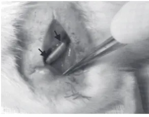
Rats were anesthetized and after skin incision, the sci-atic nerve was exposed using a muscle splitting incision. Under an operating microscope the left sciatic nerve was exposed at the mid-thigh, and a 10 mm segment of the nerve was removed. A 12 mm silicone tube (1 mm
in-ner diameter, 2 mm outer diameter) was interposed into this nerve gap (Y. S. Chen et al., 2000). Both proximal and distal ends of the nerve were anchored into the con-duit with 10-0 nylon suture (Fig.5). The silicone tube in the BMSCs group was filled with fibrin gel seeded with about 500,000 BMSCs, in the SCs group was filled with fibrin gel seeded with about 500,000 SCs, in the co-transplantation group was filled with fibrin gel seeded with about 250,000 BMSCs and 250,000 SCs; and the control group with fibrin gel without any cell. Finally the skin was sutured with 5-0 silk.
2.4. Electromyography (EMG) Study
Twelve weeks after the transplantation, rats were anes-thetized and the sciatic nerves were exposed. Electric stimulation was utilized to the proximal site of the in-jured nerve. The compound muscle action potential was recorded in the gastrocnemius with a needle electrode and a reference cap electrode inserted at the knee joint. The stainless steel needle used as the ground elec-trode was inserted into the tail skin (Chen et al., 2007; Mimura, Dezawa, Kanno, Sawada, & Yamamoto, 2004; Zarbakhsh et al., 2012).
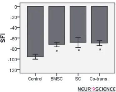
2.5. Functional Assessment
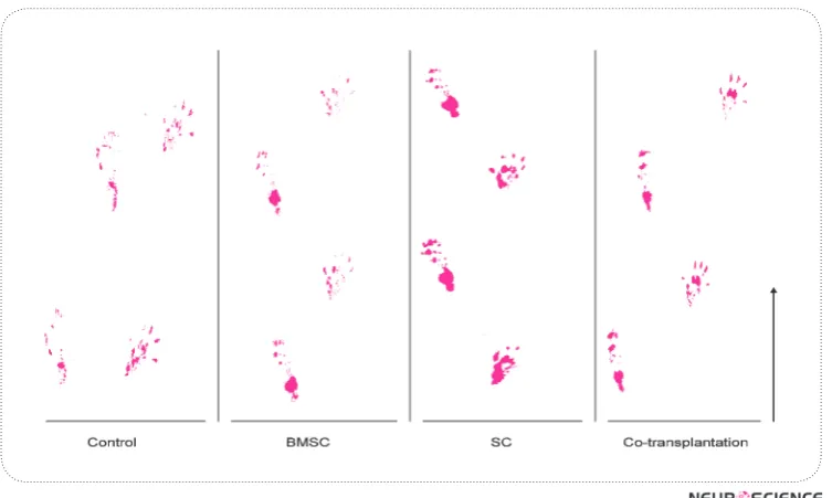
The functional assessment of the sciatic nerve regen-eration was expressed by the sciatic function index (SFI). Briefly, twelve weeks after the transplantation, rats hind feet were dipped in ink and the rats were al-lowed to walk through a plastic tunnel so that the foot-prints could be recorded on paper loaded onto the bot-tom of the tunnel. The distance among the fingers, toes
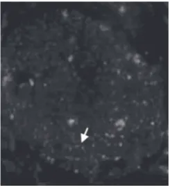
Figure 2. Cross-section of the sci-atic nerve, 4 weeks after the trans-plantation of BMSCs labeled with anti BrdU antibody shown as red spots in the sciatic nerve (arrow) (×100).
and heels was measured. The SFI was calculated as fol-low: SFI= -38.3×(EPL-NPL)/NPL+109.5×(ETS-NTS)/ NTS+13.3×(EITS-NITS)/NITS-8.8. In general, the SFI oscillates around 0 for normal nerve function, whereas around -100, the SFI indicates a total dysfunction (Chen et al., 2007; Mimura et al., 2004; Zarbakhsh et al., 2012).
2.6. Statistical Analyses
All data were analyzed by one-way analysis of vari-ance (ANOVA) followed by the Tukey test. Obtained data were presented as means ± standard error and a level of p<0.05 was considered statistically significant.
3. Results
3.1. BMSCs Culture
BMSCs obtained from the femurs and tibias of adult rats comprised heterogeneous groups of cells after seed-ing and growseed-ing in culture plates. After initial platseed-ing,
the adherent cells exhibited a small rounded-shape, a spindle-shape or a large flattened morphology (Fig.1A). Most cells grew and exhibited a fibroblast-like morphol-ogy on reaching confluence. The small rounded cells adhered to the surface of these cell layers (Fig.1B). These rounded cells disappeared after repeated passages, whereas the fibroblast-like cells became enriched. Upon the 4th passage, the fibroblast-like cells became mor-phologically homogeneous (Fig.1C).
3.2. Labeled BMSCs in the Sciatic Nerve Tissue
Immunohistochemistry technique showed the BMSCs labeled with the anti-BrdU antibody were red spots in the cross section of the sciatic nerve (Fig.2). Presence of the red color was due to the use of rhodamine as the secondary antibody. Our results confirmed not only the the presence but the viability of the transplanted cells in the silicone tube bridging the gap 4 weeks after the transplantation.
Figure 4. Cross-sections of the sciatic nerve 4 weeks after the transplantation of Schwann cells labeled with DiI shown as red spots in the sciatic nerve (arrow) (×100).
Figure 5. Nerve graft. The left sciatic nerve was exposed at the mid-thigh and a 10 mm segment of the nerve was re-moved. A 12 mm silicone tube was interposed into this nerve gap. Arrows show two heads of the nerve in the sili-cone tube.
Figure 7. The results of electromyography tests of amplitude and latency showed there were statistically significant differences between the control and experimental groups (BMSCs, SCs and co-transplantation of BMSCs and SCs)* and SCs transplanta-tionresulted in a more favorable results compared to the other groups (*p<0.05).
Figure 8. Foot print of control, BMSCs, SCs and co-transplantation groups 12 weeks after surgery. Arrow shows walking direction.
3.3. SCs Cultured
The spindle-shaped cellular morphology of the SCs seen on the culture plate was viable. Most of the cells were small, elongated, and spindle shaped (Fig.3A). Fluorescence microscopy showed the SCs were S100-positive cells. In the culture dishes, the SCs had a ten-dency to line up side by side or end to end and to form interconnected networks (Fig.3B).
3.4. Labeled SCs in the Sciatic Nerve Tissue
Histochemistry technique showed the SCs labeled with DiI as red-positive spots in the cross section of sciatic nerve (Fig.4). The results confirmed the presence and vi-ability of the transplanted cells in the silicone tube bridg-ing the gap 4 weeks after the transplantation.
3.5. Electromyography (EMG)
The results of the EMG tests comprised both amplitude and latency measures. The time calibration bar was 2 milliseconds (ms) and the amplitude calibration bar was 10 millivolts (mV). The stimulation intensity was 2.3 milliampere (mA) and the duration was 0.1 ms (Fig.6).
3.6. Functional Analysis
The results of SFI showed a statistically significant dif-ference between the control and experimental groups (BMSCs, SCs and co-transplantation), however, no sta-tistically significant difference was observed amongst the experimental groups (p<0.05) (Fig.8, 9).
4. Discussion
Peripheral nerve injury is considered as one of the most challenging microsurgical problems. These damages may lead to a considerable disability due to the loss of both motor and sensory functions. Since the auto graft procedure involves multiple surgeries, loss of function, and loss of sensation at the donor site, development of a high quality replacement for auto grafts is required (Belkas et al., 2004; Dezawa, 2005; Ishikawa et al., 2009).
In the previous study we compared the effects of trans-plantation of BMSCs and SCs on the recovery of rats’ sciatic nerve injury. We also showed that BMSCs and SCs can be effective by their own and SCs were signifi-cantly more effective than the BMSCs (Zarbakhsh et al., 2012). In this study, we compared the effects of the sole transplantation of BMSCs, SCs and the co-transplanta-tion of these cells on the recovery of rats’ sciatic nerve injury under similar condition. Several investigators have shown that BMSCs and SCs can repair the sciatic nerve injuries (Braga-Silva et al., 2006; C. J. Chen et al., 2007; Lu et al., 2006; Schlosshauer et al., 2003), howev-er, BMSCs and SCs have not been used in combination for the same purpose. Comparing the effects these cells’ co-transplantation (BMSCs and SCs) with the effect of their sole transplantation on the recovery of the injured
peripheral nerve under similar conditions may introduce a novel clinical approache in utilizing these cells to re-place peripheral nerve recovery auto grafts.
Due to the benefits of stem cells and Schwann cells in producing nerve growth factors such as NGF, BDNF and VEGF, as well as substantiating extracellular matrix proteins such as collagen IV and laminin (Braga-Silva et al., 2006; C. J. Chen et al., 2007; Fan et al., 2011; Feng, Zhou, Rush, & Ferguson, 2008; Ide, 1996), there is good evidence to support the hypothesis that transplantation of BMSCs and SCs may repair peripheral nerve injuries. Moreover, using the co-transplant of these cells may pos-sibly be an important step in the selection of a repair pro-cedure based on the peripheral nerve recovery measures.
showed there were statistically significant differences between the control and experimental groups. These re-sults are in agreement with findings of other investiga-tions (C. J. Chen et al., 2007; Hou, Zhang, Quan, Liu, & Zhu, 2006; Kim, Lee, & Lee, 2007; Nie et al., 2007), while the lack of any statistically significant difference amongst the experimental groups is a new finding and has not been previously addressed under similar condi-tions. Presumably after 12 weeks, due to the growing ax-ons, a significance difference was observed in the results of EMG amongst the experimental groups, while myelin formation was not completed. Accordingly, there was no statistically significant difference amongst the experi-mental groups with regard to the SFI results. Neverthe-less, we might possibly have observed a significance dif-ference in the results of SFI amongst the experimental groups if waited longer.
Our results suggested that the sole- or co-transplanta-tion of BMSCs and SCs have the potential to generate functional recovery of rats’ injured sciatic nerve. EMG evaluation revealed that the SCs transplantation results in a greater functional improvement in the injured nerve as compared to BMSCs transplantation or co-transplan-tation of BMSCs and SCs. Our findings support use of SCs transplant as opposed to BMSCs or combination of BMSCs and SCs transplant for the clinical repair of the injured peripheral nerves.
References
Al, J., Epstein, P. N., Gozal, D., Yang, B., Wurster, R., & Cheng, Z., J. (2007). Morphology and topography of nucleus am-biguus projections to cardiac ganglia in rats and mice. Neu-roscience, 149, 845-860.
Azizi, S. A., Stokes, D., Augelli, B. J., Digirolamo, C., & Prockop, D. J. (1998). Engraftment, and migration of human bone mar-row stromal cells implanted in the brains of albino rats simi-larities to astrocytes grafts. Proc Natl Acad Sci USA, 95(7), 3908-3913.
Bakhtyari, M., Abootaleb, H., & Mansouri, K. (2009). Electro-physiological study of sciatic nerve regeneration through tubes seeded with schwann cells. Iranian Journal of Neuro-science, 1(3), 49-56.
Belkas, J. S., Shoichet, M. S., & Midha, R. (2004). Peripheral nerve regeneration through tubes. Neural Res, 26(2), 151-160.
Braga-Silva, J., Gehlen, D., Roman, J. A., Menta, C., Atkinson, E. A., Machado, D. C., et al. (2006). Bone marrow stem cells and platelet-rich plasma effects on nervous regeneration and functional recovery in an acute defect model of rats` periph-eral nerve. Acta Ortop Bras, 14(5), 273-275.
Chen, C. J., Ou, Y. C., Liao, S. L., Chen, W. Y., Chen, S. Y., Wu, C. W., et al. (2007). Transplantation of bone marrow stromal cells for peripheral nerve repair. Exp. Neurology, 204(1), 443-453.
Chen, Y. S., Hsieh, C. L., Tasi, C. C., Chen, T. H., Cheng, W. C., Hu, C. L., et al. (2000). Peripheral nerve regeneration using silicone rubber chambers filled with collagen, laminin and fibronectin. Biomaterials, 21(15), 1541-1547.
Dezawa, M. (2005). Future views and challengges to the pe-ripheral nerve regeneration by cell based therapy. Rinsho Shinkeigaku (Japanes), 45(11), 877-879.
Fan, W., Crawford, R., & Xiao, Y. (2011). The ratio of VEGF/ PEDF expression in bone marrow mesenchymal stem cells regulates neovascularization. Differentiation, 81(3), 181-191.
Feng, S. Q., Zhou, X. F., Rush, R. A., & Ferguson, I. A. (2008). Graft of pre-injured sural nerve promotes regeneration of corticospinal tract, and functional recovery in rats with chronic spinal cord injury. Brain Res, 1209, 40-48.
Haastert, K., Lipokatic, E., Fischer, M., Timmer, M., & Grothe, C. (2006). Differentially promoted peripheral nerve regenera-tion by grafted Schwann cells over-expressing different FGF-2 isoforms. Neurobiology of Disease, FGF-21(1), 138-153.
Hou, S. Y., Zhang, H. Y., Quan, D. P., Liu, X. L., & Zhu, J. K. (2006). Tissue engineered peripheral nerve grafting by dif-ferentiated bone marrow stromal cells. Neuroscience, 140(1), 101-110.
Ide, C. (1996). Peripheral nerve regeneration. Neurosci. Res., 25(2), 101-121.
Ishikawa, N., Suzuki, Y., Dezawa, M., Kataoka, K., Ohta, M., Cho, H., et al. (2009). Peripheral nerve regeneration by trans-plantation of BMSC-derived schwann cells as chitosan gel sponge scaffolds. Willey Periodicals, Inc. J Biomed Mater Res., 89(4), 1118-1124.
Kim, S. M., Lee, S. K., & Lee, J. H. (2007). Peripheral nerve regen-eration using a three dimensionally cultured Schwann cell conduit. The Journal of Craniofacial Surgery, 18(3), 475-488.
Li, P., Akimoto, T., Zhang, M., Williams, R. S., & Yan, Z. (2006). Resident stem cells are not required for exercise-induced fiber-type switching and angiogenesis but are necessary for activity-dependent muscle growth. Am J Physiol Cell Phys-iol, 290(6), 1461-8.
Li, N., Yang, H., Lu, L., Duan, C., Zhao, C., & Zhao, H. (2008). Comparison of the labeling efficiency of BrdU, DiI and FISH labeling techniques in bone marrow stromal cells. Brain Re-search, 1215, 11-19.
Liao, G., Wu, F. Y., & Hayward, S. D. (2001). Interaction with the Epstein-Barr virus helicase target Zta to DNA replication compartments. J Virol, 75(18), 8792-802.
Millesi, H. (1984). Nerve grafting. Clin Plast Surg 11(1), 105-113.
Mimura, T., Dezawa, M., Kanno, H., Sawada, H., & Yamamoto, I. (2004). Peripheral nerve regeneration by transplantation of bone marrow stromal cell-derived Schwann cells in adult rats. J. Neurosurg, 101(5), 806-812.
Murakami, T., Fujimoto, Y., Yasunaga, Y., Ishida, O., Tanaka, N., Ikuta, Y., et al. (2003). Transplanted neuronal progenitor cells in a peripheral nerve gap promote nerve repair. Brain Res, 974(1-2), 17-24.
Nie, X., Zhang, Y. J., Tian, W. D., Jiang, M., Dong, R., Chen, J. W., et al. (2007). Improvement of peripheral nerve regenera-tion by a tissue-engineered nerve filled with ectomesenchy-mal stem cells. Int. J. Oral Maxillofac. Surg., 36(1), 32-38.
Pourheydar, B., Joghataei, M. T., Bakhtiari, M., Mehdizadeh, M., Yekta, Z., & Najafzadeh, N. (2012). Co-transplantation of bone marrow stromal cells with schwann cells evokes me-chanical allodynia in the contusion model of spinal cord in-jury in rat. Cell Journal (yakhteh), 13(4), 213-222.
Rodriguez, F. J., Verdu, E., Ceballos, D., & Navarro, X. (2000). Nerve guides seeded with autologous Schwann cells im-prove nerve regeneration. Exp. Neurology, 161(2), 571-584.
Schlosshauer, B., Muller, E., Schroder, B., Planck, H., & Muller, H. (2003). Rat schwann cells in bioresorbable nerve guides to promote, and accelerate axonal regeneration. Brain Res, 963(1-2), 321-326.
Wang, D., Liu, X. L., Zhu, J. K., Jiang, L., Hu, J., Zhang, Y. J., et al. (2008). Bridging small-gap peripheral nerve defects us-ing acellular nerve allograft implanted with autologous bone marrow stromal cells in primates. Brain Res, 1188, 44-53.
Zarbakhsh, S., Bakhtiari, M., Faghihi, A., Joghataie, M. T., Mehdizadeh, M., Khoei, S., et al. (2012). The effects of Schwann and bone marrow stromal stem cells on sciatic nerve injury in rat: A comparison of functional recovery. Cell Journal(Yakhteh) 14(1), 39-46.
Zurita, M., Bonilla, C., Otero, L., Aguayo, C., & Vaquero, J. (2008). Neural transdifferentiation of bone marrow stromal cells obtained by chemical agents is a short-time reversible phenomenon. Neuroscience Research, 60(3), 275-280.
Antiepileptic and Antioxidant Effect of Hydroalcoholic
Extract of Ferula Assa Foetida Gum on
Pentylentetrazole-induced Kindling in Male Mice
Zahra Kiasalari1, Mohsen Khalili1*, Mehrdad Roghani1, Hamid Heidari2, Yaser Azizi3
1. Neurophysiology Research Centre, Shahed University, Tehran, Iran.
2. Department of Physiology. Faculty of Medical Sciences, Ahvaz Jundishapur University of Medical Sciences, Ahvaz; Iran. 3. Department of Physiology. Tehran University of Medical Sciences, Tehran Iran.
* Corresponding Author:
Mohsen Khalili , PhD
Neurophysiology Research Centre, Shahed University, Tehran, Iran. Tel: +98 21 88963849
E-mail: [email protected]
Introduction: Considering the prevalence of epilepsy and the failure of available treatments for many epileptic patients, finding more effective drugs in the treatment of epilepsy seems necessary. Oxidative stress has a special role in the pathogenesis of epileptic syndrome. Therefore, in the present study, we have examined the anti-epileptic and anti-oxidant properties of the Ferula Assa Foetida gum extract, using the pentylentetrazole (PTZ) kindling method. In this experimental study, sixty male Albino mice weighing 25-30 g were selected and were randomly divided into 6 groups. 1- the control group, 2- PTZ-kindled mice, 3- positive control group which received valproate (100 mg/kg) as anti-convulsant drug, 4-5 & 6- the groups of kindled mice that pretreated with 25, 50 and 100 mg/kg doses of Ferula Assa Foetida gum extract.
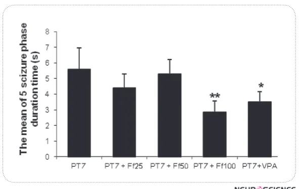
Methods: Kindling has been induced in all groups, except for the control group via 11 PTZ injections (35 mg /kg; ip) every other day for 22 days. In the 24th day, the PTZ challenge dose was injected (75 mg / kg) to all groups except the control group. The intensity of seizures were observed and noted until 30 minutes after PTZ injection. At list, the mice were decapitated and the brains of all the mice were removed.. and their biochemical factors levels including malondialdehyde (MDA), superoxide dismutase (SOD) and nitric oxide (NO) were determined. Results: Results of this study show that Ferula Assa Foetida gum extract is able to reduce seizure duration and its intensity. In addition, this extract has reduced MDA and NO levels and increased the level of SOD in the brain tissue compared to the PTZ- kindled mice.
Discussion: It can be concluded that Ferula Assa Foetida gum extract, in specific doses, is able to show an anti-epileptic effect because of its antioxidant properties, probably acting through an enzyme activity mechanism.
A B S T R A C T
Article info:
Received: 20 January 2013 First Revision: 26 February 2013 Accepted: 22 May 2013
Key Words:
Ferula Assa Foetida, Epilepsy,
PTZ, Nitric Oxide,
Superoxide Dismutase, Malondialdehyde.
1. Introduction
pilepsy is one of the most serious neurologi-cal disorders, which is induced by a sudden increment of stimulatory factors in the cor-tical neurons. About 0.5-3% of people ex-perience it during their lifetime (Theodore & Fisher, 2007; Levav, Stephenson & Theodore, 1999). Epilepsy refers to a functional disorder of the brain with