Original Article
Computed tomogram image encoding for
internet of things based systems
Ali Akbar Siddique1, M. Tahir Qadri2 & Zia Mohy-Ud-din3
1Department of Telecommunication, Sir Syed University of Engineering & Technology
2Department of Electronics, Sir Syed University of Engineering & Technology
3Department of Biomedical, Sir Syed University of Engineering & Technology
Abstract
Background: In recent times, transmission of information over a wireless channel is increasing at an exponential rate. Applications based on IOT will exceed all form of data available over the internet, it can be in the form of data based on videos and images which alone size up to 70% of the global traffic.
Methodology: In this paper, an encoding technique based on discrete cosine transform (DCT) have been utilized to reduce information of a CT image that is unwanted or a human eye is unable to perceive. For medical images, preserving maximum information in order to properly diagnose any disease is important and for such application, the amount of quantization needed to gain an image with minimum error is to observe the Correlation index (CI).
Results: CI at 50% Quality Quantization Table (QQT) for image 1 was found to be 0.9981 and its size reduced to 0.403 MB, it means that the compressed image is 99.8% similar to the original image. But its similarity reduces with the increase in QQT, its size will also decrease by at this point quality will degrade too.
Conclusion: In order to store images on the drive, it is imperative to apply a compression technique. In this paper, a compression technique is proposed utilizing a DCT algorithm to reduce an image size with different QQT provided for quality assessment. Each image has a different set of information and to cater their differences this QQT can be manipulated for best possible quality with acceptable size on disk.
Keywords
Internet of Things, Image compression, Discrete Cosine Transform, Quantization, Encoding.
DOI:10.29052/IJEHSR.v6.i4.2018.09-19 Corresponding Author Email:
[email protected] & [email protected] Received 04/06/2018
Introduction
Daily huge amount of information is transferred or uploaded from different sensory sources including cameras, sensors or any other equipment. If the data needs to be transmitted to a server location containing a data hub for multiple user interfaces, such a process can be considered to be IOT1. For medical applications such as CT images, produced a bulk amount of information that needs to be stored and transmitted at a very high rate. CT creates detailed images that provide information inside the body including organs and even bones and blood vessels2.
Due to the sample size, some hurdles might occur during transmission such as lack of storage capacity at the data storage hub, high bandwidth utilization and also time consumption for uploading information to the main server. In some hospitals, it is required to keep the record of the CT scan for about 10 years, hence a huge amount of storage capacity is required but this can be avoided if the size of those images were to reduce by applying efficient compression algorithm without compromising its overall quality3. However, in some areas of medical imaging, it is required to maintain a sufficient amount of image quality for better analysis.
Many researchers have implemented adaptive algorithms using lossy compression techniques in order to reduce the image size without degrading the overall quality4. Lossless techniques for compression using differential transfer was proposed by Ridenour and his colleagues5. Legendre polynomial was also used for lossy and lossless compressions6. Compression is performed by utilizing the DCT algorithm to distinguish between the Low and high-frequency componnts7-12. The process of noise filtration by implementing DCT is also utilized13. Discrete Wavelet
Transform (DWT) implemented with DCT in satellite images for compression14&15.
DCT is also implemented using a grayscale images16. Compression ratio is a technique that is used to determine the amount of information reduced using DCT and MSVQ, this information stack up to the space required to store an image. Its size varies according to its resolution and using DCT, it is possible to determine the level of compression that can be performed on an image17. Adaptive Huffman coding for lossless compression was introduced in order to store image information without using the concept of quantization which may lead to a distorted or corrupted image if over compressed18. Compression is a process of reducing information and has been used in the field of medicine for many years 19-23.
In the current study, a process of compression algorithm is implemented based on DCT in order to reduce the unwanted information that a human eye is unable to perceive, these components are basically high-frequency components and a human eye can only perceive low-frequency components. For lossy compression, the process quantization was utilized to eliminate these high-frequency components and then apply the basic Huffman lossless encoding algorithm to produce a binary bit stream for transmission, this bit stream is transmitted to the main server and also to the data hub for storage.
Methodology
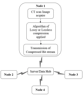
Block Representation of an IOT System
allocated channel. Allocated bandwidth is of 18 Mbps (megabits per second), and sometimes images captured or acquired are large enough to surpass this it. In such application, compression becomes necessary in order to transmit that information.
The main server is embedded with a decoder to decode an incoming bit stream and store the image information for future use. In a system based on IOT possess multiple nodes for such purpose to store and transmit information.
Figure 1: Block diagram of Internet of Things (IOT) system
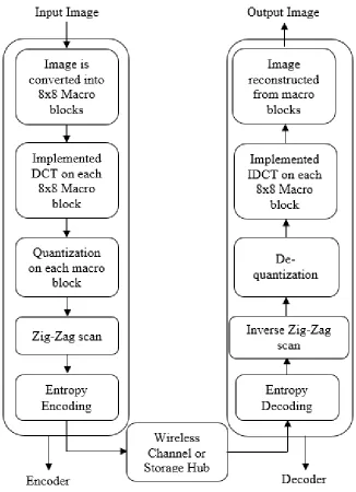
Block Representation of DCT Based Compression
Figure 2: Encoder and Decoder Block Representation
Figure 3: Macro Block of 64 Pixels
Equation 1 represents the 2D-DCT, it is applied to each 8x8 macro blocks present in an entire image. There are two different components present in the macro block low-frequency components and high-frequency components. As we know that low-high-frequency components are the ones that we can perceive and AC components like high frequency, the human eye is unable to perceive. The purpose of DCT is not to remove the frequency components but to distinguish between the high and low-frequency components. After DCT, the process of quantization is applied to make the high-frequency values close to zero. So in order to remove these high-frequency components we employ quantization. Joint Photographic Expert Group (JPEG) used different forms of 8x8 quantization table, it is a well-known fact that quantization is actually the lossy part in the whole encoding process, so we can tinker with the quality of encoding by utilizing the different quantization matrix.
𝐹(𝑢, 𝑣) =1
4𝐶𝑢𝐶𝑣∑ ∑ 𝑃(𝑥,𝑦)cos (𝑢𝜋
2𝑥+1
2𝑁 ) cos (𝑣𝜋
2𝑦+1
2𝑁 )
𝑁−1 𝑥=0 𝑁−1
𝑦=0 (1)
Where,
Cu and Cv = { 1
√2 if u = 0 1 other wise}
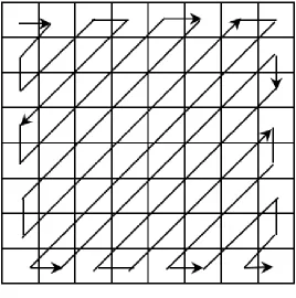
Figure 4: Zig-Zag scanning for data serialization
A zig-zag process is then applied on the macro block as shown in figure 4 to serialize its data through which we acquire a lot of zeros which can easily be coded using entropy coding. Huffman coding is utilized to convert the data into a binary bit stream which can be transmitted over a wireless channel or can be stored in the data hub.
𝑀𝑆𝐸 = √ 1
𝑛×𝑚∑(𝐾̂ − 𝐾)
2 (2)
Same for the process of encoding, if we are to retrieve the encoded bitstream stored in the data hub that information is then passed through the decoder which is literally the reverse process of the encoder. And finally we acquire an image back from the storage hub or from a wireless channel that is almost as same as the original input image with minimum Mean Squared Error (MSE) as given in equation 2, where n×m are the number of pixels in rows and column, K ̂ is compressed image and K is an original image.
Results
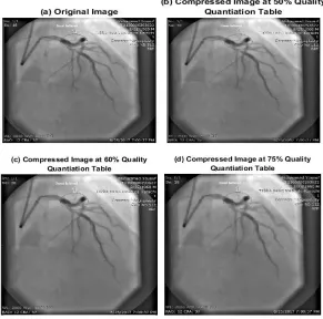
Two distinct images of coronary angiography were utilized for implementing compression algorithm. Figure 5a is an original image 1 and contains only luminance or intensity (grayscale) contents. Figure 5b represents the image quality compressed using 50% quality quantization
Figure 5(a-d): CT scan images for the original and compressed version of the implemented QQT in Percentage.
Correlation index at 50% quality quantization table (QQT) was 0.9981, which points the fact that the original image and compressed image at 50% (QQT) has a similarity index of 99.81%. Figure 5c and 5d were taken using 60% and 75% (QQT), their sizes further reduces to 0.31 Mb and 0.194 MB respectively at the cost of their quality but there is no drastic quality degradation, it is still visually not possible to distinguish between them as they retained 91.02% and 80.11% of their previous traits.
Table 1: Image sizes after different level of compression with Quality Quantization Tables (QQT) provided by JPEG
Input Image Size of an Image in MB
Actual
Size (QQT) 50% (QQT) 60% (QQT) 75%
Original Image 1 (Figure 4) 1.24 0.403 0.31 0.194
Original Image 2 (Figure 5) 1.06 0.399 0.287 0.127
* QTT= Quality Quantization Tables
Figure 6: QQT in percentage for the different images with similarity Index.
Figure 6a represents the original image 2 of coronary angiography, and the rest of the Figures-6b, 6c and 6d are the images displaying the (QQT) at 50%, 60% and 75%. Here these images also retained most of their previous traits as their correlation index or similarity index is 99.65, 90.07 and 79.07 respectively. If we eliminate the step of quantization from this process this type of compression will become lossless. In some cases, we require a lossless image for medical application, so we skip the quantization step to acquire original coded information at the decoding end. Each image is encoded in a binary bit stream using entropy coding.
Table 2. Image quality in terms of Correlation Index at a different level of compression with Quality Quantization Tables (QQT) provided by JPEG
Input Image Quality of Image in terms of Correlation Index
50%
(QQT) (QQT) 60% (QQT) 75%
Original Image 1 (Figure 4) 0.9981 0.9102 0.8011
Original Image 2 (Figure 5) 0.9965 0.9007 0.7907
Discussion
In the field of image compression, it is imperative to acquire a reconstructed image with maximum similarity index. It means that for that naked eye a viewer may not be able to distinguish between them and in order to achieve that, several algorithms such as DWT and DCT are utilized to further distinguish between the high and low-frequency components.
Bruylants and colleagues implemented a compression algorithm to reduce medical image size using Haar Discrete Wavelet Transform (DWT) as it supported by the Digital Imaging and Communications in Medicine (DICOM) image24. They utilized (JP3D) codec to tackle information handling and also with a directional wavelet transform. They achieved the result with improved accuracy of 10% in bit rate reduction, which certainly improves the transmission factor by the same amount. They utilized the algorithm on different medical images but due to the fact that it may be possible for some images to have the same directional features thus the process may not work on such images 24.
Pingzhang Zhou and colleges displayed the reducing of density based topology optimization using DCT23. They discussed the information stored in an image is composed of high and low-frequency components and only low-frequency components are needed for the optimization in order to attain high resolution. By using such technique they suggested that there is no need to apply additional filters such as density or sensitivity filters as the high-frequency
In this work same approach is applied using DCT transmit or to store the compressed image. Quality varies with a different set of images used but it is possible to regain the compressed information with minimum error using the best possible QQT. Correlation index obtained is given in table 2 representing the similarity index of the two distinct images on which the proposed algorithm is implemented. It is mandatory to retrieve every bit of information when it comes to a medical image, hence the size of an image increase exponentially. It can be reduced by selecting the best possible QQT for the image that can easily retrieve information with maximum similarity index. Each image varies from one another and in order to tackle these image, it is possible to vary QQT according to the image to achieve maximum correlation index.
Although, it is possible for an image with high contrast and sudden changes in the pixel density to lose its information at an extended scale as compared with the ones with less change in luma intensity. It is true that low-frequency components are needed to attain almost every possible information of an image that resembles the original and thus the quantization table is utilized to eliminate the high concentration of high components and reduce them to zero. For choosing a QQT for the purpose of compression, it must be kept in mind that not every reconstructed image after the process of compression will retain all of its previous traits.
Conclusion
retrieved. In our proposed model we can reduce the size of the acquired medical image and transmit it to the server using IOT. Stored information will be in the form of the binary bit stream that can be retrieved at any time if needed. Many medical institutes require their patient information and CT images to be stored for at least ten years, which may require a lot of storage capacity, so in this regard, we can compress these images to a level that they are visually indistinguishable in order to occupy less storage capacity. Correlation between the original image and a compressed image is almost unity which points the fact that is almost identical. Size of an image reduces at least three times without losing is overall quality. This will indeed reduce the time and space required to send the concerned doctor the patient information in image form with the quality that almost resembles the original. For storage purpose, it will occupy less space as compared to the original image and also preserve its overall quality. This information can be transmitted to the main server for future reference. We identified that this process can be implemented in multiple medical institutes where CT machines are present and information can directly be delivered to their doctor in an instant if needed using IOT.
Conflicts of Interest
None.Acknowledgement
We would like to acknowledge the institute & fellows of Sir Syed University of Engineering & Technology for their support.
Funding
None.References
1. Kopetz H. Internet of things. In Real-time systems. Boston: Springer, MA; 2011. 307-323.
2. Brenner DJ, Hall EJ. Computed
tomography—an increasing source of radiation exposure. N Engl J Med. 2007; 357(22):2277-2284.
3. European Society of Radiology 2009. The future role of radiology in healthcare. Insights Imaging. 2010; 1(1):2-11.
4. Johnston N, Vincent D, Minnen D, Covell M, Singh S, Chinen T, Hwang SJ, Shor J, Toderici G. Improved lossy image compression with priming and spatially adaptive bit rates for recurrent networks. Structure. 2017; 10: 23.
5. Ridenour RL, Frederiksen JE, Hendry IC, inventors; Apple Inc, assignee. Lossless image compression using differential transfer. United States patent US 9,386,318. 2016. 2018.
6. Goel N, Gabarda S. Lossy and lossless image compression using Legendre polynomials. In Advances in Signal Processing (CASP), Conference 2016;
Pune, India: IEEE; 2016. pp. 315-320. 7. Raid AM, Khedr WM, El-Dosuky MA,
Ahmed W. Jpeg image compression using discrete cosine transform-A survey. arXiv preprint arXiv:1405.6147. 2014
8. Pandey DS, Singh MP, Pandey V. Block wise image compression & Reduced Blocks
Artifacts Using Discrete Cosine
Transform. Int J Sci Res Publications. 2015; 5(3):1-10.
9. Zhou X, Bai Y, Wang C. Image
compression based on discrete cosine
transform and multistage vector
quantization. Int J Multimedia Ubiquitous Eng. 2015; 10(6):347-356.
cosine transform (DCT) compression technique on low level features of image for image retrieval system. J Fundam Appl Sci. 2017; 9(3):11-27.
11. Amiri SA, Hassanpour H, Marouzi OR. No-reference image quality assessment based on localized discrete cosine transform for JPEG compressed images. Multimedia Tools and Applications. 2018; 77(1):787-803.
12. Hasan TS. Image Compression Using Discrete Wavelet Transform And Discrete Cosine Transform. J. appl. sci. res. 2017; 13(3):1-8.
13. Zhao Y, Belkasim S. Image Compression and Denoising Algorithm based on Multi-resolution Discrete Cosine Transform. In
Proceedings of the International
Conference on Image Processing,
Computer Vision, and Pattern Recognition (IPCV) 2016; Monte Carlo Resort, Las Vegas, USA: 2016. pp. 110.
14. Shihab HS, Shafie S, Ramli AR, Ahmad F.
Enhancement of Satellite Image
Compression Using a Hybrid (DWT– DCT) Algorithm. Sensing and Imaging. 2017; 18(1):30.
15. Kumar G, Bhatia PK. Empirical analysis of image compression using wavelets, discrete cosine transform and neural network. In Computing for Sustainable Global Development (INDIACom), In 3rd International Conference 2016; New Delhi, India: IEEE; 2016. pp. 3862-3866. Available from: IEEE Xplore.
16. Landge MA, Bagal MS, Lichade MS. Grayscale Image Compression using Discrete Cosine Transform. IJEDR, 2016; 4(3): 1359-1366.
17. Krishna V, Rao VP, Naresh P,
Compression Ratio of an image. Int Res J Engineering Tech, 2016; 3(3):686-689. 18. Jain DK, Gaur D, Gaur K, Jain N. Image
compression using discrete cosine
transform and adaptive Huffman coding. Int J Emer T Tech Com Sci. 2014; 3(1): 90-94.
19. Bui V, Chang LC, Li D, Hsu LY, Chen MY. Comparison of lossless video and image compression codecs for medical computed tomography datasets. In Big Data (Big Data), 2016 IEEE International Conference; Washington, DC, USA: IEEE; 2016. pp. 3960-3962. Available from: IEEE Xplore.
20. Vaishnav M, Kamargaonkar C, Sharma MM. Medical Image Compression Using Dual Tree Complex Wavelet Transform and Arithmetic Coding Technique. Int J Scientific Res Comp Sci Eng Info Tech. 2017; 2(3): 172-176.
21. Jinimole CG, Harsha A. Comparative Study of Different Enhancement Techniques for Computed Tomography Images. World Academy of Science, Engineering and Technology, Int J Med Hea Biomed, Bioeng Pharma Engineering. 2017; 11(9):524-527.
22. Jindal R. A Review on Recent
Development of Image Compression Techniques. Int J Adv Res Development. 2017; 1(1):1-10.
23. Zhou P, Du J, Lü Z. Highly efficient density-based topology optimization using DCT-based digital image compression. Struc Multidisciplinary Optim. 2018; 57(1):463-467.