R E S E A R C H A R T I C L E
Open Access
Comparison of sterols and fatty acids in two
species of
Ganoderma
Guang-ping Lv
1,2, Jing Zhao
1,2*, Jin-ao Duan
3, Yu-ping Tang
3and Shao-ping Li
1,2*Abstract
Background:Two species of Ganoderma, G. sinenseandG. lucidum, are used asLingzhi in China. Howerver, the content of triterpenoids and polysaccharides, main actives compounds, are significant different, though the extracts of bothG. lucidumandG. sinensehave antitumoral proliferation effect. It is suspected that other compounds contribute to their antitumoral activity. Sterols and fatty acids have obvious bioactivity. Therefore, determination and comparison of sterols and fatty acids is helpful to elucidate the active components ofLingzhi.
Results:Ergosterol, a specific component of fungal cell membrane, was rich inG. lucidumandG. sinense. But its content inG. lucidum(median content 705.0μg·g-1, range 189.1-1453.3μg·g-1, n = 19) was much higher than that inG. sinense(median content 80.1μg·g-1, range 16.0-409.8 μg·g-1, n = 13). Hierarchical clustering analysis based on the content of ergosterol showed that 32 tested samples ofGanodermawere grouped into two main clusters,G. lucidumandG. sinense. Hierarchical clustering analysis based on the contents of ten fatty acids showed that two species ofGanodermahad no significant difference though two groups were also obtained. The similarity of two species ofGanodermain fatty acids may be related to their antitumoral proliferation effect.
Conclusions:The content of ergosterol is much higher inG. lucidumthan inG. sinense. Palmitic acid, linoleic acid, oleic acid, stearic acid are main fatty acids inGanodermaand their content had no significant difference between G. lucidumandG. sinense, which may contribute to their antitumoral proliferation effect.
Background
Ganoderma (Lingzhi in Chinese) is a genus of well-known medicinal mushrooms with multiple benefits to human health. So far, more than 120 species of Gano-derma have been reported in the world, 98 species of which could be found in China [1]. However, only two species ofGanoderma (Figure 1),G. lucidum (Leyss.ex Fr.) Karst. andG. sinenseZhao, Xu et Zhang, are docu-mented asLingzhiin Chinese Pharmacopoeia (2010) [2]. Modern studies have revealed that Lingzhi contain a variety of bioactive ingredients, including triterpenoids, polysaccharides, sterols, fatty acids, nucleosides and alkaloids [3], and possess multiple pharmacological activities, such as antitumor [1,4], immunomodulation [5,6], anti-inflammatory [7], antiviral [8], anti-aging [9] and anti-diabetic [10] effects. It is usually considered that triterpenoids and polysaccharides are the main
active components inLingzhi[3]. However, our previous study has revealed that the contents of triterpenes are significantly different betweenG. lucidumandG. sinense [11]. Actually, little or no triterpene was detected inG. sinense [1,11,12]. It is very interesting that ethanol extracts of bothG. lucidum andG. sinense, at the same cytotoxicity concentration, have similar antitumoral pro-liferation effect through both apoptosis pathway and cell cycle arrest effect [1], which indicates that some other compounds may contribute to their activity besides tri-terpenoids and polysaccharides.
To date, several sterols and fatty acids, which have sig-nificant bio-functions in fungi [13,14], were separated or identified fromGanoderma[7,8,15-17]. Ergosterol and its analogues contained inGanodermahad multiple pharma-cological effects such as anti-HIV-1 [7], anti-complement [8], anti-aging [9] activities, and protecting cerebral corti-cal neurons from hypoxia/reoxygenation injury [18]. On the other hand, free fatty acids (FFAs) are not only essen-tial nutritional ingredients but also significant to many cel-lular functions through receptors of FFAs [19]. Especially, * Correspondence: [email protected]; [email protected]
1
State Key Laboratory of Quality Research in Chinese Medicine, University of Macau, Macao SAR 999078, China
Full list of author information is available at the end of the article
long chain fatty acids in the spores ofG. lucidumcould triggers tumor cell death through induction of apoptosis [20]. Therefore, analysis of sterols and fatty acids in two species ofGanodermais not only important for evaluation of their quality but also beneficial to the elucidation of their pharmacological activities.
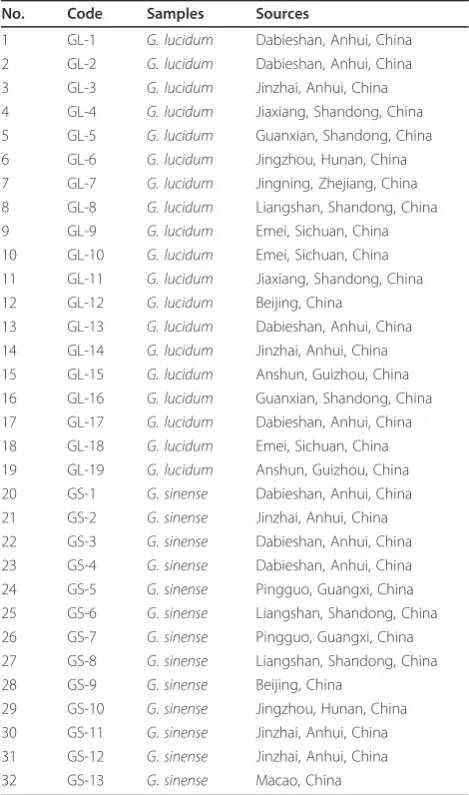
Up to date, TLC [21-23], HPLC [11,24-28], GC [16,29-31], MEKC [32] have been developed for analysis of triterpenoids [11,21-24,26,27], polysaccharides [25,28-31] and nucleosides [32] in Ganoderma. How-ever, few researches focus on the analysis of sterols or fatty acids in Ganoderma [15,16,33]. In present study, ergosterol and ten free fatty acids, including lauric acid (C12:0), myristic acid (C14:0), pentadecanoic acid (C15:0), palmitic acid (C16:0, PA), palmitoleic acid (C16:1), stearic acid (C18:0, SA), oleic acid (C18:1, OA), linoleic acid (C18:2, LoA), docosanoic acid (C22:0) and lignoceric acid (C24:0), in two species of Ganoderma were determined by using pressurized liquid extraction (PLE) and GC-MS analysis after one-step TMS derivati-zation. The contents of the free sterol and fatty acids in G. lucidumandG. sinensewere also compared.
Experimental
Materials and chemicals
Lauric acid (C12:0, dodecanoic acid), myristic acid (C14:0, tetradecanoic acid), pentadecanoic acid (C15:0), palmitic acid (C16:0, hexadecanoic acid), palmitoleic acid (C16:1, (Z)-hexadec-9-enoic acid), stearic acid (C18:0, octadecanoic acid), oleic acid (C18:1, (Z)-octa-dec-9-enoic acid), linoleic acid (C18:2, (9Z,12Z)-octa-deca-9,12-dienoic acid), docosanoic acid (C22:0), lignoceric acid (C24:0, tetracosanoic acid) and ergosterol were purchased from Sigma (St. Louis, MO, USA). Pet-roleum ether (60-90°C) andn-hexane were purchased from Merck (Darmstadt, Germany). Derivatization reagent BSTFA (N, O-bis (trimethylsilyl) trifluoroaceta-mide) containing 1% TMCS (trimethylchlorosilane) was purchased from Supelco (St. Louis, MO, USA). Reagents not mentioned here were from standard sources. The materials of Ganoderma were obtained from different locations in China (Table 1). Voucher specimens of
these samples were deposited at the Institute of Chinese Medical Sciences, University of Macau, Macao, China.
Sample preparation
Pressurized liquid extractions were performed on a Dio-nex ASE 200 (DioDio-nex Corp., Sunnyvale, CA, USA) sys-tem. In brief, 0.2 g powder of Ganodermawere mixed with diatomaceous earth in a proportion (1:1) and placed into an 11 ml stainless steel extraction cells, respectively. The extraction with petroleum ether was performed under optimized conditions: temperature, 160°C; static extraction time, 10 min; pressure, 1500 psi; flush volume, 40%; static cycle 1 and one for the num-ber of extraction. The extract (approximate 15 mL) was dried under N2 (Organomation Associates, Inc., Berlin, MA, USA), then 100 μL derivatization agent (BSTFA) and 400 μL n-hexane were added to the residue and reacted at 70°C for 30 min. The derivative mixture was
1cm
B A
1cm
Figure 1 Raw materials of (A)Ganoderma lucidumand (B)
Ganoderma sinense.
Table 1 Summary for the tested samples ofGanoderma
No. Code Samples Sources
1 GL-1 G. lucidum Dabieshan, Anhui, China
2 GL-2 G. lucidum Dabieshan, Anhui, China
3 GL-3 G. lucidum Jinzhai, Anhui, China
4 GL-4 G. lucidum Jiaxiang, Shandong, China
5 GL-5 G. lucidum Guanxian, Shandong, China
6 GL-6 G. lucidum Jingzhou, Hunan, China
7 GL-7 G. lucidum Jingning, Zhejiang, China
8 GL-8 G. lucidum Liangshan, Shandong, China
9 GL-9 G. lucidum Emei, Sichuan, China
10 GL-10 G. lucidum Emei, Sichuan, China 11 GL-11 G. lucidum Jiaxiang, Shandong, China 12 GL-12 G. lucidum Beijing, China
13 GL-13 G. lucidum Dabieshan, Anhui, China 14 GL-14 G. lucidum Jinzhai, Anhui, China 15 GL-15 G. lucidum Anshun, Guizhou, China 16 GL-16 G. lucidum Guanxian, Shandong, China 17 GL-17 G. lucidum Dabieshan, Anhui, China 18 GL-18 G. lucidum Emei, Sichuan, China 19 GL-19 G. lucidum Anshun, Guizhou, China
20 GS-1 G.sinense Dabieshan, Anhui, China
21 GS-2 G.sinense Jinzhai, Anhui, China
22 GS-3 G.sinense Dabieshan, Anhui, China
23 GS-4 G.sinense Dabieshan, Anhui, China
24 GS-5 G.sinense Pingguo, Guangxi, China
25 GS-6 G.sinense Liangshan, Shandong, China
26 GS-7 G.sinense Pingguo, Guangxi, China
27 GS-8 G.sinense Liangshan, Shandong, China
28 GS-9 G.sinense Beijing, China
dried under N2 to remove the excess BSTFA, and subse-quently re-dissolved in 1 mL n-hexane. The solution was filtered through a 0.45 μm Econofilter (Agilent Technologies, Palo Alto, CA, USA) before GC-MS analysis.
GC-MS analysis
GC-MS was performed with an Agilent 6890 gas chro-matography instrument coupled to an Agilent 5973 mass spectrometer and an Agilent ChemStation software (Agilent Technologies, Palo Alto, CA). A capillary col-umn (30 m × 0.25 mm i.d.) coated with 0.25 μm film 5% phenyl methyl siloxane was used for separation. High purity helium was used as carrier gas with flow-rate at 1 ml·min-1. The column temperature was set at 100°C and held for 5 min for injection, then pro-grammed at 20°C min-1to 200°C and held for 10 min, then at 10°C min-1 to 230°C, and finally, at 5°C min-1to 320°C, and held for 5 min. Split injection (2μL) with a split ratio of 1:10 (or 1:100 if the content of analytes beyond the upper limit of linearity ranges) was used, and injection temperature was set at 260°C.
The spectrometers were operated in electron-impact (EI) mode, the scan range was 40-550 amu, the ioniza-tion energy was 70 eV and the scan rate was 0.34 s per scan. The quadrupole, ionization source temperature were 150°C and 280°C, respectively.
Calibration curves
Stock solutions containing 11 reference compounds were prepared and diluted into appropriate concentra-tions with n-hexane (Table 2) for the construction of calibration curves using SIM mode of GC-MS, and the fragment ions m/z 257, 285, 299, 313, 311, 341, 339, 337, 397, 425 and 363 were used for lauric acid (C12:0), myristic acid (C14:0), pentadecanoic acid (C15:0),
palmitic acid (C16:0, PA), palmitoleic acid (C16:1), stea-ric acid (C18:0, SA), oleic acid (C18:1, OA), linoleic acid (C18:2, LoA), docosanoic acid (C22:0), lignoceric acid (C24:0) and ergosterol, respectively. At least six concen-trations of the solution were analyzed in duplicates, and then the calibration curves were constructed by plotting the peak area versus the amount (ng) of each analyte.
LOD and LOQ
Stock solution containing 11 reference compounds was diluted to a series of appropriate concentrations with n-hexane, and an aliquot of the diluted solutions was injected into GC-MS for analysis. The limits of detec-tion (LOD) and quantificadetec-tion (LOQ) under the present chromatographic conditions were determined at the ratio of signal to noise (S/N) equal to 3 and 10, respectively.
Precision, repeatability and accuracy
Intra-day, inter-day variations were chosen to determine the precision of the developed GC-MS assay. A certain concentration (about at the middle of linear range) solu-tion of 11 reference compounds was tested. For intra-day variability, the samples were analyzed for six times within one day, while for inter-day variability, the sam-ples were examined in duplicate for consecutive three days. Variations were expressed by the relative standard deviations (RSD).
To test the repeatability of derivatization, PLE extract of sample GL-1 was divided into three and respectively derivatized under the optimum conditions then analyzed by GC-MS as mentioned above. Variations were expressed by RSD.
The recovery was used to evaluate the accuracy of the method. Accurate amounts of individual standards were added into a certain amount (0.1 g) of GL-1. The
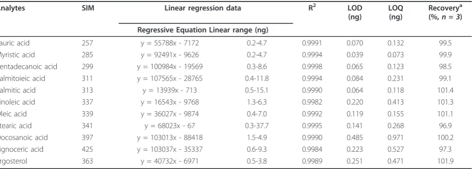
Table 2 SIM, regression data, LOD, LOQ and recovery of 11 investigated compounds analyzed by GC-MS
Analytes SIM Linear regression data R2 LOD
(ng)
LOQ (ng)
Recoverya (%,n = 3) Regressive Equation Linear range (ng)
Lauric acid 257 y = 55788x - 7172 0.2-4.7 0.9991 0.070 0.132 99.5
Myristic acid 285 y = 92491x - 9626 0.2-4.7 0.9994 0.039 0.073 99.9
Pentadecanoic acid 299 y = 100984x - 19569 0.3-8.6 0.9998 0.065 0.123 98.5 Palmitoieic acid 311 y = 107565x - 28765 0.4-11.8 0.9994 0.084 0.231 99.1
Palmitic acid 313 y = 13939x - 713 0.5-15.1 0.9990 0.064 0.118 101.4
Linoleic acid 337 y = 16543x - 9768 1.3-6.3 0.9982 0.220 0.413 101.3
Oleic acid 339 y = 36027x - 9874 0.4-7.0 0.9992 0.119 0.155 101.1
Stearic acid 341 y = 68023x - 67 0.3-37.7 0.9995 0.141 0.268 96.9
Docosanoic acid 397 y = 103013x - 88418 1.5-4.9 0.9990 0.485 0.971 100.2 Lignoceric acid 425 y = 103037x - 35337 0.6-9.3 0.9984 0.223 0.527 97.3
Ergosterol 363 y = 40732x - 6971 0.5-3.8 0.9989 0.251 0.471 101.9
a
10
10.00 20.00 30.00 40.00 min 0 2 4 6 8 10 12 14 16 18 20h104
Time--> Abundance 257 285 299 311 313 337 339 397 341 425 363 1 2 34 5 6 7 8 9 11
10.00 20.00 30.00 40.00 min 0 1 2 3 4 5 6 7 8 9h105
Time--> Abundance 257 285 311 313 337 341 339 397 425 363 299 1 2 3 4 5 6 7 8 9 10 11 1 2 3 4 5 6 7 8 9 10 11
10.00 20.00 30.00 40.00 min 0 2 4 6 8 10 12 14 16 18 20h104
Time--> Abundance 257 285 299 311 313 337 339 397 341 425 363
10.00 20.00 30.00 40.00 min 0 1 2 3 4 5 Time--> Abundance
6h106
1 2 34 5 6 8 7 9 10 11
10.00 20.00 30.00 40.00 min 0 1 2 3 4 5 6 7 8 9h106
Time--> Abundance
1 2 34 5 6 8 7 910 11 4
10.00 20.00 30.00 40.00 min 0 1 2 3 4 5 6 7 8 9 10 11 12 13 14 h106
Time--> Abundance
1
235
6 78 9 11 10
C
B
A
mixture was extracted and analyzed using the devel-oped method. Three replicates were performed for the test.
Data analysis
Hierarchical clustering analysis was performed by SPSS 18.0 for windows (SPSS Inc., Chicago, IL, USA), which comprise a number of“procedures”graphical, statistical, reporting, processing and tabulating procedures that enable simple and rapid data evaluation. Hierarchical cluster analysis was performed based on ergosterol and ten fatty acids characteristics from GC-MS profiles of 32 tested samples, respectively. Ward’s method, a very effi-cient method for the analysis of variance between
clusters was applied, and Euclidean Distance was selected as measurement.
Result and discussion
Optimization of derivatization conditions
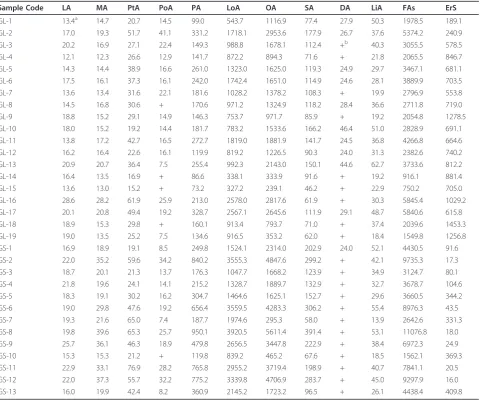
The derivatization conditions including derivatization temperature (50, 70, 90°C) and the reaction time (30, 60, 90 min) for the investigated compounds were opti-mized. On the other hand, the sufficient amount of deri-vatization agent is necessary for good derideri-vatization efficiency. The amount of BSTFA (50, 100 and 200 μL) were also investigated using univariate approach. Peak area of each investigated compound in Ganoderma extract was used as the marker for evaluation of the Table 3 The contents (μg·g-1) of ten investigated fatty acids and ergosterol inGanoderma
Sample Code LA MA PtA PoA PA LoA OA SA DA LiA FAs ErS
GL-1 13.4a 14.7 20.7 14.5 99.0 543.7 1116.9 77.4 27.9 50.3 1978.5 189.1 GL-2 17.0 19.3 51.7 41.1 331.2 1718.1 2953.6 177.9 26.7 37.6 5374.2 240.9
GL-3 20.2 16.9 27.1 22.4 149.3 988.8 1678.1 112.4 +b 40.3 3055.5 578.5
GL-4 12.1 12.3 26.6 12.9 141.7 872.2 894.3 71.6 + 21.8 2065.5 846.7
GL-5 14.3 14.4 38.9 16.6 261.0 1323.0 1625.0 119.3 24.9 29.7 3467.1 681.1 GL-6 17.5 16.1 37.3 16.1 242.0 1742.4 1651.0 114.9 24.6 28.1 3889.9 703.5
GL-7 13.6 13.4 31.6 22.1 181.6 1028.2 1378.2 108.3 + 19.9 2796.9 553.8
GL-8 14.5 16.8 30.6 + 170.6 971.2 1324.9 118.2 28.4 36.6 2711.8 719.0
GL-9 18.8 15.2 29.1 14.9 146.3 753.7 971.7 85.9 + 19.2 2054.8 1278.5
GL-10 18.0 15.2 19.2 14.4 181.7 783.2 1533.6 166.2 46.4 51.0 2828.9 691.1 GL-11 13.8 17.2 42.7 16.5 272.7 1819.0 1881.9 141.7 24.5 36.8 4266.8 664.6 GL-12 16.2 16.4 22.6 16.1 119.9 819.2 1226.5 90.3 24.0 31.3 2382.6 740.2 GL-13 20.9 20.7 36.4 7.5 255.4 992.3 2143.0 150.1 44.6 62.7 3733.6 812.2
GL-14 16.4 13.5 16.9 + 86.6 338.1 333.9 91.6 + 19.2 916.1 881.4
GL-15 13.6 13.0 15.2 + 73.2 327.2 239.1 46.2 + 22.9 750.2 705.0
GL-16 28.6 28.2 61.9 25.9 213.0 2578.0 2817.6 61.9 + 30.3 5845.4 1029.2
GL-17 20.1 20.8 49.4 19.2 328.7 2567.1 2645.6 111.9 29.1 48.7 5840.6 615.8
GL-18 18.9 15.3 29.8 + 160.1 913.4 793.7 71.0 + 37.4 2039.6 1453.3
GL-19 19.0 13.5 25.2 7.5 134.6 916.5 353.2 62.0 + 18.4 1549.8 1256.8
GS-1 16.9 18.9 19.1 8.5 249.8 1524.1 2314.0 202.9 24.0 52.1 4430.5 91.6
GS-2 22.0 35.2 59.6 34.2 840.2 3555.3 4847.6 299.2 + 42.1 9735.3 17.3
GS-3 18.7 20.1 21.3 13.7 176.3 1047.7 1668.2 123.9 + 34.9 3124.7 80.1
GS-4 21.8 19.6 24.1 14.1 215.2 1328.7 1889.7 132.9 + 32.7 3678.7 104.6
GS-5 18.3 19.1 30.2 16.2 304.7 1464.6 1625.1 152.7 + 29.6 3660.5 344.2
GS-6 19.0 29.8 47.6 19.2 656.4 3559.5 4283.3 306.2 + 55.4 8976.3 43.5
GS-7 19.3 21.6 65.0 7.4 187.7 1974.6 295.3 58.0 + 13.9 2642.6 331.3
GS-8 19.8 39.6 65.3 25.7 950.1 3920.5 5611.4 391.4 + 53.1 11076.8 18.0
GS-9 25.7 36.1 46.3 18.9 479.8 2656.5 3447.8 222.9 + 38.4 6972.3 24.9
GS-10 15.3 15.3 21.2 + 119.8 839.2 465.2 67.6 + 18.5 1562.1 369.3
GS-11 22.9 33.1 76.9 28.2 765.8 2955.2 3719.4 198.9 + 40.7 7841.1 20.5
GS-12 22.0 37.3 55.7 32.2 775.2 3339.8 4706.9 283.7 + 45.0 9297.9 16.0
GS-13 16.0 19.9 42.4 8.2 360.9 2145.2 1723.2 96.5 + 26.1 4438.4 409.8
LA, lauric acid; MA, myristic acid; PtA, pentadecanoic acid; PoA, palmitoleic acid; PA, palmitic acid; LoA, linoleic acid; OA, oleic acid; SA, stearic acid; DA, docosanoic acid; LiA, lignoceric acid; ErS, ergosterol; FAs: total content of ten fatty acids
a
The data are presented as average of duplicates.
b
derivatization efficiency. Finally, the optimum derivatiza-tion condiderivatiza-tions, which had highest efficiency in short reaction time under mild temperature, were as follows: 100μL BSTFA reacted with free sterol and fatty acids in 400μL of n-hexane at 70°C for 30 min.
Validation of method
SIM method of GC-MS analysis for simultaneous inves-tigation of the analytes was used for the quantification and the fragment ions for simultaneous monitoring of analytes were listed in Table 2.
All the calibration curves showed good linearity within test ranges. The overall LODs and LOQs were less than 0.485 ng and 0.971 ng, respectively (Table 2). The optimized method showed good precision for the quantification of 11 investigated components with intra- and inter-day variations less than 2.7% and 4.3%, and the repeatability of derivatization detected in PLE extract of test sample (GL-1) was less than 3.2%. The developed method also had good accuracy with overall recovery of 96.9%-101.9% for the analytes (Table 2). The results showed that the developed GC-MS method was sensitive, precise and accurate for quantitative determination of 11 investigated components in Ganoderma.
Quantification of the investigated compounds in Ganoderma by GC-MS
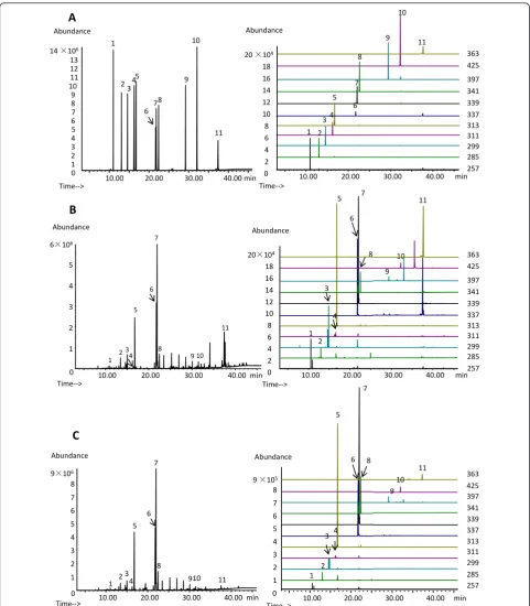
Typical TIC and SIM chromatograms of TMS derivatives of mixture of 11 reference compounds and PLE extracts of two species ofGanodermawere shown in Figure 2. The identification of investigated compounds was carried out by comparison of their retention time and mass spec-tra with those obtained by injecting standards in the same conditions. By using the calibration curves of each analyte, the contents (μg·g-1) of ten fatty acids and one sterol inGanodermawere determined, which were sum-marized in Table 3. The data showed that ergosterol, a characteristic compound in fungi, was abundant in both G. lucidumandG. sinense. PA, LOA, OA and SA were the main fatty acids inGanoderma, which were in accor-dance to the results inG. lucidumspore [15].
Comparison of free sterol and fatty acids in two species of Ganoderma
In China, G. lucidum and G. sinense is recorded as Lingzhiin Chinese Pharmacopoeia. Ergosterol, as a spe-cific component of fungal cell membrane, was also rich inG. lucidumandG. sinense. But the content of ergos-terol inG. lucidum (median content 705.0μg·g-1, range 189.1-1453.3 μg·g-1, n = 19) was much higher than that in G. sinense(median content 80.1 μg·g-1, range 16.0-409.8 μg·g-1, n = 13), which is similar to triterpenoids and polysaccharides [1,11,34]. However, it was much
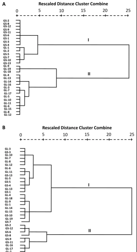
lower than that reported before [11,35], which might attribute to the different sample and/or peak overlap-ping during HPLC analysis. Hierarchical clustering ana-lysis based on the contents of ergosterol also showed that 32 tested samples ofGanodermawere grouped into two main clusters, G. lucidum and G. sinense, except twoG. lucidumsamples (Figure 3).
On the other hand, hierarchical clustering analysis based on the contents of ten fatty acids was also per-formed. The results showed that two species of Gano-dermahad no significant difference though two groups were also obtained (Figure 3). Actually, the total content
GS-2 GS-8 GS-12 GS-9 GS-11 GS-6 GS-1 GS-3 GS-4 GL-1 GL-2 GS-5 GS-7 GS-10 GS-13 GL-9 GL-19 GL-18 GL-4 GL-13 GL-14 GL-16 GL-3 GL-7 GL-17 GL-5 GL-10 GL-11 GL-6 GL-15 GL-8 GL-12
I
II
+ + + + + +
0 5 10 15 20 25
Rescaled Distance Cluster Combine
GL-3 GS-3 GL-10 GL-7 GL-8 GL-12 GL-6 GL-11 GS-13 GL-5 GS-5 GS-4 GL-13 GS-1 GL-4 GL-18 GL-9 GL-1 GL-14 GL-15 GS-10 GL-19 GS-7 GS-2 GS-12 GS-6 GS-8 GS-9 GS-11 GL-16 GL-17 GL-2
+ + + + + +
0 5 10 15 20 25
Rescaled Distance Cluster Combine
I
II
A
B
Figure 3Dendrograms of hierarchical cluster analysis resulting from (A) ergosterol and (B) ten fatty acids in 32 tested
of fatty acids was higher in cluster II. Palmitic acid, lino-leic acid, olino-leic acid and stearic acid were the main fatty acids in Ganoderma, which were in accordance with the previous reports for G. lucidum spore [15,36]. It was reported that fatty acids from the spores ofG. lucidum could inhibit tumor cell proliferation. Nonadecanoic acid (C19:0) showed the highest inhibitory activity, fol-lowed by heptadecanoic acid (C17:0), stearic acid (C18:0, SA) and palmitic acid (C16:0, PA) [20]. Actually, saturated fatty acids including SA and PA are proapop-totic agents [37]. Unsaturated fatty acids LoA and OA have been revealed their anticancer activities [38-42] by activating GPR40 [38] and inducing oxidant stress and mitochondrial dysfunction [42] in cancer cell lines. Therefore, the similarity of two species ofGanoderma in fatty acids may be related to their antitumoral prolif-eration effect.
Conclusion
The content of ergosterol is much higher inG. lucidum than inG. sinense. Palmitic acid, linoleic acid, oleic acid, stearic acid are main fatty acids inGanoderma and their content had no significant difference betweenG. luci-dumand G. sinense, which may contribute to their anti-tumoral proliferation effect.
Abbreviations
DA: docosanoic acid; ErS: ergosterol; GC-MS: gas chromatography-mass spectrometry; LA: lauric acid; LoA: linoleic acid; LiA: lignoceric acid; MA: myristic acid; OA: oleic acid; PLE: pressurized liquid extraction; PtA: pentadecanoic acid; PoA: palmitoleic acid; PA: palmitic acid; SA: stearic acid; SIM: Selected ion monitoring; TIC: Total ion chromatogram; TMS:
trimethylsilyl.
Acknowledgements
This study was partially supported by grants from the National Natural Science Foundation of China (No.30928033) and University of Macau (UL015A).
Author details
1
State Key Laboratory of Quality Research in Chinese Medicine, University of Macau, Macao SAR 999078, China.2Institute of Chinese Medical Sciences, University of Macau, Macao SAR 999078, China.3Jiangsu Key Laboratory for TCM Formulae Research, Nanjing University of Chinese Medicine, Nanjing, Jiangsu, China.
Authors’contributions
SPL initiated and all authors designed the study. The extraction and method developments were conducted by GPL who drafted the manuscript. All authors contributed to data analyses and to finalizing the manuscript. All authors have read and approved the final version.
Competing interests
The authors declare that they have no competing interests.
Received: 5 December 2011 Accepted: 31 January 2012 Published: 31 January 2012
References
1. Liu YW, Gao JL, Guan J, Qian ZM, Feng K, Li SP:Evaluation of
antiproliferative activities and action mechanisms of extracts from two
species of ganoderma on tumor cell lines.J Agric Food Chem2009, 57:3087-3093.
2. Chinese Pharmacopoeia Commission:Pharmacopoeia of the People’s Republic of China 2010Chemical Industry Press: Beijing; 2010. 3. Paterson RRM:Ganoderma - A therapeutic fungal biofactory.
Phytochemistry2006,67:1985-2001.
4. Zhao S, Ye G, Fu G, Cheng JX, Yang BB, Peng C:Ganoderma lucidum exerts anti-tumor effects on ovarian cancer cells and enhances their sensitivity to cisplatin.Int J Oncol2011,38:1319-1327.
5. Xu Z, Chen X, Zhong Z, Chen L, Wang Y:Ganoderma lucidum
polysaccharides: Immunomodulation and potential anti-tumor activities.
Am J Chin Med2011,39:15-27.
6. Seo HW, Hung TM, Na M, Jung HJ, Kim JC, Choi JS, Kim JH, Lee HK, Lee I, Bae K, Hattori M, Min BS:Steroids and triterpenes from the fruit bodies of Ganoderma lucidum and their anti-complement activity.Arch Pharmacal Res2009,32:1573-1579.
7. Dudhgaonkar S, Thyagarajan A, Sliva D:Suppression of the inflammatory response by triterpenes isolated from the mushroom Ganoderma lucidum.Int Immunopharmacol2009,9:1272-1280.
8. Sato N, Zhang Q, Ma CM, Hattori M:Anti-human immunodeficiency virus-1 protease activity of new lanostane-type triterpenoids from Ganoderma sinense.Chem Pharm Bull2009,57:1076-1080.
9. Weng Y, Xiang L, Matsuura A, Zhang Y, Huang Q, Qi J:Ganodermasides A and B, two novel anti-aging ergosterols from spores of a medicinal mushroom Ganoderma lucidum on yeast via UTH1 gene.Bioorg Med Chem2010,18:999-1002.
10. Seto SW, Lam TY, Tam HL, Au ALS, Chan SW, Wu JH, Yu PHF, Leung GPH, Ngai SM, Yeung JHK, Leung PS, Lee SMY, Kwan YW:Novel hypoglycemic effects of Ganoderma lucidum water-extract in obese/diabetic (+db/ +db) mice.Phytomedicine2009,16:426-436.
11. Zhao J, Zhang XQ, Li SP, Yang FQ, Wang YT, Ye WC:Quality evaluation of Ganoderma through simultaneous determination of nine triterpenes and sterols using pressurized liquid extraction and high performance liquid chromatography.J Sep Sci2006,29:2609-2615.
12. Wang XM, Yang M, Guan SH, Liu RX, Xia JM, Bi KS, Guo DA:Quantitative determination of six major triterpenoids in Ganoderma lucidum and related species by high performance liquid chromatography.J Pharm Biomed Anal2006,41:838-844.
13. Pedrini N, Crespo R, Juárez MP:Biochemistry of insect epicuticle degradation by entomopathogenic fungi.Comp Biochem Physiol C Toxicol Pharmacol2007,146:124-137.
14. Roberts CW, McLeod R, Rice DW, Ginger M, Chance ML, Goad LJ:Fatty acid and sterol metabolism: potential antimicrobial targets in apicomplexan and trypanosomatid parasitic protozoa.Mol Biochem Parasitol2003,126:129-142. 15. Liu X, Xu SP, Wang JH, Yuan JP, Guo LX, Li X, Huang XN:Characterization of ganoderma spore lipid by stable carbon isotope analysis: Implications for authentication.Anal Bioanal Chem2007,388:723-731.
16. Shen M, Xie M, Nie S, Wan Y, Xie J:Discrimination of different Ganoderma species and their region based on GC-MS profiles of sterols and pattern recognition techniques.Anal Lett2011,44:863-873.
17. Smania EFA, Delle Monache F, Smania A Jr, Yunes RA, Cuneo RS: Antifungal activity of sterols and triterpenes isolated from Ganoderma annulare.Fitoterapia2003,74:375-377.
18. Zhao HB, Wang SZ, He QH, Yuan L, Chen AF, Lin ZB:Ganoderma total sterol (GS) and GS1 protect rat cerebral cortical neurons from hypoxia/ reoxygenation injury.Life Sci2005,76:1027-1037.
19. Hirasawa A, Hara T, Katsuma S, Adachi T, Tsujimoto G:Free fatty acid receptors and drug discovery.Biol Pharm Bull2008,31:1847-1851. 20. Fukuzawa M, Yamaguchi R, Hide I, Chen Z, Hirai Y, Sugimoto A, Yasuhara T,
Nakata Y:Possible involvement of long chain fatty acids in the spores of Ganoderma lucidum (Reishi Houshi) to its anti-tumor activity.Biol Pharm Bull2008,31:1933-1937.
21. Huang SM, Yang XL, Zhu HS:Antitumor effects of triterpene acids extracted from Ganoderma lucidum.Envir Trans Engin2011,11:7398-7401. 22. Cai Y, Cai TG, Shi Y, Cheng XL, Ma LY, Ma SC, Lin RC, Feng W:
Simultaneous determination of eight PDE5-IS potentially adulterated in herbal dietary supplements with TLC and HPLC-PDA-MS methods.J Liq Chromatogr Related Technol2010,33:1287-1306.
24. Chen TQ, Zhao XY, Wu JZ, Yu DY, Wu YB:Supercritical fluid CO2 extraction, simultaneous determination of components in ultra-fine powder of Ganoderma sinense by HPLC-ESI-MS method.J Taiwan Inst Chem Eng2011,42:428-434.
25. Guan J, Li SP:Discrimination of polysaccharides from traditional Chinese medicines using saccharide mapping-Enzymatic digestion followed by chromatographic analysis.J Pharm Biomed Anal2010,51:590-598. 26. Huang SQ, Wei G, Yao SJ, Liu CL, Ning ZX:Optimization and comparison
of fingerprints of triterpenoids from Ganoderma lucidum and Ganoderma sinense.J South China Univ Tec2010,38:121-125. 27. Keypour S, Rafati H, Riahi H, Mirzajani F, Moradali MF:Qualitative analysis
of ganoderic acids in Ganoderma lucidum from Iran and China by RP-HPLC and electrospray ionisation-mass spectrometry (ESI-MS).Food Chem2010,119:1704-1708.
28. Yang Q, Wang S, Xie Y, Sun J, Wang J:HPLC analysis of Ganoderma lucidum polysaccharides and its effect on antioxidant enzymes activity and Bax, Bcl-2 expression.Int J Biol Macromol2010,46:167-172. 29. Chen Y, Xie MY, Wang YX, Nie SP, Li C:Analysis of the monosaccharide
composition of purified polysaccharides in Ganoderma atrum by capillary gas chromatography.Phytochemical Anal2009,20:503-510. 30. Huang SQ, Li JW, Li YQ, Wang Z:Purification and structural
characterization of a new water-soluble neutral polysaccharide GLP-F1-1 from Ganoderma lucidum.Int J Biol Macromol2011,48:165-169. 31. Ye L, Zheng X, Zhang J, Yang Y, Meng Y, Li J, Chen W, Li A, Pan YJ:
Composition analysis and immunomodulatory capacity of peptidoglycan from ling zhi or reishi medicinal mushroom, ganoderma lucidum (W. Curt.: Fr.) P. Karst. Strain 119 (Aphyllophoromycetideae).Inter J Med Mushrooms2010,12:157-165.
32. Cheung HY, Ng CW, Hood DJ:Identification and quantification of base and nucleoside markers in extracts of Ganoderma lucidum, Ganoderma japonicum and Ganoderma capsules by micellar electrokinetic chromatography.J Chromatogr A2001,911:119-26.
33. Pedneault K, Angers P, Gosselin A, Tweddell RJ:Fatty acid profiles of polar and neutral lipids of ten species of higher basidiomycetes indigenous to eastern Canada.Mycol Res2008,112:1428-1434.
34. Ding P, Cai H, Liu Y, Lin L, Xu H:Comparison of the Components of Ganoderma lueidum and Ganoderma japonicum.J Chin Med Mater1999, 22:433-434.
35. Liu JJ, Huang WH, Lv ML, Si JP, Guo BL, Li SJ:Determination of Ergosterol in Ganoderma lucidum from Different Varieties and Cultured Tree Species by HPLC.J Chin Med Mater2011,34:187-190.
36. Wang YJ, Zhang L, Ding AW:Quantification of five kinds of organic acids in Ganoderma Lucidum Spores Oil by GC-MS.J Nanjing Univ Trad Chin Med2009,25:136-139.
37. Hardy S, El-Assaad W, Przybytkowski E, Joly E, Prentki M, Langelier Y: Saturated fatty acid-induced apoptosis in MDA-MB-231 breast cancer cells. A role for cardiolipin.J Biol Chem2003,278:31861-31870. 38. Yonezawa T, Katoh K, Obara Y:Existence of GPR40 functioning in a
human breast cancer cell line, MCF-7.Biochem Biophys Res Commun2004, 314:805-809.
39. Hasanzadeh GK, Latiffah AL, Hanachi P, Hj Lajis N:Effect of Linoleic acid of Nigella sativa on MDA-MB-231 human breast cancer cells.Iran J Cancer Prev2011,4:65-70.
40. Ohmori H, Sasahira T, Fujii K, Yi L, Shimomoto T, Kuniyasu H: Linoleic-acid-induced growth suppression induces quiescent cancer cell nests in nude mice.Pathobiology2008,75:226-232.
41. Carrillo C, Cavia MD, Alonso-Torre SR:Oleic acid inhibits store-operated calcium entry in human colorectal adenocarcinoma cells.Eur J Nutr2011. 42. Lu X, Yu H, Ma Q, Shen S, Das UN:Linoleic acid suppresses colorectal
cancer cell growth by inducing oxidant stress and mitochondrial dysfunction.Lipids Health Dis2010,9:106-116.
doi:10.1186/1752-153X-6-10
Cite this article as:Lvet al.:Comparison of sterols and fatty acids in two species ofGanoderma.Chemistry Central Journal20126:10.
Open access provides opportunities to our colleagues in other parts of the globe, by allowing
anyone to view the content free of charge.
Publish with
Chemistry
Central and every
scientist can read your work free of charge
W. Jeffery Hurst, The Hershey Company.
available free of charge to the entire scientific community peer reviewed and published immediately upon acceptance cited in PubMed and archived on PubMed Central yours you keep the copyright
Submit your manuscript here: