PRIMARY RESEARCH
Long noncoding RNA RHPN1-AS1 promotes
colorectal cancer progression via targeting
miR-7-5p/OGT axis
Wei Zheng
1, Hui Li
2, Hui Zhang
1, Chao Zhang
1, Zhonglin Zhu
1, Hong Liang
1and Yifeng Zhou
2*Abstract
Background: Rhophilin Rho GTPase binding protein 1 antisense RNA 1 (RHPN1-AS1) is a newly discovered oncogene in several diseases, such as breast cancer, non-small cell lung cancer and uveal melanoma. Nevertheless, its molecular role in colorectal cancer (CRC) remains unknown. This paper explored the role of RHPN1-AS1 in CRC progression.
Methods: qRT-PCR was used to detect relevant RNAs expression. CCK-8, EdU, flow cytometry, Transwell and western blot assays were performed to investigate the function of RHPN1-AS1 in CRC cells. Xenograft model was constructed to evaluate the effects of RHPN1-AS1 on tumor growth in vivo. Mechanical experiments were performed to investi-gate the relationship between relative genes.
Results: RHPN1-AS1 was significantly overexpressed in CRC cell lines. Knockdown of RHPN1-AS1 could inhibit cell proliferation, while stimulating cell apoptosis in vitro. Cell migration and invasion abilities were greatly suppressed after silencing RHPN1-AS1. Besides, signal transducer and activator of transcription 3 (STAT3) served as transcription factor of RHPN1-AS1. Moreover, miR-7-5p was identified as a target of RHPN1-AS1 and was negatively regulated by RHPN1-AS1 in CRC. MiR-7-5p inhibition rescued the oncogenic function of RHPN1-AS1. Additionally, O-GlcNAcylation transferase (OGT) was the downstream target of miR-7-5p. OGT overexpression could abrogate the anti-tumor effects of RHPN1-AS1 knockdown on CRC.
Conclusion: RHPN1-AS1 regulates CRC by mediating OGT through sponging miR-7-5p, suggesting that RHPN1-AS1 might be a potential therapeutic target for CRC.
Keywords: RHPN1-AS1, miR-7-5p, OGT, Colorectal cancer
© The Author(s) 2020. This article is licensed under a Creative Commons Attribution 4.0 International License, which permits use, sharing, adaptation, distribution and reproduction in any medium or format, as long as you give appropriate credit to the original author(s) and the source, provide a link to the Creative Commons licence, and indicate if changes were made. The images or other third party material in this article are included in the article’s Creative Commons licence, unless indicated otherwise in a credit line to the material. If material is not included in the article’s Creative Commons licence and your intended use is not permitted by statutory regulation or exceeds the permitted use, you will need to obtain permission directly from the copyright holder. To view a copy of this licence, visit http://creat iveco mmons .org/licen ses/by/4.0/. The Creative Commons Public Domain Dedication waiver (http://creat iveco mmons .org/publi cdoma in/ zero/1.0/) applies to the data made available in this article, unless otherwise stated in a credit line to the data.
Background
On a global scale, colorectal cancer (CRC) is one of the
most common malignant [1]. Annually, approximately
1.2,000,000 new cases were diagnosed and 60,000 death
cases happened [2]. The overall occurrence and mortality
are declining due to the advance in new techniques and therapeutic methods for CRC predication and treatment
[3]. However, the overall 5-year survival rate is still lower than anticipation as metastatic CRC frequently occurred in patients at the time of diagnosis [4, 5]. Hence, it is paramount to reveal the underlying mechanism behind the progression of CRC, so as to identify potential novel diagnostic and prognostic biomarkers.
The newly-identified non-coding RNAs (ncRNAs) are a cluster of RNAs with the ability of transcriptional con-trol and post-transcriptional mediation, but they lack the potential to encode proteins [5–8]. Long non-coding RNAs (lncRNAs) are a kind of ncRNAs containing over 200 nucleotides in length. They have been extensively studied in the carcinogenesis of many diseases, including
Open Access
*Correspondence: [email protected]
2 Department of Gastroenterology, Affiliated Hangzhou First People’s
Hospital, Zhejiang University School of Medicine, No. 261 Huanshan Road, Hangzhou 310006, Zhejiang, China
cancer. Moreover, recent findings manifested that lncR-NAs are involved in a series of biological processes, such as cell proliferation, apoptosis, invasion and migration
[9, 10]. Furthermore, lncRNAs have been identified as
new biomarkers of many malignancies, since they are associated with the complicated pathogenesis of
malig-nancies [11, 12]. In addition, the prevalent competing
endogenous RNA (ceRNA) mechanism has revealed the crucial regulatory role of lncRNAs on downstream RNAs, consequently impacting various physiological and
pathophysiological activities within cells [13]. To date,
the pathologic roles of most lncRNAs remain undiscov-ered, which indicates the extensive application potential of lncRNAs in the prediction and treatment of different cancers.
In CRC, the expression status and functional role of some lncRNAs were investigated. For instance, lncRNA XIRP2-AS1 was discovered significantly lowly expressed in CRC tissues and could serve as a favorable biomarker
for CRC patients [14]. Silencing lncRNA AWPPH
signifi-cantly curbed CRC cell proliferation via down-regulating
the expression of GLUT-1 [15]. FEZF1-AS1 has been
reported to accelerate the progression of CRC via up-reg-ulating the expression of NT5E through sponging miR-30a-5p [16].
RHPN1-AS1, as a newly identified lncRNA, has been revealed to promote the carcinogenesis of head and neck
squamous cell carcinoma [17]. Furthermore, it has been
reported as a potential clinical biomarker and partici-pated in important biological processes and pathways in
non-small cell lung cancer (NSCLC) [18]. Nevertheless,
the molecular role of RHPN1-AS1 behind the carcino-genesis and development of CRC has not been explored yet. Therefore, the aim of present study is to explore the cellular function of RHPN1-AS1 in CRC, which might contribute to providing some novel thoughts about find-ing effective treatment targets for CRC patients.
Materials and methods Cell culture
Normal human colorectal mucosal cell (FHC) and CRC cells (SW620, SW480, HCT-116, HT29) were bought from Chinese Academy of Sciences (Beijing, China). Human kidney cell (293T) was gained from Genechem (Shanghai China). Cells were grown in RPMI-1640 medium (Invitrogen, Carlsbad, CA, USA) adding 10% fetal bovine serum (FBS; Invitrogen), 1% penicillin/strep-tomycin (Sigma-Aldrich, Milan, Italy) and cultured in a 5% CO2 incubator at 37 °C.
Cell transfection
HCT-116 and HT29 cells were transfected with specific shRNAs against RHPN1-AS1 (sh-RHPN1-AS1#1/#2),
STAT3 (sh-STAT3), OGT (sh-OGT#1/#2) and their cor-responding control group (sh-Ctrl), and pcDNA3.1/ STAT3, pcDNA3.1/OGT and the empty pcDNA3.1 vec-tor, respectively. The miR-7-5p inhibivec-tor, miR-7-5p mim-ics, NC mimics and NC inhibitor were synthesized by GenePharma (Shanghai, China). Lipofectamine 2000 (Invitrogen) was used for executing transfection experi-ments for 48 h.
Quantitative real time polymerase chain reaction (qRT‑PCR)
TRIzol reagent (Invitrogen) was employed to obtain total RNA from cells. Reverse Transcription Kit (Applied Biosystems, CA, USA) or Taqman Advanced miRNA cDNA Synthesis Kit (Thermo Fisher Scientic) was used to reverse transcribe total RNA into cDNA. qRT-PCR was performed on Bio-Rad CFX96 System utilizing the ABI StepOnePlus system (Applied Biosystems). Fold
expres-sion changes were calculated with 2−∆∆Ct method, and
GAPDH/U6 was endogenous control.
Cell counting kit‑8 (CCK‑8) assay
1 × 10³ cells were added to 96-well plates with culture
media. The medium of every well was replaced by a fresh culture media containing 10 µL CCK8 at specific time points. The cells were cultivated for additional 4 h. The absorbance at 450 nm was measured via a microplate reader (Olympus, Tokyo, Japan).
EdU incorporation assay
EdU (5-ethynyl-2′-deoxyuridine) assay was performed
to detect cell proliferation ability using the Click-iT EdU Imaging Kit (Invitrogen) in line with manufacturer’s directions. Transfected HT29 and HCT-116 were main-tained into 96-well plate. After incubation of 48 h, 100 μl medium with 50 μM EdU was added into all wells. Cells underwent 2 h incubation at 37 °C and were fixed with 4% paraformaldehyde. Nuclei were dyed in DAPI for 30 min. EdU-positive cells were visualized using a micro-scope (Olympus). Proliferation rate was determined by calculating the ratio of the fluorescent positive cells to total cells.
Flow cytometry analysis
HCT-116 and HT29 cells were reaped and cultured in 6-well plates after transfection. Followed by rinsing with PBS, Annexin V-FITC and Propidium iodide (PI) were used for double staining. Apoptosis rate was measured via a flow cytometry (BD Biosciences, Beijing, China).
Transwell assay
Matrigel (Becton–Dickinson, San Jose, CA, USA) for invasion or migration analysis. 5 × 10⁴ cells with serum-free medium were added into the top compartment, while medium containing 10% FBS was placed into the bottom compartment later. After incubation for 24 h (migration) or 48 h (invasion), cells on the underside of membrane were immobilized and dyed in methanol or crystal violet. Finally, cells were counted in 5 random chosen fields under the microscope (Olympus).
Western blot
Total proteins were extracted with RIPA lysis buffer (Invitrogen). Protein concentrations were examined by BCA kit. There was 10% SDS-PAGE for the isola-tion of proteins. And then proteins were moved on PVDF membranes. After being blocked with PVDF, membranes were cultured with primary antibodies for E-cadherin (ab194982), N-cadherin (ab202030), Vimen-tin (ab193555), Slug (ab51772), Twist (ab187008), OGT (ab96718) and GAPDH (ab8245) from Abcam (Cam-bridge, CA, USA). Then, secondary antibodies were added for 1 h cultivation. The amount of protein was determined using chemiluminescence detection system.
Subcellular fractionation
Both nuclear and cytoplasmic RNA from CRC cells were separated with PARIS Kit (Ambion, Austin, TX, USA). Subsequently, qRT-PCR was employed for detecting the relative expression level of RHPN1-AS1. GAPDH/U6 was used as cytoplasmic/nuclear control.
Fluorescence in situ hybridization (FISH) assay
RHPN1-AS1 FISH probe was constructed by Ribo Bio Technology (Guangzhou, China). Cells were fixed in 4% paraformaldehyde, and then permeabilized in PBS which was mixed with 0.5% Triton X-100. After that, cells were hybridized with Cy3 labeled RNA FISH probe at 37 °C. Subsequently, cells were stained by the use of Hoechst. The presence of RHPN1-AS1 was visualized using confo-cal microscopy (Olympus).
Luciferase reporter assay
The wild-type (WT) and mutant (MUT) binding sites
of miR-7-5p in RHPN1-AS1 sequence or OGT 3′-UTR
was sub-cloned into pmirGLO dual-luciferase vector to construct RHPN1-AS1-WT/MUT, OGT-WT/MUT, and then co-transfected with miR-7-5p mimics or NC mimics into HCT-116, HT29 and HEK-293T cells, respectively. The pGL3-RHPN1-AS1 promoter was co-transfected with pcDNA3.1/STAT3, pcDNA3.1, sh-STAT3 or sh-Ctrl into HEK-293T cell. Dual-Luciferase Reporter Assay Sys-tem (Promega, USA) detected the luciferase activity.
RNA pull down assay
The RHPN1-AS1 WT, RHPN1-AS1 MUT or NC was biotin labeled into RHPN1-AS1 WT, Biotin-RHPN1-AS1 MUT or Bio-NC, severally. Afterwards, cell lysates were incubated with biotinylated RNA over-night and M-280 streptavidin magnetic beads (Sigma-Aldrich) were added for cultivation for 48 h. Relative enrichment of miR-7-5p was assayed by RT-qPCR.
Chromatin immunoprecipitation (ChIP) assay
Cells were subjected to chromatin immunoprecipitation based upon an EZ-ChIP chromatin immunoprecipita-tion kit (Millipore, Bedford, MA, USA). Briefly, cells were cross-linked by formaldehyde. Cell lysates were gathered and sonicated to shear DNA. After that, chromatin was immunoprecipitated using anti-STAT3 (Millipore), while IgG was the negative control. The relative enrichment of RHPN1-AS1 was evaluated by qRT-PCR.
RNA immunoprecipitation (RIP) assay
EZ-Magna RIP kit (Millipore) was applied for conduct-ing RIP assays. HCT-116 and HT29 cells were lysed in RIP lysis buffer, and incubated with anti-Ago2 (Mil-lipore) and IgG (Millpore). The relative expression lev-els of miR-7-5p, RHPN1-AS1 and OGT were examined using qRT-PCR analysis.
Tumor xenograft experiment
Female nude mice were gained from Shi Laike Com-pany (Shanghai, China). Cells infected with sh-RHPN1-AS1#1 or sh-Ctrl was injected subcutaneously into the hip back of mice. 4 days later, tumor growth was recorded by measuring their volume. After 4 weeks, nude mice were euthanized to weigh the tumors. This study was progressed with the approval of the Ethics Committee of Henan Provincial People’s Hospital.
Statistical analysis
Experimental data were manifested as mean ± SD. All
statistical analyses were achieved using the GraphPad software 6.0 (GraphPad Inc., San Diego, CA, USA). Each experiment was done at least three times. The var-iance among groups was analyzed via Student’s t-test or one-way ANOVA. Differences were seen as statistically significant when P < 0.05.
Results
RHPN1‑AS1 is aberrantly up‑regulated in CRC
and knockdown of RHPN1‑AS1 suppresses the malignant behaviors of CRC
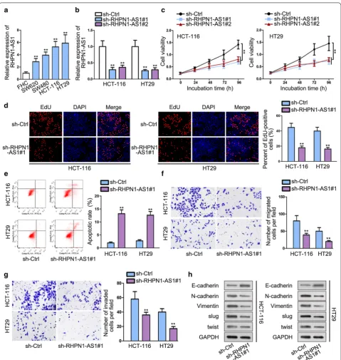
RHPN1-AS1 was predominantly up-regulated in CRC cell lines (SW620, SW480, HCT-116 and HT29) than healthy colon cell line (FHC). HCT-116 and HT29 cell lines contained the most obviously high expression of
RHPN1-AS1 and were chose to apply in the
follow-ing assays (Fig. 1a). Subsequently, loss-of-function
assay was adopted to investigate the role of RHPN1-AS1 in CRC cells. qRT-PCR result manifested that the
Fig. 1 RHPN1-AS1 is aberrantly up-regulated in CRC and knockdown of RHPN1-AS1 suppresses the malignant behaviors of CRC. a qRT-PCR assay was used to examine RHPN1-AS1 expression in CRC cell lines and non-tumor cell line. b The transfection efficiency of sh-RHPN1-AS1#1/#2 plasmid was detected by qRT-PCR. c, d CCK-8 and EdU were performed to investigate cell proliferation. e Flow cytometry was used to determine apoptosis.
inhibiting effect of sh-RHPN1-AS1#1 on RHPN1-AS1 in HCT-116 and HT29 cell lines was more visually than sh-RHPN1-AS1#2. Accordingly, sh-RHPN1-AS1#1 was
used in next researches (Fig. 1b). Then, CCK-8 and EdU
assays were performed to determine cell proliferation after knocking down RHPN1-AS1. We observed that silencing RHPN1-AS1 significantly detered cell
prolif-eration than control group (Fig. 1c, d). Flow cytometry
assay was conducted to assess cell apoptosis ability. We found a significant increase in the fraction of apoptosis cells after silencing RHPN1-AS1, which indicated that knockdown of RHPN1-AS1 promoted CRC cell
apop-tosis (Fig. 1e). Besides, transwell assays showed that
knockdown of RHPN1-AS1 diminished migrated and
invaded cells compared with control group (Fig. 1f, g).
(Epithelial-mesenchymal transition) EMT is a crucial
phase in CRC metastasis process [19]. Western blot
assay was conducted to investigate the expression sta-tus of EMT-related proteins. We found an increase of E-cadherin, while a reduction of N-cadherin, Vimentin,
slug and twist after RHPN1-AS1 knockdown (Fig. 1h).
This result further proved the migration and anti-invasion effects of silencing RHPN1-AS1 on CRC cells. Above all, RHPN1-AS1 is aberrantly up-regulated in CRC and knockdown of RHPN1-AS1 suppresses the malignant behaviors of CRC.
STAT3 stimulates the transcription of RHPN1‑AS1
Accumulating evidences manifested that some epige-netic regulators and key transcription factors
contrib-uted to lncRNAs dysregulation in human cancers [20,
21]. We found that the transcription factor STAT3 might
associate with RHPN1-AS1 promoter region on UCSC (http://genom e.ucsc.edu/) and Jaspar (http://jaspa r.gener eg.net/) databases. STAT3 has been revealed to
func-tion as oncogene in CRC [22]. Additionally, it has been
found to engage in the activation of IL-6/STAT3
path-way in CRC [23]. To investigate the effects of STAT3 on
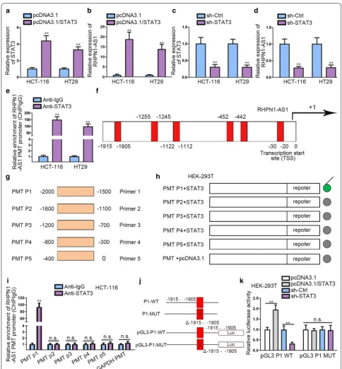
RHPN1-AS1, the overexpression efficiency of STAT3
was measured in the first place (Fig. 2a). Then qRT-PCR
result manifested that the expression of RHPN1-AS1 was increased after transfecting pcDNA3.1/STAT3 plasmids
into CRC cells (Fig. 2b). Afterwards, we knocked down
STAT3 and the expression of STAT3 declined greatly
(Fig. 2c). Besides, we noticed a significant
down-regula-tion of RHPN1-AS1 after STAT3 knockdown (Fig. 2d).
These results indicated that STAT3 could play a crucial role in regulating the expression of RHPN1-AS1. ChIP assay was performed to determine the binding relation between STAT3 and RHPN1-AS1 promoter (PMT) using anti-IgG and anti-STAT3. The results manifested that RHPN1-AS1 PMT was significantly enriched in anti-STAT3 group than anti-IgG group, which indicated that
STAT3 could bind to RHPN1-AS1 PMT region (Fig. 2e).
Additionally, the potential binging sites of STAT3 in RHPN1-AS1 PMT promoter area were obtained by
uti-lizing UCSC and Jaspar (Fig. 2f). To determine the
spe-cific binding segments, we divided RHPN1-AS1 PMT into five segments according to the putative binding sites (Fig. 2g). Furthermore, luciferase reporter assay was performed to investigate the promoter activity after the five truncations of pGL3-RHPN1-AS1 promoter were co-transfected with pcDNA3.1 or pcDNA3.1/STAT3 in HEK-293T cells. We observed that STAT3 overexpres-sion could significantly enhance the transcription activity of RHPN1-AS1 after co-transfecting with
pGL3-RHPN1-AS1 PMT P1, as illustrated in Fig. 2h. Furthermore, we
noticed that STAT3 could bind to P1 truncation from
ChIP data (Fig. 2i). These findings revealed that STAT3
bound to RHPN1-AS1 promoter at about − 2000 to
− 1500 bp upstream transcription start site (TSS). To
determine whether STAT3 bound to RHPN1-AS1 pro-moter at the putative binding site existing in P1, we con-structed P1-WT (-1915 to - 1905) and P1-MUT (Δ-1915 to -1905) and transfected them into pGL3 vector respec-tively (Fig. 2j). We observed that the promoter activity of P1-WT was significantly enhanced after co-transfecting with pcDNA3.1/STAT3, but weakened in sh-STAT3 group in HEK-293T. Conversely, P1-MUT (Δ-1915 to -1905), which lacked -1915 to -1905 sites, showed barely
no promoter activity change (Fig. 2k). To sum up, these
results demonstrated that STAT3 plays a positive regula-tory role in the transcription of RHPN1-AS1 via binding to -1915 to -1905 site upstream TSS in RHPN1-AS1 pro-moter region.
RHPN1‑AS1 directly interacts with miR‑7‑5p and inhibits miR‑7‑5p expression
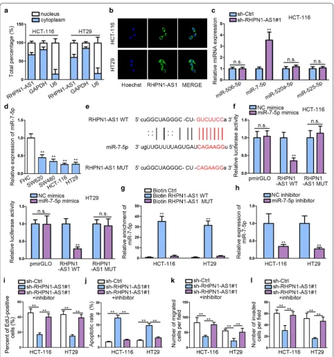
We determined the location of RHPN1-AS1 via sub-cellular fractionation assay and FISH analysis. Both results indicated that RHPN1-AS1 was largely situated
in cytoplasm (Fig. 3a, b). We screened out the
combin-able miRNA via Starbase database (http://starb ase.sysu.
edu.cn/) according to several restricted binding
condi-tions (class ≥ 7 mer-m8; Ago ExpNum ≥ 1, target gene:
RHPN1-AS1). And only four miRNAs were qualified. Subsequently, qRT-PCR was performed to study the effects on these miRNA after silencing RHPN1-AS1. The expression of miR-7-5p increased most significantly after knockdown RHPN1-AS1 compared with control group, while other miRNAs showed no significant
up-regula-tion (Fig. 3c). Besides, miR-7-5p has been validated to
be down-regulated in CRC, and served as a tumor
sup-pressor by targeting oncogene KLF4 in CRC [24].
Fig. 3 RHPN1-AS1 directly interacts with miR-7-5p and inhibits miR-7-5p expression. a, b Subcellular fractionation and FISH assays were performed to detect the subcellular location of RHPN1-AS1. c qRT-PCR analysis was performed to evaluate miRNA expression after silencing RHPN1-AS1. d
miR-7-5p in CRC cell lines compared with normal colon
cell line (Fig. 3d). Putative binding site of miR-7-5p in
the sequence of RHPN1-AS1 was predicted and mutated
accordingly, as shown on Fig. 3e. Next, dual luciferase
reporter assays were performed in HCT-116 and HT19 to examine the physical association between RHPN1-AS1 and miR-7-5p. We observed that miR-7-5p mimics transfection evidently attenuated the luciferase activity of RHPN1-AS1-WT, but didn’t significantly change that
of RHPN1-AS1-MUT (Fig. 3f). qRT-PCR based on RNA
pull down findings demonstrated a significant enrich-ment of miR-7-5p by biotinylated RHPN1-AS1-WT in
contrast to negative control groups (Fig. 3g), which
fur-ther proved that miR-7-5p was the downstream target of RHPN1-AS1.
To determine the influence of miR-7-5p in CRC, we performed rescue experiments after knocking down it
(Fig. 3h). EdU result showed that miR-7-5p inhibition
could greatly abolish the anti-proliferation role of
sh-RHPN1-AS1 in cell proliferation (Fig. 3i). Flow
cytom-etry results demonstrated that miR-7-5p inhibitor could reverse the promoting effects of silencing RHPN1-AS1
on cell apoptosis (Fig. 3j). Transwell assays manifested
that miR-7-5p inhibitor could recover CRC migration and invasion abilities repressed by silencing RHPN1-AS1 (Fig. 3k, l). Based on these results, we identify that RHPN1-AS1 plays an oncogenic role in CRC via spong-ing miR-7-5p.
OGT is the potential direct target gene of miR‑7‑5p We found five downstream targets of miR-7-5p on Star-base dataStar-base after strictly limiting binding conditions
(Clip data:high stringency ≥ 3; Degradome data: medium
stringency ≥ 2; Program Number: 1; Predicted program:
microT, miRanda, miRmap, PITA and PicTar; Ago
Exp-Num ≥ 12). qRT-PCR was performed to detect the effects
of transfecting miR-7-5p mimics on mRNAs. OGT expression decreased dramatically after
overexpress-ing miR-7-5p compared with control group (Fig. 4a).
Besides, OGT was discovered to be an early biomarker for poor prognosis in esophageal squamous cell
car-cinoma (ESCC) [25]. In this study, OGT was also
aber-rantly up-regulated in CRC cell lines (Fig. 4b), which was contrary to the expression profile of miR-7-5p in CRC cell lines. We found the putative miR-7-5p binding site
in the 3′ UTR of OGT by utilizing Starbase and mutated
the binding site accordingly, as shown on Fig. 4c.
Lucif-erase reporter assay performed on HEK-293T showed that miR-7-5p mimics could dramatically impair the
luciferase activity of OGT-WT (Fig. 4d). RIP assay was
performed to further ensure this interaction. The result showed that miR-7-5p, OGT and RHPN1-AS1 were
significant enriched in anti-Ago2 group compared with IgG control group (Fig. 4e).
Next, loss-of-function experiments were performed to explore the role of OGT in CRC. We noticed that shOGT#1 exhibited better inhibiting effect on OGT by
qRT-PCR analysis (Fig. 4f). EdU results demonstrated
that cell proliferation was dampened after transfecting
sh-OGT#1 in CRC cells (Fig. 4g). Also, flow cytometry
showed that silencing OGT could accelerate CRC cell apoptosis (Fig. 4h, Additional file 1: Fig. S1A). In addi-tion, knockdown OGT exerted negative effects on CRC
migration and invasion, as shown on Fig. 4i, j. Western
blot detected the expression level of EMT-related pro-teins, revealing that OGT knockdown suppressed EMT process. The level of N-cadherin, Vimentin, slug and twist was decreased, yet that of E-cadherin was increased (Fig. 4k). Collectively speaking, these experiment findings demonstrate that OGT plays an oncogenic role in CRC.
OGT overexpression abolishes the tumor suppressor effects of RHPN1‑AS1 knockdown
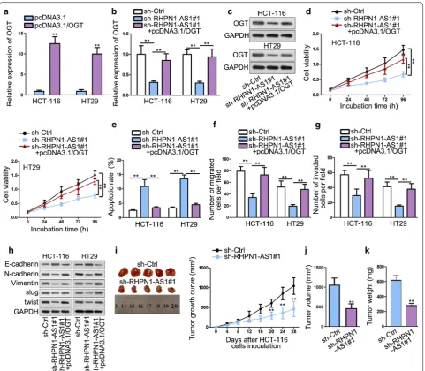
To determine whether RHPN1-AS1 regulated CRC development by sponging miR-7-5p and up-regulating OGT, functional rescue experiments were performed. To begin with, qRT-PCR analysis results ensured the
transfection efficiency of pcDNA3.1/OGT (Fig. 5a).
qRT-PCR analysis also studied the effects on OGT after knocking down RHPN1-AS1. The results showed that knockdown of RHPN1-AS1 could inhibit OGT expression significantly compared with control group, but then the expression level of OGT was recovered by
overexpressing OGT (Fig. 5b). Western blot detected
that the change tendency of OGT protein level was
same as that of OGT mRNA expression (Fig. 5c).
CCK-8 showed that cell viability was inhibited after silencing RHPN1-AS1, but recovered again after
overexpressing OGT (Fig. 5d). Flow cytometry
mani-fested that knockdown of RHPN1-AS1 could increase apoptotic cells, but this effect was abolished by OGT
overexpression (Fig. 5e). Transwell results indicated
that OGT enrichment significantly abrogated the anti-migration and anti-invasion role of RHPN1-AS1
knockdown (Fig. 5f, g). Furthermore, western blot
assay further validated this phenomenon, with the results showing that OGT overexpression neutralized the suppressing effects on EMT exerted by
RHPN1-AS1 knockdown (Fig. 5h). In general, these results
RHPN1-AS1 could slow down xenograft tumor growth
than control group (Fig. 5i). Tumor volume and weight
were also obviously lessened in sh-RHPN1-AS1 group
compared with negative control (Fig. 5j, k). These
results suggested that RHPN1-AS1 knockdown could suppress CRC xenograft tumor growth in vivo. In a summary, RHPN1-AS1 serves as miR-7-5p sponge to up-regulate the expression of OGT in CRC cells. MiR-7-5p inhibition and OGT enrichment contribute to neutralizing the suppressing role of RHPN1-AS1 depletion in the malignant behaviors of CRC.
Discussion
Mounting evidence has demonstrated that lncRNAs are closely associated with normal physiological progresses
and pathological change [26]. The aberrantly expressed
lncRNAs could be applied as prediction biomarkers for
many diseases, including cancers [27–29]. In present
study, we found that RHPN1-AS1, as a newly discov-ered lncRNA, was aberrantly up-regulated in CRC cells than non-tumor one. On the one hand, knockdown of RHPN1-AS1 curbed CRC cell proliferation and promoted apoptosis. On the other hand, RHPN1-AS1 knockdown
greatly inhibited cell migration and invasion capacities. These results indicated that RHPN1-AS1 played an onco-genic role in the development of CRC in vitro. STAT3 signal transduction pathway has been reported to be an
important pathway in CRC [30]. In present study,
tran-scription factor STAT3 was found to be closely related to the abnormal up-regulation of RHPN1-AS1 in CRC. We identified that STAT3 could activated the transcription of RHPN1-AS1 via ChIP and luciferase reporter assays.
The competing endogenous RNAs (ceRNAs) network has revealed that lncRNAs could restore the expression and biological function of mRNAs via competitively binding to shared miRNAs. This lncRNA-miRNA-mRNA regulatory mechanism exerted significant impacts on
malignant tumor progression [31]. In this study, we
found that miR-7-5p could interact with RHPN1-AS1. Contrary to the expression status of RHPN1-AS1, miR-7-5p was aberrantly down-regulated in CRC cell lines
compared with normal colorectal epithelial cell line. Furthermore, miR-7-5p inhibition could abrogate the suppressing effects of RHPN1-AS1 knockdown on CRC cell proliferation, apoptosis, migration and invasion. This indicated that RHPN1-AS1 was a sponge of miR-7-5p in CRC and its oncogenic function was fulfilled by sponging miR-7-5p expression.
MiRNAs can bind to 3′UTR of target gene with micro
response elements (MREs) and further inhibit the
cel-lular function of target gene [32]. OGT was identified
a target gene of miR-7-5p by multiple means, including qRT-PCR, luciferase reporter and RIP assays. We found that OGT expression down-regulated most apparently by miR-7-5p overexpression compared with other potential combinable mRNAs. It has been found that OGT is sig-nificantly upregulate in colon tissues and may play cru-cial role in colon cancer carcinogenesis and progression
[33]. Furthermore, OGT has been found to promote CRC
metastasis by negatively regulating miR-101 and
increas-ing EZH2 stability [34]. In this study, OGT was also
sig-nificantly up-regulated in CRC cell lines compared with normal colon cell line. Silencing OGT inhibited CRC malignant progresses, including cell proliferation, migra-tion and invasion. Besides, silencing OGT facilitated CRC apoptosis. Rescue experiments revealed that OGT overexpression could abrogate the repressing effects of RHPN1-AS1 down-regulation on CRC cell proliferation, migration and invasion. Overall, these findings initially revealed that lncRNA RHPN1-AS1 promoted CRC pro-gression via acting as ceRNA to up-regulating OGT by sponging miR-7-5p.
Conclusion
In summary, we initially discovered the underlying reg-ulatory role of RHPN1-AS1/miR-7-5p/OGT network in CRC. Additionally, whether RHPN1-AS1 could be applied as a potential therapeutic target in CRC waits to be further explored in future researches.
Supplementary information
Supplementary information accompanies this paper at https ://doi.
org/10.1186/s1293 5-020-1110-9.
Additional file 1: Figure S1. (A) The apoptosis of sh-OGT#1 or sh-Ctrl transfected CRC cells was measured by flow cytometry assay.
Abbreviations
ANOVA: analysis of variance; Bio: biotinylated; CCK-8: cell counting kit-8; ceRNA: competing endogenous RNA; ChIP: chromatin immunoprecipitation; CRC : colorectal cancer; EdU: 5-ethynyl-2′-deoxyuridine; EMT: epithelial–mes-enchymal transition; ESCC: esophageal squamous cell carcinoma; FBS: fetal bovine serum; FEZF1-AS1: FEZ family zinc finger 1 antisense RNA 1; FISH: fluo-rescence in situ hybridization; GLUT-1: glucose transporter 1; IL-6: Interleukin-6; lncRNAs: long non-coding RNAs; mRNA: Messenger RNA; miRNA: microRNA; MREs: micro response elements; MUT: mutant; ns: no significance; ncRNAs:
non-coding RNAs; NSCLC: non-small cell lung cancer; OGT: O-GlcNAcylation transferase; PBS: phosphate buffer saline; PI: Propidium iodide; PMT: promoter; PVDF: polyvinylidene fluoride; qRT-PCR: quantitative real time polymerase chain reaction; RHPN1-AS1: Rhophilin Rho GTPase binding protein 1 antisense RNA 1; RIP: RNA immunoprecipitation; RIPA: radio immunoprecipitation assay; SD: standard deviation; SDS-PAGE: sodium dodecyl sulfate polyacrylamide gel electrophoresis; STAT3: signal transducer and activator of transcription 3; WT: wild-type; XIRP2-AS1: xin actin binding repeat containing 2 antisense RNA 1.
Acknowledgements
This study was supported by research members for their substantial experi-ment results.
Authors’ contributions
Conceptualization: WZ. Methodology: YZ. Software: HL. Validation: WZ. Formal analysis: YZ. Investigation: CZ. Data curation: ZZ. Writing—original draft preparation: HL and YZ. Writing—review and editing: WZ. Visualization: HZ. Supervision: WZ. Project administration: WZ. All authors read and approved the final manuscript.
Funding
None.
Availability of data and materials
Research data are not shared.
Ethics approval and consent to participate
This study was progressed with the approval of the Ethics Committee of Henan Provincial People’s Hospital.
Consent for publication
We hope it to be considered for publication in your journal. All authors agreed upon the publication of this manuscript.
Competing interests
The authors declare that they have no competing interests.
Author details
1 Department of Gastrointestinal Surgery, Henan Provincial People’s Hospital,
Zhengzhou University People’s Hospital, Zhengzhou 450003, Henan, China.
2 Department of Gastroenterology, Affiliated Hangzhou First People’s Hospital,
Zhejiang University School of Medicine, No. 261 Huanshan Road, Hang-zhou 310006, Zhejiang, China.
Received: 14 October 2019 Accepted: 16 January 2020
References
1. Siegel R, Desantis C, Jemal A. Colorectal cancer statistics, 2014. Cancer J Clin. 2014;64(2):104–17.
2. Miller KD, Siegel RL, Lin CC, Mariotto AB, Kramer JL, Rowland JH, Stein KD, Alteri R, Jemal A. Cancer treatment and survivorship statistics, 2016. Cancer J Clin. 2016;66(4):271–89.
3. Ladabaum U, Dominitz JA, Kahi C, Schoen RE. Strategies for colorectal cancer screening. Gastroenterology. 2020;158(2):418–32.
4. Han Y, Yang YN, Yuan HH, Zhang TT, Sui H, Wei XL, Liu L, Huang P, Zhang WJ, Bai YX. UCA1, a long non-coding RNA up-regulated in colorectal cancer influences cell proliferation, apoptosis and cell cycle distribution. Pathology. 2014;46(5):396–401.
5. Migliore L, Migheli F, Spisni R, Coppede F. Genetics, cytogenetics, and epigenetics of colorectal cancer. J Biomed Biotechnol. 2011;2011:792362. 6. Ling H, Fabbri M, Calin GA. MicroRNAs and other non-coding RNAs
as targets for anticancer drug development. Nat Rev Drug Discov. 2013;12(11):847–65.
7. Chang W. Non-coding RNAs and berberine: a new mechanism of its anti-diabetic activities. Eur J Pharmacol. 2017;795:8–12.
•fast, convenient online submission
•
thorough peer review by experienced researchers in your field
• rapid publication on acceptance
• support for research data, including large and complex data types
•
gold Open Access which fosters wider collaboration and increased citations maximum visibility for your research: over 100M website views per year
•
At BMC, research is always in progress.
Learn more biomedcentral.com/submissions
Ready to submit your research? Choose BMC and benefit from: 9. Xu Y, Li J, Wang P, Zhang Z, Wang X. LncRNA HULC promotes lung
squa-mous cell carcinoma by regulating PTPRO via NF-kappaB. J Cell Biochem. 2019;20(12):19415–21.
10. Yao N, Fu Y, Chen L, Liu Z, He J, Zhu Y, Xia T, Wang S. Long non-coding RNA NONHSAT101069 promotes epirubicin resistance, migration, and invasion of breast cancer cells through NONHSAT101069/miR-129-5p/ Twist1 axis. Oncogene. 2019;38(47):7216–33.
11. Ramnarine VR, Kobelev M, Gibb EA, Nouri M, Lin D, Wang Y, Buttyan R, Davicioni E, Zoubeidi A, Collins CC. The evolution of long noncoding RNA acceptance in prostate cancer initiation and progression, and its clinical utility in disease management. Eur Urol. 2019;76(5):546–59.
12. Chang Y, Li N, Yuan W, Wang G, Wen J. LINC00997, a novel long noncod-ing RNA, contributes to metastasis via regulation of S100A11 in kidney renal clear cell carcinoma. Int J Biochem Cell Biol. 2019;116:105590. 13. Taucher V, Mangge H, Haybaeck J. Non-coding RNAs in pancreatic
cancer: challenges and opportunities for clinical application. Cell Oncol (Dordrecht). 2016;39(4):295–318.
14. Zhou F, Shen F, Zheng Z, Ruan J. The LncRNA XIRP2-AS1 predicts favora-ble prognosis in colon cancer. OncoTargets Ther. 2019;12:5767–78. 15. Bai J, Xu J, Zhao J, Zhang R. Downregulation of lncRNA AWPPH inhibits
colon cancer cell proliferation by downregulating GLUT-1. Oncol Lett. 2019;18(2):2007–12.
16. Li J, Zhao LM, Zhang C, Li M, Gao B, Hu XH, Cao J, Wang GY. The lncRNA FEZF1-AS1 promotes the progression of colorectal cancer through regu-lating OTX1 and targeting miR-30a-5p. Oncol Res. 2019;28(1):51–63. 17. Qiu X, Lei Z, Wang Z, Xu Y, Liu C, Li P, Wu H, Gong Z. Knockdown of
LncRNA RHPN1-AS1 inhibits cell migration, invasion and proliferation in head and neck squamous cell carcinoma. J Cancer. 2019;10(17):4000–8. 18. Li X, Zhang X, Yang C, Cui S, Shen Q, Xu S. The lncRNA RHPN1-AS1
downregulation promotes gefitinib resistance by targeting miR-299-3p/TNFSF12 pathway in NSCLC. Cell Cycle (Georgetown, Tex). 2018;17(14):1772–83.
19. Matsuyama T, Ishikawa T, Takahashi N, Yamada Y, Yasuno M, Kawano T, Uetake H, Goel A. Transcriptomic expression profiling identifies ITGBL1, an epithelial to mesenchymal transition (EMT)-associated gene, is a promis-ing recurrence prediction biomarker in colorectal cancer. Mol Cancer. 2019;18(1):19.
20. Xu TP, Liu XX, Xia R, Yin L, Kong R, Chen WM, Huang MD, Shu YQ. SP1-induced upregulation of the long noncoding RNA TINCR regulates cell proliferation and apoptosis by affecting KLF2 mRNA stability in gastric cancer. Oncogene. 2015;34(45):5648–61.
21. Wang Y, Zhou J, Wang Z, Wang P, Li S. Upregulation of SOX2 activated LncRNA PVT1 expression promotes breast cancer cell growth and inva-sion. Biochem Biophys Res Commun. 2017;493(1):429–36.
22. Chung SS, Dutta P, Chard N, Wu Y, Chen QH, Chen G, Vadgama J. A novel curcumin analog inhibits canonical and non-canonical functions of tel-omerase through STAT3 and NF-kappaB inactivation in colorectal cancer cells. Oncotarget. 2019;10(44):4516–31.
23. Wang J, Zhou J, Jiang C, Zheng J, Namba H, Chi P, Asakawa T. LNRRIL6, a novel long non-coding RNA, protects colorectal cancer cells by activating the IL-6-STAT3 pathway. Mol Oncol. 2019;13(11):2344–60.
24. Dong M, Xie Y, Xu Y. miR-7-5p regulates the proliferation and migration of colorectal cancer cells by negatively regulating the expression of Kruppel-like factor 4. Oncol Lett. 2019;17(3):3241–6.
25. Cui XB, Zhang SM, Xu YX, Dang HW, Liu CX, Wang LH, Yang L, Hu JM, Liang WH, Jiang JF, et al. PFN2, a novel marker of unfavorable prognosis, is a potential therapeutic target involved in esophageal squamous cell carcinoma. J Transl Med. 2016;14(1):137.
26. Zhang Y, Xu Y, Feng L, Li F, Sun Z, Wu T, Shi X, Li J, Li X. Comprehensive characterization of lncRNA-mRNA related ceRNA network across 12 major cancers. Oncotarget. 2016;7(39):64148–67.
27. Choi RS, Lai WYX, Lee LTC, Wong WLC, Pei XM, Tsang HF, Leung JJ, Cho WCS, Chu MKM, Wong EYL, et al. Current and future molecular diagnos-tics of gastric cancer. Exp Rev Mol Diagn. 2019;19(10):863–74. 28. Wei L, Wang X, Lv L, Zheng Y, Zhang N, Yang M. The emerging role of
noncoding RNAs in colorectal cancer chemoresistance. Cell Oncol (Dordrecht). 2019;42(6):757–68.
29. Oehme F, Krahl S, Gyorffy B, Muessle B, Rao V, Greif H, Ziegler N, Lin K, Thepkaysone ML, Polster H, et al. Low level of exosomal long non-coding RNA HOTTIP is a prognostic biomarker in colorectal cancer. RNA Biol. 2019;16:1–7.
30. Tan H, Li X, Yang WH, Kang Y. A flavone, Wogonin from Scutellaria baicalensis inhibits the proliferation of human colorectal cancer cells by inducing of autophagy, apoptosis and G2/M cell cycle arrest via modulating the PI3K/AKT and STAT3 signalling pathways. J BUON. 2019;24(3):1143–9.
31. Sharma S, Munger K. Expression of the cervical carcinoma expressed PCNA regulatory (CCEPR) long noncoding RNA is driven by the human papillomavirus E6 protein and modulates cell proliferation independent of PCNA. Virology. 2018;518:8–13.
32. Yu Y, Yin W, Yu ZH, Zhou YJ, Chi JR, Ge J, Cao XC. miR-190 enhances endo-crine therapy sensitivity by regulating SOX9 expression in breast cancer. J Exp Clin Cancer Res. 2019;38(1):22.
33. Mi W, Gu Y, Han C, Liu H, Fan Q, Zhang X, Cong Q, Yu W. O-GlcNAcylation is a novel regulator of lung and colon cancer malignancy. Biochem Biophys Acta. 2011;1812(4):514–9.
34. Jiang M, Xu B, Li X, Shang Y, Chu Y, Wang W, Chen D, Wu N, Hu S, Zhang S, et al. O-GlcNAcylation promotes colorectal cancer metastasis via the miR-101-O-GlcNAc/EZH2 regulatory feedback circuit. Oncogene. 2019;38(3):301–16.
Publisher’s Note