P R I M A R Y R E S E A R C H
Open Access
The mitochondrial C16069T polymorphism, not
mitochondrial D310 (D-loop) mononucleotide
sequence variations, is associated with bladder
cancer
Nasser Shakhssalim
1, Massoud Houshmand
2,4*, Behnam Kamalidehghan
3, Abolfazl Faraji
4, Reza Sarhangnejad
1,
Sepideh Dadgar
4, Maryam Mobaraki
4, Rozita Rosli
5and Mohammad Hossein Sanati
3Abstract
Background:Bladder cancer is a relatively common and potentially life-threatening neoplasm that ranks ninth in terms of worldwide cancer incidence. The aim of this study was to determine deletions and sequence variations in the mitochondrial displacement loop (D-loop) region from the blood specimens and tumoral tissues of patients with bladder cancer, compared to adjacent non-tumoral tissues.
Methods:The DNA from blood, tumoral tissues and adjacent non-tumoral tissues of twenty-six patients with bladder cancer and DNA from blood of 504 healthy controls from different ethnicities were investigated to deter-mine sequence variation in the mitochondrial D-loop region using multiplex polymerase chain reaction (PCR), DNA sequencing and southern blotting analysis.
Results:From a total of 110 variations, 48 were reported as new mutations. No deletions were detected in tumoral tissues, adjacent non-tumoral tissues and blood samples from patients. Although the polymorphisms at loci 16189, 16261 and 16311 were not significantly correlated with bladder cancer, the C16069T variation was significantly present in patient samples compared to control samples (p < 0.05). Interestingly, there was no significant difference (p > 0.05) of C variations, including C7TC6, C8TC6, C9TC6 and C10TC6, in D310 mitochondrial DNA between patients and control samples.
Conclusion:Our study suggests that 16069 mitochondrial DNA D-Loop mutations may play a significant role in the etiology of bladder cancer and facilitate the definition of carcinogenesis-related mutations in human cancer.
Keywords:Mitochondrial DNA displacement loop, 16069 D-Loop mutation, Urinary bladder neoplasm
Introduction
Human mitochondrial DNA (mtDNA) is a 16569-bp closed circular, double-stranded molecule approximately 1000 copies per cell. mtDNA contains 37 genes, includ-ing 13 subunits involved in the electron transport chain, 22 tRNAs, the 12S and 16S rRNAs, and a non-coding region (D-loop) located at nucleotide position 16024– 576 (MITOMAP, 2011) [1]. The D-loop region regulates the replication and transcription of mtDNA, where
mutations in this region might lead to copy number and/or change in mtDNA gene expression [2].
Bladder cancer is the ninth most common cancer worldwide [3]. According to the latest American Cancer Society statistics, bladder cancer accounts for 7% of all cancers and 3% of all cancer deaths [4,5]. In Iran, blad-der cancer accounts for 7.04% of all cancers [6].
Many attempts have been made to develop an urothelial cancer biomarker test to complement or replace urine cy-tology, including NMP22, BTA stat, BTA TRAK, and FISH. Most studies on the molecular genetics of the bladder can-cer focus on changes in genomic DNA, including onco-genes and tumor suppressor onco-genes, such asHRAS, ERBB2, TP53andRB,and subsequent cellular events [7,8].
* Correspondence:[email protected] 2
National Institute for Genetic Engineering and Biotechnology, Tehran, Iran 4Medical Genetics Department, Special Medical Center, Tehran, Iran
Full list of author information is available at the end of the article
Mitochondrial function and DNA attract less interest in studies on bladder carcinoma. Mitochondrial dysfunc-tion has been linked to a wide range of degenerative and metabolic diseases, cancer, and even aging. mtDNA, which has a very high mutation rate, results in three classes of clinically relevant phenotypes: deleterious germline mtDNA mutations, which are linked to mito-chondrial diseases; mtDNA polymorphisms, which are related to environmental adaptation in human evolution; and mtDNA somatic mutations, which are associated with aging and cancer. Mitochondrial defects were first associated with carcinogenesis several decades before, when Warburg reported“injury of the respiratory chain” and high glycolytic rate as typical of cancer [9-12].
Mitochondrial DNA is thought to accumulate more mutations than nuclear DNA (nDNA) to some extent, because the protective histones as well as the highly effi-cient DNA repair mechanisms do not exist in the mito-chondrial nucleus. Certain tumors have been shown to result from mutations in nDNA-encoded mitochondrial proteins, which may result in increased reactive oxygen species (ROS) production. Mitochondrial dysfunction does appear to be a factor in cancer etiology. Alterations in mitochondrial DNA (mtDNA), including point muta-tions, delemuta-tions, insertions and genome copy number changes, are believed to be responsible for carcinogen-esis [13-15]. For example, many reports have identified a mtDNA 4977-bp deletion in lung [16], breast [17] and endometrial carcinomas [18].
The use of mtDNA mutation and/or polymorphism patterns as a biomarker is rapidly expanding in disci-plines, ranging from rare metabolic diseases and aging to cancer and the tracing of human migration patterns, population characterization and human identification in forensic science. In this study, we examined the presence of mutations in the mitochondrial D-Loop sequences of tumoral tissues as compared with adjacent non-tumoral tissues from Iranian patients with bladder cancer.
Table 1 Age and histological type of primary urothelial bladder neoplasm sybtypes
No. of Male patients
Age Histological type
1 62 Carcinoma in situ+
2 60 Papilloma
3 58 Papillary Urothelial carcinoma- low grade
4 63 Neoplasm of low malignant potential Papillary urothelial
5 73 Carcinoma in situ+
6 80 Papillary urothelial carcinoma–high grade
7 69 Papillary Urothelial carcinoma- low grade
8 68 Neoplasm of low malignant potential Papillary urothelial
9 53 Non-papillary urothelial carcinoma–high grade
10 55 Papillary urothelial carcinoma–high grade
11 75 Non-papillary urothelial carcinoma–high grade
12 78 Papillary Urothelial carcinoma- low grade
13 73 Neoplasm of low malignant potential Papillary urothelial
14 69 Papillary Urothelial carcinoma- low grade
15 68 Non-papillary urothelial carcinoma–high grade
16 57 Neoplasm of low malignant potential Papillary urothelial
17 53 Non-papillary urothelial carcinoma–high grade
18 50 Papillary urothelial carcinoma–high grade
19 49 Papillary Urothelial carcinoma- low grade
20 45 Non-papillary urothelial carcinoma–high grade
21 29 Papillary urothelial carcinoma–high grade
22 70 Papillary Urothelial carcinoma- low grade
23 66 Non-papillary urothelial carcinoma–high grade
24 58 Papillary urothelial carcinoma–high grade
25 59 Papillary Urothelial carcinoma- low grade
26 74 Neoplasm of low malignant potential Papillary urothelial
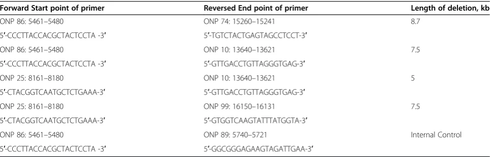
Table 2 Primers used for detection of four deletions
Forward Start point of primer Reversed End point of primer Length of deletion, kb
ONP 86: 5461–5480 ONP 74: 15260–15241 8.7
5′-CCCTTACCACGCTACTCCTA -3′ 5′-TGTCTACTGAGTAGCCTCCT-3′
ONP 86: 5461–5480 ONP 10: 13640–13621 7.5
5′-CCCTTACCACGCTACTCCTA -3′ 5′-GTTGACCTGTTAGGGTGAG-3′
ONP 25: 8161–8180 ONP 10: 13640–13621 5
5′-CTACGGTCAATGCTCTGAAA-3′ 5′-GTTGACCTGTTAGGGTGAG-3′
ONP 25: 8161–8180 ONP 99: 16150–16131 7.5
5′-CTACGGTCAATGCTCTGAAA-3′ 5′-GTGGTCAAGTATTTATGGTA-3′
ONP 86: 5461–5480 ONP 89: 5740–5721 Internal Control
Table 3 List of variations in both healthy controls and bladder cancer patients
NO. Variations Controls Patients
1 15968
2 15969
3 15996
4 16004
5 16017 *
6 16021
7 16026
8 16033 *
9 16051 *
10 16067 *
11 16069
12 16071
13 16075
14 16082
15 16085
16 16086
17 16092 *
18 16093
19 16095
20 16111 *
21 16114
22 16124
23 16126
24 16129 *
25 16140
26 16145
27 16147 *
28 16148
29 16150 *
30 16153
31 16155
32 16162
33 16163
34 16167
35 16169
36 16172
37 16173
38 16174 *
39 16176 *
40 16179
41 16183 *
42 16184
43 16187 *
Table 3 List of variations in both healthy controls and bladder cancer patients(Continued)
44 16188 *
45 16189
46 16192
47 16193
48 16201 *
49 16203
50 16207 *
51 16209
52 16213
53 16217 *
54 16220
55 16222
56 16223
57 16224 *
58 16227 *
59 16230 *
60 16234 *
61 16239 *
62 16242 *
63 16243
64 16245
65 16247 *
66 16248 *
67 16249
68 16256 *
69 16261
70 16263
71 16264
72 16265 *
73 16266
74 16270 *
75 16274 *
76 16278 *
77 16286
78 16287 *
79 16288
80 16290 *
81 16292
82 16294
83 16295
84 16296
85 16298 *
86 16304 *
Materials and Methods
Twenty-six men with primary urothelial bladder cancer with a mean age of 62.5 years were enrolled in this study (Table 1). The patients’written consent was obtained and the institutional review board approved this study. Tu-moral tissues were obtained from transurethral resection of the bladder tumor (TURBT) or radical cystectomy specimens. Tumoral tissues and adjacent non-tumoral tis-sues were immediately frozen in liquid nitrogen and kept at −80°C, while blood samples from patients were ob-tained before surgery.
Urothelial bladder cancer diagnosis was done via histological analysis. Blood samples from healthy con-trols with a mean age of 57.5 years were obtained from 404 individuals of 17 ethnicities and 100 random indi-viduals, all from the Tehran Special Medical Center. The exclusion criterion for the control group was any history of cancer, metabolic diseases and mitochondrial DNA related diseases that may affect the mtDNA. Ethics ap-proval and patient informed consent including consent to participate in the study and consent to publish was obtained for the present study in accordance to the Tehran Special Medical Center and Medical Ethics Committee (Approval No. MS-16-2007).
DNA extraction and sequencing
Genomic DNA (DNA fast, QIAGEN, Cat. No. 51204) was isolated from the tumoral tissues, adjacent non-tumoral tissues and blood samples of patients, as well as from the blood samples of controls, according to the manufacturer’s protocol. Two pairs of primers designed to amplify the mtDNA D-loop region are as follows: ONP 98 F )1579-15810(: 5′-ATC ATT GGA CAA GTA
Table 3 List of variations in both healthy controls and bladder cancer patients(Continued)
88 16311
89 16318 *
90 16318
91 16319
92 16320 *
93 16324 *
94 16325 *
95 16327 *
96 16342
97 16343
98 16352 *
99 16354
100 16355 *
101 16356 *
102 16362
103 16390 *
104 16391 *
105 16399 *
106 16413
107 16468
108 16482 *
109 16497 *
110 16527 *
*Indicates novel mutation has not been reported before.
GCA TC -3′ and ONP 79R )780-761(: 5′-GAG CTG CAT TGC TGC GTG CT-3′. Polymerase chain reaction (PCR) was carried out with the following protocol: pre-denaturation at 95°C for 5 min, then 35 cycles of 94°C for 30 sec, 60°C for 45 sec and 72°C for 1 min, and a final extension step of 72°C for 6 min. Each amplified fragment was purified using a Agarose Gel DNA Frag-ment Recovery Kit, Ver.2.0 (TaKaRa, Japan) and subse-quently sequenced using a ABI PRISM 3730 sequence analyzer (gene Fanavaran, Macrogene Seoul, Korea). The quality of the obtained chromatograms was assessed by FinchTV® software Version 1.4.0 (Geospiza, Inc., USA).
Multiplex PCR
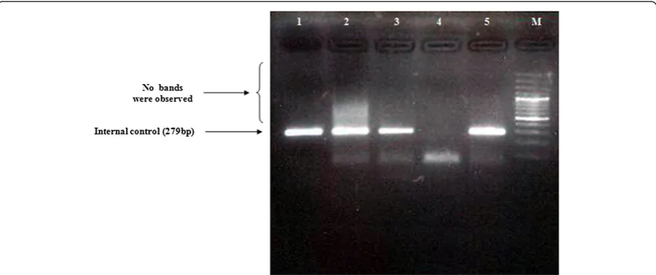
The PCR reactions were performed for 35 cycles of the following steps: 94°C for 10 min, 55°C for 10 min, and 72°C for 35 sec. Using the primers ONP 86, ONP 89, ONP 10, ONP 74, ONP 25 and ONP 99, the deletion-prone region between 5461 nt of the light strand and 15000 nt of the heavy strand was investigated in all the patients. The distances between the primers were long enough to allow amplification only if a part of the DNA between each respective primers was deleted. As a con-trol in PCR analysis, a normal internal mtDNA fragment in a region which is seldom affected by deletions was Figure 2Long range PCR amplification of mtDNA using Phusion Flash high-fidelity PCR Master Mix, Thermo Scientific.A two-step long-range PCR was carried out on the major arc of the mitochondrial genome using the Expand Long Template PCR System to detect mitochondrial deletions. DNA products were separated using a 0.7% agarose gel containing ethidium bromide and viewed under UV light. Lanes 1 and 3: negative control; Lanes 2 and 4: an amplified 11 Kb fragment, indicating no deletions were observed in mtDNA; lane M: 1 kb DNA ladder marker.
amplified using the primer pair of ONP 86 and ONP 89 (Table 2). Polymerase chain reaction products were sepa-rated on 2% agarose gels and run in 0.5× Tris/Borate/ EDTA buffer at 110 V for 50 min, stained in 0.002μg/mL ethidium bromide, and visualized by means of an ultravio-let light.
Southern blot analysis
primer (5′-GAGCTGCATTGCTGCGTGCT-3′), located at 780–761 bp of the mtDNA, were used to amplify a 1558-bp fragment from the D-loop region. This fragment was used as a mtDNA probe. Southern blot analysis was performed using the DIG DNA Labeling and Detection Kit (Cat. #11093657910, Roche).
Statistical analysis
Sequences were edited and aligned using ClustalX. The revised Cambridge Reference Sequence was used as a reference (GI: 251831106) (MITOMAP, 2009). The Chi-square test was used with SPSS (Statistical Package for the Social Sciences, version: 13) to examine the associ-ation of variassoci-ations with control and patient samples. P-values < 0.05 were regarded as statistically significant.
Results
Samples from a total of 26 patients with sporadic bladder cancer were screened for mitochondrial deletions and var-iations. Sequence analysis found a total of 110 variations (Cambridge Mitochondrial Sequences), of which 62 muta-tions were previously reported (MITOMAP). However, 48 of these mutations were reported as new mutations, which are summarized in Table 3. In this study, almost all of the variations were homoplasmic, but in 6 (16.6%) cases, a C nucleotide insertion was seen in locus 16194. No mito-chondrial deletions were found in the patient samples (Figures 1 and 2), as confirmed by Southern blotting (Figure 3).
Four common variations, 16069, 16189, 16261 and 16311, were found in the tumoral tissues, adjacent non-tumoral tissues and blood samples of both patients and controls from different ethnicities. The polymorphisms at 16189, 16261 and 16311 were not significantly corre-lated with bladder cancer. However, the D-loop C16069T polymorphism (Figure 4) was significantly correlated with bladder cancer (P< 0.05). Analysis of control samples by ethnicities for these 4 variations is summarized in Table 4. No significant difference (p> 0.05) in D310 C variations was observed between the patient and control samples (Table 5).
Discussion
Our sequencing analysis focused on the mtDNA D-loop region, which is highly polymorphic and contains two hypervariable regions, HV1 (16024–16383) and HV2 (57–333), that was considered as a somatic mutation “hot spot”in many types of cancer [19]. In this study, no deletions were seen in the mitochondrial genome. One hundred and sixteen variations were observed in the D-Loop region, where 48 of them were not previously re-ported. Wada et al.[20] also reported that the majority of somatic mutations were homoplasmic, suggesting that the mutant mtDNA became dominant in tumor cells.
Table 4 Comparison of 4 common variations in bladder cancer patients and controls
Ethnicity NO. 16069 16189 16261 16311
Arab 23 0 (0%) 7 (30.4%) 4 (17.4%) 4 (17.4%)
Armenian 18 0 (0%) 5 (27.7%) 2 (11%) 3 (16.7%)
Asurian 19 1 (5.2%) 7 (36.8%) 3 (15.8%) 1 (5.3%)
Azari 22 0 (0%) 6 (27.3%) 0 (0%) 4 (18.2%)
Turkmen 37 1 (2.7%) 5 (13.5%) 1(2.7%) 6 (16.2%)
Baluch 13 0 (0%) 2 (15.4%) 1 (7.7%) 0 (0%)
Bandari 31 0 (0%) 13 (42%) 2 (6.5%) 5 (13.5%)
Guilani 24 0 (0%) 2 (8.3%) 3 (12.5%) 3 (12.5%)
Jews 37 1 (2.7%) 6 (16.2%) 4 (10.8%) 4 (10.8%)
Kurd 24 2 (8.3%) 3 (12.5%) 4 (16.7%) 6 (25%)
Lur 22 0 (0%) 1 (4.5%) 8 (36.4%) 8 (36.4%)
Mazani 23 0 (0%) 4 (17.4%) 4 (17.4%) 1 (4.3%)
Persian Isfahan 16 0 (0%) 6 (37.5%) 2 (12.5%) 5 (31.2%)
Persian Kerman 25 0 (0%) 7 (28%) 1 (4%) 5 (20%)
Persian Mashhad 23 0 (0%) 5 (21.7%) 2 (8.7%) 2 (8.7%)
Persian Shiraz 23 0 (0%) 4 (17.4%) 2 (8.7%) 3 (3.2%)
Persian Yazd 24 0 (0%) 5 (20.8%) 1 (4.1%) 2 (8.3%)
Mixed Tehran 100 8 (8%) 9 (9%) 9 (9%) 12 (12%)
Total (controls) 504 13 (2.6%) 95 (18.8%) 53 (10.5%) 78 (15.5%)
Patients 26 5 (19%)* 4 (15.4%) 4 (15.4%) 8 (31%)
*Shows statistically significant,p< 0.05.
Table 5 Association of the mtDNA D310 variation in bladder cancer patients and controls
Ethnicity NO. C 7TC6 C 8TC6 C 9TC6 C 10TC6
Arab 23 12 (52.2%) 8 (34.7%) 2 (8.7%) 1 (4.3%)
Armenian 18 7 (38.9%) 11 (61.1%) 0 (0%) 0 (0%)
Azari 22 8 (36.4%) 12 (54.5%) 2 (9%) 0 (0%)
Turkmen 37 17 (45.9%) 16 (43%) 4 (10.8%) 0 (0%)
Bandari 31 10 (32%) 15 (48.4%) 6 (19.4%) 0 (0%)
Persian Isfahan 16 5 (31.3%) 9 (56.3%) 2 (6.5%) 0 (0%)
Persian Mashhad 23 14 (60.9%) 8 (34.8%) 1 (4.3%) 0 (0%)
Persian Shiraz 23 6 (26%) 16 (69.6) 1 (4.3%) 0 (0%)
Persian Yazd 24 9 (37.5%) 9 (37.5%) 5 (20.8%) 1 (4.1%)
Guilani 24 8 (33%) 12 (50%) 4 (16.6%) 0 (0%)
Jews 37 16 (43%) 17 (45.9) 4 (10.8%) 0 (0%)
Kurd 24 3 (12.5%) 14 (58%) 7 (29%) 0 (0%)
Lur 22 9 (41%) 9 (41%) 4 (18%) 0 (0%)
Total (controls) 324 124 (38.3) 156 (48.1%) 42 (13%) 2 (0.6%)
Patients 21 9 (42.9%) 10 (47.6%) 2 (9.5%) 0 (0%)
Fliss et al.[21] screened 14 urinary bladder cancers for somatic mutations in the D-loop region, and found mu-tations in 4 (29%) samples.
Polymorphism 16189, which is highly polymorphic, was the previous focus of oncological research because carriers with the T16189C polymorphism were appar-ently more susceptible to breast cancer and ganglioma development. Interestingly, the T16189C polymorphism was found in 14% of endometrial cancers [22] and type II diabetes mellitus [23,24].
In this study, in contrast to 16189, 16194, 16261 and 16311 variations, the C16069T polymorphism of the D-loop indicated significant correlation with bladder cancer (P < 0.05), which has not been studied in bladder cancer before. However, the C16069T polymorphism has been reported in prostate cancer [25], pancreatic cancer [26], endometrial cancer [27], breast cancer [28,29], repeated pregnancy loss [30] and age-related macular degener-ation [31]. This result supports our hypothesis, which shows the potential of specific mitochondrial 16069 polymorphism involvement in carcinogenesis.
Many studies reported that the C150T polymorphism is correlated with longevity (MITOMAP, 2009). The pos-sible function of the C150T transition was investigated in a previous study [32], suggesting that the C150T tran-sition functions in remodeling mtDNA replication. How-ever, in our study, no significant differences were found between C150T mutations in patients and control sam-ples from different ethnicities.
Large-scale mtDNA deletions have been demonstrated in several cancers. Kamalidehghanet al. [33] found that the common mtDNA4977 deletion was less frequent in gastric cancer tissues compared to the normal adjacent tissues. While in another study, a deletion of approxi-mately 8.9 kb was more frequent in gastric carcinoma tissues than adjacent normal tissue samples [34]. How-ever, in the present study, no deletions were detected in bladder carcinoma tissues nor adjacent non-tumoral tis-sues. Therefore, the pattern of mitochondrial deletions may differ among different carcinomas.
Marchingtonet al.[35] first used the term D310 to de-scribe a highly polymorphic mononucleotide tract of poly (C) that varies from 12 to 18 Cs, located between nucleotide positions 303 and 318 in CSB II, that forms a RNA–DNA hybrid known as an R-loop. This poly(C) re-gion is interrupted at nucleotide position 310 by a T (CCCCCCCTCCCCC), in which the number of Cs before the T can vary between 7 to 9 in normal poly-morphic variants [35]. D310 has been reported as a mutational hot-spot in a large panel of tumors includ-ing gastric, head and neck, breast, colorectal, lung and bladder cancers, where head and neck cancer has the highest rate of D310 variants (37%), followed by breast (29%) and colorectal (28%) cancers. However, no D310
alterations were detected in prostate and ovarian can-cers [36,37].
The D310 region of mtDNA plays an important role in mitochondrial biogenesis, where somatic insertions or deletions of one or two base pairs in this region are thought to have negligible effects on cancers. However, major deletions or insertions of up to ten bases in the D310 region could interfere with mtDNA biogenesis [38]. Mutations in the D-loop, mostly at D310, have been found in 21% of all head and neck squamous cell carcin-omas [39]. However, in our study, the D310 mtDNA se-quence variations, including C7TC6, C8TC6, C9TC6 and C10TC6, were not significantly different (p > 0.05) be-tween bladder cancer patients and controls of different ethnicities.
In conclusion, our study suggests that the mitochon-drial DNA D-Loop 16069 mutation may play a signifi-cant role in the etiology of bladder cancer and facilitate the definition of carcinogenesis-related mutations in hu-man mtDNA.
Abbreviations
mtDNA:mitochondrial DNA; D-Loop: Displacement loop; PCR: Polymerase chain reaction; nDNA: nuclear DNA; ROS: Reactive oxygen species; TURBT: Transurethral resection of the bladder tumor; SPSS: Statistical package for the social sciences; HV: Hypervariable; mtMSI: Mitochondrial microsatellite instability.
Authors’contributions
NS, AF, SD, MM and RS carried out the experimental procedures. BK and RR wrote and edited the manuscript and performed the statistical analysis. MH conceived the project and supervised the study. All authors read and approved the final manuscript.
Competing of interests
The authors declare that they have no competing of interests.
Acknowledgements
We are thankful to the“Urology and Nephrology Research Center (UNRC) of Tehran”for giving the grant for this project and the“National Institute for Genetic Engineering and Biotechnology (NIGEB) of Tehran”, Project 187.
Author details 1
Urology and Nephrology Research Center (UNRC), Shahid Labbafinejad Medical Center, Shahid Beheshti University of Medical Sciences, Tehran, Iran. 2
National Institute for Genetic Engineering and Biotechnology, Tehran, Iran. 3Department of Pharmacy, Faculty of Medicine, University of Malaya (UM),
Kuala Lumpur 50603, Malaysia.4Medical Genetics Department, Special Medical Center, Tehran, Iran.5UPM-MAKNA Cancer Research Laboratory, Institute of Bioscience, Universiti Putra Malaysia, UPM Serdang, Selangor 43400, Malaysia.
Received: 19 June 2013 Accepted: 27 November 2013 Published: 5 December 2013
References
1. Suzuki M, Toyooka S, Miyajima K, Iizasa T, Fujisawa T, Bekele NB, Gazdar AF: Alterations in the mitochondrial displacement loop in lung cancers.Clin Cancer Res2003,9(15):5636–5641.
2. Yu M, Zhou Y, Shi Y, Ning L, Yang Y, Wei X, Zhang N, Hao X, Niu R: Reduced mitochondrial DNA copy number is correlated with tumor progression and prognosis in Chinese breast cancer patients.IUBMB life 2007,59(7):450–457.
4. Jemal A, Siegel R, Ward E, Hao Y, Xu J, Murray T, Thun MJ:Cancer statistics, 2008.CA Cancer J Clin2008,58(2):71–96.
5. Ferlay J, Autier P, Boniol M, Heanue M, Colombet M, Boyle P:Estimates of the cancer incidence and mortality in Europe in 2006.Ann Oncol2007, 18(3):581.
6. Shakhssalim N, Hosseini SY, Basiri A, Eshrati B, Mazaheri M, Soleimanirahbar A:Prominent bladder cancer risk factors in Iran.Asian Pac J Cancer Prev 2010,11:601–606.
7. Ørntoft TF, Wolf H:Molecular alterations in bladder cancer.Urol Res1998, 26(4):223–233.
8. Brandau S, Böhle A:Bladder cancer.Eur Urol2001,39(5):491–497. 9. Warburg O:On the origin of cancer cells.Science1956,123(3191):309–314. 10. Baysal BE, Ferrell RE, Willett-Brozick JE, Lawrence EC, Myssiorek D, Bosch A,
Mey A, Taschner PEM, Rubinstein WS, Myers EN:Mutations in SDHD, a mitochondrial complex II gene, in hereditary paraganglioma.Science 2000,287(5454):848.
11. Gambhir SS:Molecular imaging of cancer with positron emission tomography.Nat Rev Cancer2002,2(9):683–693.
12. Lima J, Teixeira-Gomes J, Soares P, Máximo V, Honavar M, Williams D, Sobrinho-Simões M:Germline succinate dehydrogenase subunit D mutation segregating with familial non-RET C cell hyperplasia.J Clin Endocrinol Metab2003,88(10):4932.
13. Clayton DA, Doda JN, Friedberg EC:The absence of a pyrimidine dimer repair mechanism in mammalian mitochondria.Proc Natl Acad Sci1974, 71(7):2777.
14. LeDoux SP, Wilson GL, Beecham EJ, Stevnsner T, Wassermann K, Bohr VA: Repair of mitochondrial DNA after various types of DNA damage in Chinese hamster ovary cells.Carcinogenesis1992,13(11):1967. 15. Croteau DL, Bohr VA:Repair of oxidative damage to nuclear and
mitochondrial DNA in mammalian cells.J Biol Chem1997, 272(41):25409–25412.
16. Dai JG, Xiao YB, Min JX, Zhang GQ, Yao K, Zhou RJ:Mitochondrial DNA 4977 BP deletion mutations in lung carcinoma.Indian J Cancer2006, 43(1):20.
17. Ye C, Shu XO, Wen W, Pierce L, Courtney R, Gao YT, Zheng W, Cai Q: Quantitative analysis of mitochondrial DNA 4977-bp deletion in sporadic breast cancer and benign breast diseases.Breast Cancer Res Treat2008, 108(3):427–434.
18. Futyma K, Putowski L, Cybulski M, Miotla P, Rechberger T, Semczuk A:The prevalence of mtDNA4977 deletion in primary human endometrial carcinomas and matched control samples.Oncol Rep2008,20(3):683–688. 19. Akouchekian M, Houshmand M, Hemati S, Ansaripour M, Shafa M:High rate of mutation in mitochondrial DNA displacement loop region in human colorectal cancer.Dis Colon Rectum2009,52(3):526.
20. WADA T, TANJI N, OZAWA A, WANG J, SHIMAMOTO K, SAKAYAMA K, YOKOYAMA M:Mitochondrial DNA mutations and 8-hydroxy-2′ -deoxyguanosine Content in Japanese patients with urinary bladder and renal cancers.Anticancer Res2006,26(5A):3403.
21. Fliss MS, Usadel H, Caballero OL, Wu L, Buta MR, Eleff SM, Jen J, Sidransky D: Facile detection of mitochondrial DNA mutations in tumors and bodily fluids.Science2000,287(5460):2017.
22. Liu VWS, Wang Y, Yang HJ, Tsang PCK, Ng TY, Wong LC, Nagley P, Ngan H: Mitochondrial DNA variant 16189 T > C is associated with susceptibility to endometrial cancer.Hum Mutat2003,22(2):173–174.
23. Poulton J, Luan JA, Macaulay V, Hennings S, Mitchell J, Wareham NJ:Type 2 diabetes is associated with a common mitochondrial variant: evidence from a population-based case–control study.Hum Mol Genet2002, 11(13):1581–1583.
24. Chinnery P, Elliott H, Patel S, Lambert C, Keers S, Durham S, McCarthy M, Hitman G, Hattersley A, Walker M:Role of the mitochondrial DNA 16184–16193 poly-C tract in type 2 diabetes.Lancet2005, 366(9497):1650–1651.
25. Ashtiani ZO, Heidari M, Hasheminasab S-M, Ayati M, Rakhshani N: Mitochondrial D-Loop polymorphism and microsatellite instability in prostate cancer and benign hyperplasia patients.Asian Pac J Cancer Prev 2012,13:3863–3868.
26. Lesina M, Kurkowski MU, Ludes K, Rose-John S, Treiber M, Klöppel G, Yoshimura A, Reindl W, Sipos B, Akira S:Stat3/Socs3 activation by IL-6 transsignaling promotes progression of pancreatic intraepithelial neoplasia and development of pancreatic cancer.Cancer Cell2011, 19(4):456–469.
27. Czarnecka AM, Klemba A, Semczuk A, Plak K, Marzec B, Krawczyk T, Kofler B, Golik P, Bartnik E:Common mitochondrial polymorphisms as risk factor for endometrial cancer.Int Arch Med2009,2(1):33.
28. Rahmani B, Azimi C, Omranipour R, Raoofian R, Zendehdel K, Saee-Rad S, Heidari M:Mutation screening in the mitochondrial D-loop region of tumoral and non-tumoral breast cancer in Iranian patients.Acta Med Iran 2012,50(7):447–453.
29. Rosson D, Keshgegian AA:Frequent mutations in the mitochondrial control region DNA in breast tissue.Cancer Lett2004,215(1):89–94. 30. Seyedhassani SM, Houshmand M, Kalantar SM, Modabber G, Aflatoonian A:
No mitochondrial DNA deletions but more D-loop point mutations in repeated pregnancy loss.J Assist Reprod Genet2010,27(11):641–648. 31. Mueller EE, Schaier E, Brunner SM, Eder W, Mayr JA, Egger SF, Nischler C,
Oberkofler H, Reitsamer HA, Patsch W:Mitochondrial haplogroups and control region polymorphisms in age-related macular degeneration: a case–control study.PLoS One2012,7(2):e30874.
32. Zhang J, Asin-Cayuela J, Fish J, Michikawa Y, Bonafé M, Olivieri F, Passarino G, De Benedictis G, Franceschi C, Attardi G:Strikingly higher frequency in centenarians and twins of mtDNA mutation causing remodeling of replication origin in leukocytes.Proc Natl Acad Sci2003,100(3):1116. 33. Kamalidehghan B, Houshmand M, Panahi MSS, Abbaszadegan MR, Ismail P,
Shiroudi MB:Tumoral Cell mtDNA 8.9 kb deletion is more common than other deletions in gastric cancer.Arch Med Res2006,37(7):848–853. 34. Kamalidehghan B, Houshmand M, Ismail P, Panahi MSS, Akbari MHH: ΔmtDNA < sup > 4977</sup > is more common in non-tumoral cells from gastric cancer sample.Arch Med Res2006,37(6):730–735. 35. Marchington D, Hartshorne G, Barlow D, Poulton J:Homopolymeric tract
heteroplasmy in mtDNA from tissues and single oocytes: support for a genetic bottleneck.Am J Hum Genet1997,60(2):408.
36. Sanchez-Cespedes M, Parrella P, Nomoto S, Cohen D, Xiao Y, Esteller M, Jeronimo C, Jordan RCK, Nicol T, Koch WM:Identification of a mononucleotide repeat as a major target for mitochondrial DNA alterations in human tumors.Cancer Res2001,61(19):7015. 37. Schwartz S Jr, Alazzouzi H, Perucho M:Mutational dynamics in human
tumors confirm the neutral intrinsic instability of the mitochondrial D‐loop poly‐cytidine repeat.Genes Chromosomes Cancer2006, 45(8):770–780.
38. Dakubo GD:Mitochondrial Genetics and Cancer.Heidelberg Dordrecht London New York: Springer; 2010:14–123.
39. Lievre A, Blons H, Houllier A, Laccourreye O, Brasnu D, Beaune P, Laurent-Puig P:Clinicopathological significance of mitochondrial D-Loop mutations in head and neck carcinoma.Br J Cancer2006, 94(5):692–697.
doi:10.1186/1475-2867-13-120
Cite this article as:Shakhssalimet al.:The mitochondrial C16069T polymorphism, not mitochondrial D310 (D-loop) mononucleotide sequence variations, is associated with bladder cancer.Cancer Cell International201313:120.
Submit your next manuscript to BioMed Central and take full advantage of:
• Convenient online submission
• Thorough peer review
• No space constraints or color figure charges
• Immediate publication on acceptance
• Inclusion in PubMed, CAS, Scopus and Google Scholar
• Research which is freely available for redistribution


![(E) 4 [(1,5 Dimethyl 3 oxo 2 phenyl 2,3 dihydro 1H pyrazol 4 yl)iminomethyl]phenyl 4 bromobenzenesulfonate](data:image/gif;base64,R0lGODlhAQABAIAAAP///wAAACH5BAEAAAAALAAAAAABAAEAAAICRAEAOw==)