Open Access
Research
Echocardiographic AV-interval optimization in patients with
reduced left ventricular function
C Melzer*
1, AC Borges
1, F Knebel
1, WS Richter
2, W Combs
3, G Baumann
1and H Theres
1Address: 1I Medizinische Klinik mit Schwerpunkt Kardiologie, Angiologie und Pulmologie, Charité, Campus Mitte, Berlin Germany, 2Klinik für
Nuklearmedizin, Charité, Campus Mitte, Berlin Germany and 3Medtronic Inc., Minneapolis, USA
Email: C Melzer* - [email protected]; AC Borges - [email protected]; F Knebel - [email protected]; WS Richter - [email protected]; W Combs - [email protected]; G Baumann - [email protected]; H Theres - [email protected]
* Corresponding author
Abstract
Background: Ritter's method is a tool used to optimize AV delay in DDD pacemaker patients with normal left ventricular function only. The goal of our study was to evaluate Ritter's method in AV delay-interval optimization in patients with reduced left ventricular function.
Methods: Patients with implanted DDD pacemakers and AVB III° were assigned to one of two groups according to ejection fraction (EF): Group 1 (EF > 35%) and Group 2 (EF < 35%). AV delay optimization was performed by means of radionuclide ventriculography (RNV) and application of Ritter's method.
Results: For each of the patients examined, we succeeded in defining an optimal AV interval by means of both RNV and Ritter's method. The optimal AV delay determined by RNV correlated well with the delay found by Ritter's method, especially among those patients with reduced EF. The intra-class correlation coefficient was 0.8965 in Group 1 and 0.9228 in Group 2. The optimal AV interval in Group 1 was 190 ± 28.5 ms, and 180 ± 35 ms in Group 2.
Conclusion: Ritter's method is also effective for optimization of AV intervals among patients with reduced left ventricular function (EF < 35%). The results obtained by RNV correlate well with those from Ritter's method. Individual programming of the AV interval is fundamentally essential in all cases.
Background
Since introduction of the DDD pacemaker in the early 1980s, researchers have repeatedly attempted to optimize the atrioventricular (AV) interval, for the purpose of max-imizing patient hemodynamic performance. Cannon waves may be induced by programming excessively short AV intervals, and diastolic mitral regurgitation may occur
with excessively long programmed AV intervals. The AV interval is considered optimal (AVopt) if it allows maxi-mum cardiac output.
The duration of the optimal AV interval varies throughout a wide range among individuals, primarily the result of appreciable differences in interatrial conduction [1-4]. Published: 17 December 2004
Cardiovascular Ultrasound 2004, 2:30 doi:10.1186/1476-7120-2-30
Received: 05 October 2004 Accepted: 17 December 2004
This article is available from: http://www.cardiovascularultrasound.com/content/2/1/30
© 2004 Melzer et al; licensee BioMed Central Ltd.
An extensive variety of techniques has been employed to optimize AV delay, including acquisition and analysis of essential hemodynamic parameters by means of aortic-valve Doppler signals, impedance cardiography [9-11], Swan-Ganz catheterization [12-15], and especially the stroke volume [5-8]. Leman et al. [16] have demonstrated that it is also possible to utilize measurement of left ven-tricular ejection fraction and stroke volume by myocardial thallium scintigraphy as a means of AV interval optimiza-tion. A further possibility involves detection of left atrial depolarization by an esophageal electrode recording [17,18]. During recent years, the use of Doppler echocar-diography in conjunction with the mitral valve inflow profile has been investigated as means of AV interval opti-mization: i.e., Ritter's method [19]. Previous investiga-tions have evaluated Ritter's method in patients with normal left ventricular ejection fractions. During recent years, cardiac resynchronization therapy (CRT) has increasingly gained in significance for patients with chronic heart failure (CHF) [20]. In cases without ven-tricular desynchronicity, normal DDD pacemakers (or ICDs with DDD pacemaker function) will in future con-tinue to be implanted in patients with reduced left ven-tricular ejection fraction. The goal of our study was accordingly to apply Ritter's method – until now validated only for patients with normal EF – for patients with reduced left ventricular ejection fraction (EF < 35%).
Methods
We studied 20 DDD pacemaker patients within the con-text of in-office follow-up. Table 1 shows the baseline characteristics and Table 2, the inclusion criteria. We clas-sified patients into two groups, according to left ventricu-lar ejection fraction results obtained by echocardiography. Group 1 consisted of 10 patients with normal left ven-tricular ejection fraction, or with moderately reduced EF (EF > 35%). Group 2 comprised 10 patients with appreci-ably reduced left ventricular ejection fraction (EF < 35%).
We performed ejection fraction analysis by RNV and Rit-ter's method to achieve AV optimization, for 5 AV inter-vals in the range of 100 to 250 ms. We performed all measurements within 15 minutes of AV interval program-ming for every patient. All patients were permanently stimulated in the right atrium and right ventricle (binodal disease). Patient heart rate remained constant during the measurement period at programmed pacemaker lower rate (60 – 70 beats/min).
Analysis of left ventricular ejection fraction by RNV
We performed radionuclide ventriculography (RNV) after in vivo marking of erythrocytes with tin DTPA and 10 MBq/kg KG Tc-99m, using a single-head Gamma camera (CGR gammatome 2, General Electrics, Paris, France) with a high-resolution, medium-energy collimator. For RNV we applied the equilibrium technique at 16 frames per cycle with patients at rest, and ventricular pacing at programmed AV intervals. We acquired a minimum of one million counts per image, and stored the data in a 64 × 64 matrix.
We calculated left ventricular ejection fraction (LVEF) semi-automatically after spatial and temporal smoothing and background subtraction. After Fourier analysis of ven-tricular stimulation progression, we recorded (with exam-iner definition) a region of interest (ROI) around the
end-Age 68.5 ± 4.5 65.7 ± 6.3
Male sex (%) 60 100
Left ventricular ejection fraction (%) 58 ± 9.7 22 ± 7.4 Left ventricular end-diastolic dimension (mm) 47 61 Coronary artery disease (%) 30 50
Dilated cardiomyopathy (%) 0 50
Hypertension (%) 30 0
Pharmacologic therapy (%)
ACE inhibitor 40 100
Beta-blocker 50 90
Loop diuretic 0 100
Spironolactone 0 70
Table 2: Inclusion criteria
DDD pacemaker by AVB III° with permanent atrial and ventricular pacing
diastolic contour of the left ventricle and calculated the LVEF as follows:
Ritter's method
By 1994 a method developed by Ritter et al. had become established for optimizing the AV interval [19]. A prereq-uisite for application of Ritter's method is Doppler-echocardiographic measurement of the mitral inflow profile.
Ritter's method employs the following formula for calcu-lation of the optimal AV interval:
AVopt = AVlong - (a - b)
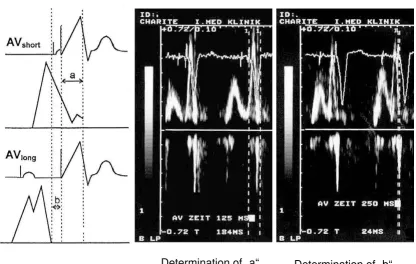
We applied the following procedure in application of this formula (see Fig. 1):
Step 1
The first step involves programming for the pacemaker a nonphysiologically short AV interval, followed by deter-mination of "a". This value "a" is the temporal interval between the ventricular contraction spike and the end of the A wave. "a" designates the electromechanical delay between right ventricular stimulation and the beginning
Ritter's method: The first step is determination of "a" for a nonphysiologically short AV interval (e.g. 125 ms), followed be determination of "b" for a nonphysiologically long AV interval (e.g. 250 ms)
Figure 1
Ritter's method: The first step is determination of "a" for a nonphysiologically short AV interval (e.g. 125 ms), followed be determination of "b" for a nonphysiologically long AV interval (e.g. 250 ms).
Determination of „a“
Determination of „b“
LVEF end diastolic counts end systolic counts end diastolic count
= −
Step 2
The next step is programming for the pacemaker a long AV interval (AVlong), followed by determination of "b". This value "b" is the temporal interval between the ventricular contraction spike and the end of the A wave. AVlong - b defines the duration of the undisturbed maximal diastolic left ventricular filling.
The purpose of AV interval optimization in accordance with Ritter is to allow the ventricular systole to begin immediately subsequent to maximum, undisturbed diastolic ventricular filling and, in turn to prevent the occurrence of Cannon waves as well as diastolic mitral regurgitation.
Results
Group 1
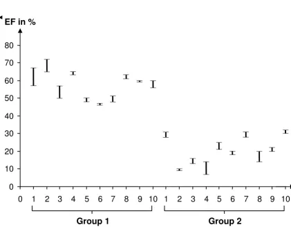
In a given patient, our results indicated that it was possible to define an optimal AV interval for every patient: both by RNV as well as by Ritter's method. The mean optimal AV interval was 190 ± 28.5 ms. The correlation between RNV and Ritter's method is good: the intra-class quotient is 0.8965 (see Fig. 2). In results calculated by RNV, the mean percent difference in left ventricular ejection fraction between the hemodynamically best and worst AV inter-vals was 11 ± 4% (see Fig. 4).
The correlation of the results of the RNV and Ritter methods, with respect to the optimal AV interval for Group 1
Figure 2
The correlation of the results of the RNV and Ritter methods, with respect to the optimal AV interval for Group 1.
120 140 160 180 200 220 240
140
160
180
200
220
IKK= 0.8965
AV opt. RNV [ms]
AV opt. Ritter [
m
Group 2
In a given patient, we likewise succeeded in defining the optimal AV interval for every patient: both by RNV as well as by Ritter's method. The mean optimal AV interval was 180 ± 35 ms. In Group 2 as well, there was good correla-tion between RNV and Ritter's method: the intra-class quotient was 0.9228 (see Fig. 3). In results calculated by RNV, the mean percent difference in left ventricular ejection fraction between the hemodynamically best and worst AV intervals was 28 ± 11% (see Fig. 4).
Discussion
A number of studies have documented the importance of AV synchronization for maximizing the left ventricular ejection fraction in pacemaker patients [21-24]. Despite
CRT, the implantation of a DDD pacemaker (or ICD with DDD-pacemaker function) is still justified for patients with reduced left ventricular function and a lack of ven-tricular asynchrony.
The goal of our study was accordingly to apply Ritter's method for patients with reduced left ventricular ejection fraction. In every subject of Groups 1 and 2, it was possi-ble on the basis of the left ventricular ejection fraction to define the optimal AV delay by means of RNV. The method of AV delay optimization by RNV has been previously verified [16]. The cost and complexity of this method, however, have hindered its extensive clinical application. On the basis of minimal inter- and
intraob-The maximum difference in left ventricular EF, determined by RNV and as a function of the programmed AV interval, for each of the patients examined
Figure 4
The maximum difference in left ventricular EF, determined by RNV and as a function of the programmed AV interval, for each of the patients examined.
0
10
20
30
40
50
60
70
80
0 1 2 3 4 5 6 7 8 9 10 1 2 3 4 5 6 7 8 9 10
EF in %
server variability this method is nevertheless very well suited as a reference method.
Our application of Ritter's method enabled definition of the optimal AV interval for all patients. We further deter-mined that Ritter's method can be reliably employed even in cases of reduced left ventricular systolic function. The AV interval calculated by Ritter's method correlated well with data obtained by RNV: both for normal (with intra-class coefficient of 0.8965) as well as for reduced left ven-tricular EF (intra-class coefficient of 0.9228).
Since Ritter's initial publication in 1994, AV interval opti-mization on the basis of the mitral valve inflow profile has been reported in one additional study [19]. In 1997 Kindermann et al. compared results calculated from
Rit-ter's formula with those obtained from impedance cardi-ography [10]. This study established a high degree of correlation between the results for the optimal AV interval determined by the two different methods. The mean devi-ation in optimal AV interval between the results from Rit-ter's formula and determination of stroke volume by impedance cardiography was ± 26 ms for the atrial-trigger-ing mode, and ± 30 ms for the AV sequential mode. Kin-dermann et al. criticized the fact that it is possible to apply Ritter's method only for patients with ventricular stimulation.
In comparison to time-consuming and expensive RNV, and AV-interval optimization by Swan-Ganz catheteriza-tion (with the associated risks of an invasive procedure), Ritter's method offers the following advantages: it is
non-The correlation of the results of the RNV and Ritter methods, with respect to the optimal AV interval for Group 2
Figure 3
The correlation of the results of the RNV and Ritter methods, with respect to the optimal AV interval for Group 2.
AV opt. RNV [ms]
AV opt. Ritter [
m
s]
100 150 200 250
100
120
140
160
180
200
220
invasive and can be quickly performed (approx. 5 min.). It does not require long years of echo experience, and it is cost-effective. Even with patients not readily amenable to sonographic detection, the mitral valve inflow profile is almost always qualitatively satisfactory enough to allow application of Ritter's method. The only noteworthy dis-advantage of this method is the necessity for continuous ventricular stimulation: which means that it can be used only for patients with a complete AV block. Patients with only intermittent high-grade AV blocks are accordingly not suited for Ritter's method.
In our patients, the mean optimal AV interval in Group 1 (EF > 35%) was 190 ± 28.5 ms. In comparison, the optimal AV interval among the patients with chronic heart failure in Group 2 was 180 ± 35 ms. Data in the literature are not consistent on the duration of the optimal AV delay. Kindermann [10] considers AVopt = 88 ms ± 35 ms with atrial triggering, and AVopt = 143 ms ± 41 ms for the AV sequential mode. Knorre [18] has determined AVopt =
100.5 ± 27.8 ms for atrial triggering, and AVopt = 169 ± 24.5 ms for the AV sequential mode. Haskel [5] has established the best AV interval to be 150 ms. Janosik [6] considers AVopt = 144 ± 48 ms with atrial triggering, and AVopt = 176 ± 44 ms for the AV sequential mode. Ishikawa [15] has determined AVopt = 161 ± 26 ms.
Our results on the length of the optimal AV delay lie within the range found in the literature. The variance in data observed in some cases emphasizes the highly indi-vidual nature of the optimal AV delay: indeed, it results from the interatrial conduction period specific to each patient, and the potential delays induced by pacing versus intrinsic depolarization and conduction in a given patient [1-4].
As a result, the mean optimal AV intervals determined by us cannot be applied to other patient cohorts with the same basic disease. Individual programming of the AV interval is therefore necessary.
Conclusion
In summary, our findings confirm that Ritter's method can be reliably applied for patients with normal and with reduced left ventricular pump function. The only prereq-uisite is a continuous ventricular stimulation.
References
1. Leier CV, Jewell GM, Magorien RD, Wepsic RA, Schaal SF:
Intera-trial conduction (activation) times. Am J Cardiol 1979,
44:442-446.
2. Ausubel K, Klementowicz P, Furman S: Interatrial conduction
during cardiac pacing.PACE 1986, 9:1026-1031.
3. Stierle U, Schmücker G, Potratz J: Das interatriale Leitungsverh-alten bei AV-sequentieller Stimulation – eine
elektrokardi-ographische Studie.Herzschr Elektrophys 1992, 3:101-109.
4. Camous JP, Raybound F, Dolisi C, Schenowitz A, Varenne A, Baudouy
M: Interatrial conduction in patients undergoing AV
stimula-tion: effects of increasing right atrial stimulation rate.PACE
1993, 16:2082-2086.
5. Haskell RJ, French WJ: Optimum AV interval in dual chamber
pacemakers.PACE 1986, 9:670-674.
6. Janosik DL, Pearson AC, Buckingham TA, Labovitz AJ, Redd RM: The hemodynamic benefit of differential atrioventricular delay intervals for sensed and paced atrial events during
physio-logic pacing.J Am Coll Cardiol 1989, 14:499-507.
7. Pearson AC, Janosik DL, Redd RM, Buckingham TA, Labovitz AJ:
Hemodynamic benefit of atrioventricular synchrony:
predic-tion from baseline Doppler-echocardiographic variables.J Am
Coll Cardiol 1989, 13:1613-21.
8. Auricchio A, Sommariva L, Salo RW, Scafuri A, Chiariello L:
Improvement of cardiac function in patients with severe congestive heart failure and coronary artery disease by dual
chamber pacing with shortened AV delay. PACE 1993,
16:2034-2042.
9. Eugene M, Lascault G, Frank R, Fontaine G, Grosgogeat Y, Teillac A:
Assessment of the optimal atrio-ventricular delay in DDD
paced patients by impedance plethysmography.Eur Heart J
1989, 10:250-255.
10. Kindermann M, Fröhlig G, Doerr T, Schieffer H: Optimizing the AV delay in DDD pacemaker patients with high degree AV block: mitral valve doppler versus impedance cardiography. PACE 1997, 20:2453-2462.
11. Ovsyshcher Eli: Toward physiological pacing: optimization of
cardiac hemodynamics by AV delay adjustment.PACE 1997,
20:861-864.
12. Wish M, Fletcher RD, Gottdiener JS, Cohen AI: Importance of left atrial timing in the programming of dual-chamber
pacemakers.Am J Cardiol 1987, 60:566-571.
13. Kataoka H: Hemodynamic effect of physiological dual cham-ber pacing in a patient with end-stage dilated
cardiomyopa-thy: a case report.PACE 1991, 14:1330-1335.
14. Nishimura RA, Hayes DL, Holmes DR, Ttajik AI: Mechanism of hemodynamic improvement by dual-chamber pacing for severe left ventricular dysfunction: an acute doppler and
catheterization hemodynamic study. J Am Coll Cardiol 1995,
25:281-8.
15. Ishikawa T, Sumita S, Kimura K, Kikuchi M, Kosuge M, Kobayaski I:
Prediction of optimal atrioventricular delay in patients with
implanted DDD pacemakers.PACE 1999, 22:1365-1371.
16. Leman RB, Kratz JM: Radionuclide evaluation of dual chamber pacing: comparison between variable AV intervals and
ven-tricular pacing.PACE 1985, 8:408-414.
17. Von Knorre GH, Petzsch M, Ismer B: Approximation of optimal atrioventricular delay in DDD pacemaker patients with atri-oventricular block by oesophageal electrocardiography.
(abstract).Eur Heart J 1996, 17(Supplement):487.
18. Von Knorre GH, Ismer B, Voss W, Petzsch M, Pulya K: What range of programmable AV delays is necessary in antibradycardia
DDD stimulation?PACE 1998, 21:264-267.
19. Ritter Ph, Dib JC, Lelievre T: Quick determination of the opti-mal AV delay at rest in patients paced in DDD mode for
complete AV block. (abstract).Eur J CPE 1994, 4(2):A163.
20. Cazeau S, Alonso C, Jauvert G, Lazarus A, Ritter P: Cardiac
resyn-chronization therapy.Europace 2004, 5(Suppl 1):42-8.
21. Raza ST, Lajos TZ, Bhayana JN: Optimizing the advantages of AV sequential pacing with afterload reduction therapy in
patients with low cardiac output. (abstract).PACE 1982, 5:302.
22. Bar-Shlomo B, Adelman AG, Goldman BS: Comparison of left ven-tricular function during venven-tricular and sequential atrioven-tricular pacing. The effect of heart rate on atrial
contribution to ventricular performance. (abstract). PACE
1982, 5:303.
23. Raza ST, Lajos TZ, Lewin AN: Hemodynamic advantages of AV sequential (DVI) pacing. Further enhancement by
optimiz-ing cardiac function. (abstract).PACE 1983, 6:A-80.
24. Linderer Th, Tyberg JV, Chatterjee K: Effects of atrial contribu-tion to ventricular filling: AV sequential pacing shifts the