O R I G I N A L R E S E A R C H
Open Access
Evaluation of SUV normalized by lean body
mass (SUL) in
68
Ga-PSMA11 PET/CT: a
bi-centric analysis
Andrei Gafita
1*, Jeremie Calais
2, Charlott Franz
1, Isabel Rauscher
1, Hui Wang
1, Andrew Roberstson
1,
Johannes Czernin
2, Wolfgang A. Weber
1and Matthias Eiber
1Abstract
Introduction:The aim of this analysis was to investigate whether the standardized uptake value (SUV) normalized by lean body mass (SUL) is a more appropriate quantitative parameter compared to the commonly used SUV normalized by patient’s weight in68Ga-PSMA11 PET/CT.
Material and methods:68Ga-PSMA11 PET/CT scans of 121 patients with prostate cancer from two institutions were evaluated. Liver SUV was measured within a 3-cm volume-of-interest (VOI) in the right hepatic lobe and corrected for lean body mass using the Janmahasatian formula. SUV and SUL repeatability between baseline and follow-up scans of the same patients were assessed.
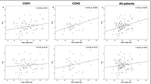
Results:SUV was significantly positively correlated with body weight (r= 0.35,p= 0.02). In contrast, SUL was not correlated with body weight (r= 0.23,p= 0.07). No significant differences were found between baseline and follow-up scan (p= 0.52).
Conclusion:The Janmahasatian formula annuls the positive correlations between SUV and body weight, suggesting that SUL is preferable to SUV for quantitative analyses of68Ga-PSMA11 PET/CT scans.
Keywords:SUL, PSMA PET, SUV
Introduction
In the last decade, positron emission tomography/com-puter tomography (PET/CT) has gradually emerged as the standard-of-care imaging modality in the diagnosis and treatment response monitoring of different onco-logical malignancies [1, 2]. Moreover, standardized up-take value (SUV), which is the commonly used quantitative parameter in PET/CT showed a high pre-dictive value for treatment outcome [3, 4]. However, quantitative SUV is still hampered by a number of physiological, technical and physical factors widely dis-cussed in the literature [5].
In 18F-FDG PET/CT, SUV normalized by patient’s weight is known to be highly dependent on body weight
[6]. Since 18F-FDG does not significantly accumulate in adipose tissue in the fasting state, the use of SUV can falsely lead to high values in patients with high body mass. Subsequently, SUL (lean body mass (LBM)– cor-rected SUV) has been proposed as a more appropriate quantitative method, with Janmahasatian formulation for LBM showing most accurate results [7].
LBM¼ 9:27103BW 6:68103þ216BMI
The liver is typically used as reference organ in PET im-aging, with a 3-cm spherical volume of interest (VOI) computed to measure the liver background activity [8]. In 18
F-FDG PET/CT, liver SUL showed only a fair repeatabil-ity between two time points in the same patient [9].
In the past 5 years, prostate-specific membrane anti-gen (PSMA), a transmembrane protein highly expressed in prostate cancer, has become a promising target for PET in prostate cancer imaging [10]. 68Ga-PSMA11
© The Author(s). 2019Open AccessThis article is distributed under the terms of the Creative Commons Attribution 4.0 International License (http://creativecommons.org/licenses/by/4.0/), which permits unrestricted use, distribution, and reproduction in any medium, provided you give appropriate credit to the original author(s) and the source, provide a link to the Creative Commons license, and indicate if changes were made.
* Correspondence:[email protected]
1Department of Nuclear Medicine, Technical University of Munich, Klinikum rechts der Isar, Munich, Germany
PET/CT has shown enhanced accuracy compared to conventional imaging modalities in lesion detection [11], with SUV being largely used as a quantitative PET-derived parameter [12, 13]. However, the effect of body weight on SUV in 68Ga-PSMA11 PET/CT has not been yet investigated. Since 68Ga-PSMA11 does not typically accumulate in adipose tissue [14], we hypothesized that liver SUV is dependent from body weight.
The aim of the present study was to investigate whether SUL is a more appropriate quantitative method compared to the commonly used SUV normalized by body weight in68Ga-PSMA11 PET/CT imaging.
Material and methods Patients
Patients from two institutions, Technical University Munich (COH1) and University of California Los Angeles (COH2), who underwent 68Ga-PSMA11 PET/ CT prior to 177Lu-PSMA radioligand therapy were in-cluded. Patients in whom it was not feasible to draw a 3-cm VOI in healthy liver tissue were excluded.
For COH1, 91 subsequent patients who received be-tween October 2014 and March 2018 were considered for this analysis. For COH2, 43 subsequent patients were prospectively enrolled in a phase 2 trial (NCT03515577). All patients signed a written consent for evaluation of their data and the institutional review board of the Technical University Munich (permit 5665/13) and Uni-versity of California Los Angeles (permit 17-000330) ap-proved this analysis.
Image acquisition
Images were obtained in accordance with the inter-national guideline [15] in conjunction with a diagnostic following application of68Ga-PSMA-11 that was synthe-sized as described previously [16]. The 68 Ga-PSMA-lig-and complex solution was applied to patients via an intravenous bolus with a mean of 146.0 ± 45.4 and 192.3 ± 19.7 MBq for COH1 and COH2, respectively. PET ac-quisition was started at a mean time of 66.5 ± 12.8 and 56.4 ± 9.7 min after tracer injection for COH1 and COH2. The PET was reconstructed by ordered subset expectation maximization (OSEM)-based algorithms. Data from the CT scan were used for attenuation correction.
Image analyses
Images were reviewed using qPSMA, an in-house devel-oped software [17]. Body weights were recorded from the patients’records.
SUV and SUL
For liver SUV computation, the VOI was semi-automatically placed using an algorithm [18] that has
shown excellent intra- and inter-reader agreement. SUL was calculated according to the Janmahasatian formula as follows:
SUL¼SUVLBMBW
Repeatability
To assess repeatability and the potential influence of tumor sink effect two 68Ga-PSMA11 PET/CT scans from a subset of patients at two different dates were in-cluded in the analysis. SUV and SUL of both scans were compared.
Statistical analysis
Values were reported as mean ± SD. Pearson correla-tions were performed to evaluate the relacorrela-tionship be-tween SUV, SUL, and body weights. Paired t-test was used when the values were considered as paired.pvalue < 0.05 was considered statistically significant. Analyses were performed using SPSS Statistics v22.0 (IBM Corp., USA).
Results Patient cohort
In total, 121 patients were included in the final analyses. Eighty patients of COH1 were eligible; as in 11 patients, the 3-cm VOI in the liver was not feasible due to severe breathing artifacts. Forty-one patients of COH2 could be included as two patients underwent 18F-DCFPyL PET/ CT prior to177LuPSMA treatment. Notably, significantly higher activity doses were injected for COH2 (mean ± SD,118 ± 25 vs. 192 ± 19 MBq, p < 0.001). Mean ± SD of body weight were 80.3 ± 11.4, 82.5 ± 17.5 and 81.0 ± 13.7 kg for COH1, COH2, and all patients, respectively. Patient characteristics are presented in Table1.
SUV, SUL, and comparisons
Mean ± SD of liver SUV were 4.30 ± 1.55, 4.58 ± 1.57, and 4.36 ± 1.56 for COH1, COH2, and all patients, re-spectively. Mean ± SD of liver SUL were 3.23 ± 1.16, 3.41 ± 1.03, and 3.29 ± 1.12 for COH1, COH2, and all patients, respectively. Both cohorts did not show any sig-nificant differences for body weights (p= 0.46), SUVs (p = 0.27), or SULs (p= 0.37).
correlations for both SUV and SUL with body weight are displayed in Fig.1.
Repeatability
Sixty patients from COH1 received a follow-up 68 Ga-PSMA11 PET/CT during 177Lu-PSMA radioligand ther-apy at a mean ± SD of 3.7 ± 0.6 months after the base-line scan. Mean ± SD-injected dose for basebase-line and follow-up scan was 118 ± 25 and 105 ± 23 MBq, respectively.
Mean liver SUV did not change significantly (p= 0.52) between the baseline (4.26 ± 1.64) and follow-up scan (4.16 ± 1.60). Mean (95%CI) relative difference was 1.69 (−7.84;11.22)% with ICC of 0.821 (0.701–0.893). Mean liver SUL did not change significantly (p= 0.72) between the baseline (3.25 ± 1.22) and follow-up scans (3.21 ± 1.22). Mean (95% CI) relative difference was 0.80 (−8.70; 10.32)%, with ICC of 0.818 (0.696–0.892). Figure 2 dis-plays the Bland-Altman plots for SUV and SUL.
Discussion
To the best of our knowledge, this is the first report evaluating the potential of using SUL as compared to the commonly used SUV as quantitative parameter in 68
Ga-PSMA11 PET/CT. Our data indicate that a weak but significant positive correlation is present between liver SUV and body weight. Contrarily, SUL as alterna-tive parameter seems to be unaffected from body weight. Since 68Ga-PSMA11 does not typically accumulate in Table 1Patients characteristics
All patients (n= 121)
Age (years) 73 ± 7.3
Body weight (kg) 81.0 ± 13.7
Time since diagnosis of prostate cancer (years) 7 ± 11
Gleason score at diagnosisa
< 8 38 (36%)
≥8 69 (64%)
PSA at the time of PET/CT imaging (ng/ml) 114 ± 671
Prior lines of systemic treatment
2 8 (7%)
≥3 113 (93%)
≥4 78 (64%)
≥5 44 (36%)
≥6 25 (21%)
Sites of disease on PSMA-PET
Bone 112 (93%)
Lymph nodes 97 (80%)
Visceralb 43 (36%)
Bone + lymph nodes 88 (73%)
Bone + lymph nodes + visceral 34 (28%)
Data are median ± standard deviation orn(%)
PSA, prostate-specific antigen;PSMA, prostate-specific membrane antigen
a
Data missing for 14 patients
b
Visceral includes liver, lungs, and adrenal glands
adipose tissue the use of SUL can be recommended to avoid any influence from patient habitus.
PET-derived parameters, such as SUVmean or SUVmax, are increasingly used for therapy response monitoring or patient outcome prediction. Therefore, highly accurate computed parameters should be addressed given their potential decisive role for the clinical image–based deci-sions. The present study attempted to reproduce a clin-ical setting where quantitative 68Ga-PSMA11 PET/CT scans are used in the framework of177Lu-PSMA radioli-gand therapy.
We investigated the relation of body weight with SUV and SUL in a patient cohort including subcohorts of both European and North-American patients. Consist-ently among the subgroups, our findings indicate that liver SUL calculated based on Janmahasatian formula annuls the body weight dependence of liver SUV. Note-worthy, even though a significant correlation was found between body weight and SUV, its strength is rather weak (r= 0.35,p= 0.02). However, since SUL (r= 0.23,
p= 0.07) annuls and lowers the strength of the positive correlations to BW, the use of SUL should be preferred over SUV. In FDG PET/CT, liver SUV showed a moder-ate correlation for women (r= 0.58,p< 0.001) and men (r = 0.54, p < 0.001) with body weight, which was an-nulled and reduced by the Janmahasatian formula, re-spectively [7].
In addition, we have to stress that the high interpatient repeatability of both liver SUV and SUL between two time points (ICC = 0.821 and 0.818) demonstrates an ac-ceptable mean difference of 1.69% and 0.80%. Despite potential changes of liver 68Ga-PSMA11 uptake during 177
Lu-PSMA therapy upon shifts of biodistribution de-pending on tumor sink effect, the use of liver as the ref-erence organ to threshold malignancy comparing subsequent timepoints is feasible [19]. Notably, it has
been shown that only high differences in tumor burden have significant implications on liver 68 Ga-PSMA11-up-take, with low vs. high tumor load exhibiting a liver SUVmeanof 4.34 vs. 3.27,p< 0.001 [13].
Androgen deprivation therapy (ADT) has shown to in-crease the PSMA-ligand uptake in the first weeks after treatment initiation in metastatic sensitive prostate-cancer [20]. Moreover, continuous long-term ADT significantly decreased lesion uptake in 68Ga-PSMA11 PET imaging [21]. However, these findings might not be valid for pa-tients with metastatic castration-resistant prostate cancer, since most of their tumor lesions are not responding properly to first-line ADT. No significant differences were noted between liver SUV in patients receiving ADT versus not receiving [22]. Same analysis further evaluated the 68
Ga-PSMA11 uptake of other tissues such blood pool (SUVmean 1.08) or muscle (SUVmean 0.50). Nevertheless, the liver showed the most feasible values (SUVmean4.73) to be used for PSMA PET quantification. The mean liver SUV obtained in the present analysis (4.36) is in concord-ance with those obtained by Jansen et al (4.73) and Gaert-ner et al. (4.25) [13].
Interestingly, in an analysis including 64 patients who received 18F-DCFPyL PET/CT no correlations were found between both liver SUV and SUL with body weight [23]. Comparing the results, for 18F-DCFPyL were obtained higher liver SUV and SUL values: 5.1 ± 0.7 vs. 4.4 ± 1.5 and 3.8 ± 0.5 vs. 3.3 ± 1.1 respectively. Similar to FDG, PSMA-ligands do not typically accumu-late in adipose tissue, therefore a positive correlation be-tween SUV and BW annulated by SUL was expected to be found for both radiopharmaceuticals.
For PSMA-targeted radioligand therapies,68GaPSMA11 PET imaging is typically used at baseline for patient selec-tion, as well as during treatment for radiographic response assessment. However, the clinical utility of 68GaPSMA11
PET in metastatic castration-resistant prostate cancer goes beyond the radioligand therapy, being increasingly used for evaluation of treatment response in patients receiving, e.g., taxanes [24]. Since the traditional SUV has shown a significant correlation with body weight, its clinical value in 68Ga-PSMA11 PET quantification remains question-able. Thus, our findings may have clinical implications es-pecially in treatment response assessment. However, further studies comparing the prognostic value of both SUV and SUL for imaging response evaluation are warranted.
Notable limitations of the current analysis are the retrospective nature of the study and the inclusion of a selected patient cohort scheduled for 177Lu-PSMA radi-oligand therapy. However, as our patients have shown both high and low tumor load the potential influence of different tumor burden is already acknowledged. Finally, our analysis is focused on the liver as most important normal organ in PSMA-ligand PET imaging severing as reference tissue. We have not investigated the influence of body weight on other normal tissues.
Conclusion
Our results indicate that the Janmahasatian formula an-nuls the positive correlations between absolute SUV and body weight, suggesting that SUL is preferable to SUV for quantitative analyses in 68Ga-PSMA11 PET. Future studies, are necessary to determine the clinical signifi-cance of the differences between SUV and SUL for dif-ferent clinical applications, such as thresholds for delineation of tumor volumes or monitoring tumor re-sponse to therapy.
Abbreviations
ADT:Androgen deprivation therapy; BW: Body weight; LBM: Lean body mass; PET: Positron emission tomography; PET/CT: Positron emission tomography/ computer tomography imaging; PSMA: Prostate-specific membrane antigen; SUL: Standardized uptake value normalized to lean body mass;
SUV: Standard uptake value; VOI: Volume of interest
Acknowledgements
The authors thank the technicians Alexandra Bartel, Coletta Kruschke, Amanda Reinhardt, Brigitte Mackert, Alida Wachendorf, and Gitti Dzewas for their excellent work on PET/CT.
Authors’contributions
AG and ME participated in the design of the study, data analysis, and drafted the manuscript. AG, JC, CF, IR, HW, and AR performed the data analysis and statistics. ME, JCZ, and WW revised the manuscript. All authors read and approved the final manuscript.
Funding
M.E. received funding from the SFB 824 (DFG Sonderforschungsbereich 824, Project B11) from the Deutsche Forschungsgemeinschaft, Bonn, Germany. Siemens Medical Solutions (Erlangen, Germany) provided the continuous bed motion option for the Biograph mCT as part of an academic collaboration.
Availability of data and materials
Please contact the corresponding author for data requests.
Ethics approval and consent to participate
All patients signed a written consent for evaluation of their data and the institutional review board of the Technical University Munich (permit 5665/ 13) and University of California Los Angeles (permit 17-000330) approved this analysis.
Consent for publication
Not applicable.
Competing interests
The authors declare that they have no competing interests.
Author details 1
Department of Nuclear Medicine, Technical University of Munich, Klinikum rechts der Isar, Munich, Germany.2Department of Molecular and Medical Pharmacology, David Geffen School of Medicine at UCLA, Los Angeles, USA.
Received: 11 September 2019 Accepted: 4 November 2019
References
1. Cheson BD. PET/CT in lymphoma: current overview and future directions. Semin Nucl Med. 2018;48:76–81.https://doi.org/10.1053/j.semnuclmed.2017. 09.007.
2. Subramaniam SM, Joyce CM, Prashanti L, Gregory R, Gustavo M, Rathan M. The role of PET/CT in the management of cervical cancer. AJR Am J Roentgenol. 2013;201(2):W192–205.https://doi.org/10.2214/AJR.12.9830. 3. van Rossum PS, Fried DV, Zhang L, Hofstetter WL, Ho L, Meijer GJ, et al. The
value of (18)F-FDG PET before and after induction chemotherapy for the early prediction of a poor pathologic response to subsequent preoperative chemoradiotherapy in oesophageal adenocarcinoma. Eur J Nucl Med Mol Imaging. 2017;44:71–80.https://doi.org/10.1007/s00259-016-3478-2. 4. Mikhaeel NG, Smith D, Dunn JT, Phillips M, Moller H, Fields PA, et al.
Combination of baseline metabolic tumour volume and early response on PET/CT improves progression-free survival prediction in DLBCL. Eur J Nucl Med Mol Imaging. 2016;43:1209–19. https://doi.org/10.1007/s00259-016-3315-7.
5. Zaidi H. Quantitative analysis in nuclear medicine imaging | Habib Zaidi | Springer. New York: Springer; 2006.
6. Zasadny KR, Wahl RL. Standardized uptake values of normal tissues at PET with 2-[fluorine-18]-fluoro-2-deoxy-D-glucose: variations with body weight and a method for correction. Radiology. 1993;189:847–50.https://doi.org/10. 1148/radiology.189.3.8234714.
7. Tahari AK, Chien D, Azadi JR, Wahl RL. Optimum lean body formulation for correction of standardized uptake value in PET imaging. J Nucl Med. 2014; 55:1481–4.https://doi.org/10.2967/jnumed.113.136986.
8. Wahl RL, Jacene H, Kasamon Y, Lodge MA. From RECIST to PERCIST: evolving considerations for PET response criteria in solid tumors. Journal of nuclear medicine: official publication. Soc Nucl Med. 2009;50(Suppl 1):122s– 50s.https://doi.org/10.2967/jnumed.108.057307.
9. Tahari AK, Paidpally V, Chirindel A, Wahl RL, Subramaniam RM. Two-time-point FDG PET/CT: liver SULmean repeatability. AJR Am J Roentgenol. 2015; 204:402–7.https://doi.org/10.2214/ajr.14.12719.
10. Eiber M, Fendler WP, Rowe SP, Calais J, Hofman MS, Maurer T, et al. Prostate-specific membrane antigen ligands for imaging and therapy. J Nucl Med. 2017;58:67s–76s.https://doi.org/10.2967/jnumed.116.186767. 11. Eiber M, Maurer T, Souvatzoglou M, Beer AJ, Ruffani A, Haller B, et al.
Evaluation of Hybrid (6)(8)Ga-PSMA Ligand PET/CT in 248 patients with biochemical recurrence after radical prostatectomy. J Nucl Med. 2015;56: 668–74.https://doi.org/10.2967/jnumed.115.154153.
12. Schmidkonz C, Cordes M, Schmidt D, Bauerle T, Goetz TI, Beck M, et al. (68)Ga-PSMA-11 PET/CT-derived metabolic parameters for determination of whole-body tumor burden and treatment response in prostate cancer. Eur J Nucl Med Mol Imaging. 2018.https://doi.org/10.1007/s00259-018-4042-z. 13. Gaertner FC, Halabi K, Ahmadzadehfar H, Kurpig S, Eppard E, Kotsikopoulos
C, et al. Uptake of PSMA-ligands in normal tissues is dependent on tumor load in patients with prostate cancer. Oncotarget. 2017;8:55094–103.https:// doi.org/10.18632/oncotarget.19049.
imaging. Nucl Med Commun. 2016;37:1169–79.https://doi.org/10.1097/ mnm.0000000000000566.
15. Fendler WP, Eiber M, Beheshti M, Bomanji J, Ceci F, Cho S, et al. (68)Ga-PSMA PET/CT: Joint EANM and SNMMI procedure guideline for prostate cancer imaging: version 1.0. Eur J Nucl Med Mol Imaging. 2017;44:1014–24.
https://doi.org/10.1007/s00259-017-3670-z.
16. Eder M, Neels O, Muller M, Bauder-Wust U, Remde Y, Schafer M, et al. Novel preclinical and radiopharmaceutical aspects of [68Ga]Ga-PSMA-HBED-CC: a new PET tracer for imaging of prostate cancer. Pharmaceuticals. 2014;7:779– 96.https://doi.org/10.3390/ph 7070779.
17. Gafita A, Bieth M, Kroenke M, Tetteh G, Guenther E, Menze B, et al. qPSMA: a semi-automatic software for whole-body tumor burden assessment in prostate cancer using (68)Ga-PSMA11 PET/CT. J Nucl Med. 2019.https://doi. org/10.2967/jnumed.118.224055.
18. Hirata K, Kobayashi K, Wong KP, Manabe O, Surmak A, Tamaki N, et al. A semi-automated technique determining the liver standardized uptake value reference for tumor delineation in FDG PET-CT. PLoS One. 2014;9:e105682.
https://doi.org/10.1371/journal.pone.0105682.
19. Eiber M, Herrmann K, Calais J, Hadaschik B, Giesel FL, Hartenbach M, et al. Prostate cancer molecular imaging standardized evaluation (PROMISE): proposed miTNM classification for the interpretation of PSMA-Ligand PET/ CT. J Nucl Med. 2018;59:469–78.https://doi.org/10.2967/jnumed.117.198119. 20. Hope TA, Truillet C, Ehman EC, Afshar-Oromieh A, Aggarwal R, Ryan CJ,
et al. 68Ga-PSMA-11 PET imaging of response to androgen receptor inhibition: first human experience. J Nucl Med. 2017;58:81–4.https://doi.org/ 10.2967/jnumed.116.181800.
21. Afshar-Oromieh A, Debus N, Uhrig M, Hope TA, Evans MJ, Holland-Letz T, et al. Impact of long-term androgen deprivation therapy on PSMA ligand PET/CT in patients with castration-sensitive prostate cancer. European journal of nuclear medicine and molecular imaging. 2018;45:2045–54.
https://doi.org/10.1007/s00259-018-4079-z.
22. Jansen BHE, Kramer GM, Cysouw MCF, Yaqub MM, de Keizer B, Lavalaye J, et al. Healthy tissue uptake of (68)Ga-prostate specific membrane antigen (PSMA), DCFPyL, Fluoromethylcholine (FCH) and (18)F-Dihydrotestosterone (FDHT). J Nucl Med. 2019.https://doi.org/10.2967/ jnumed.118.222505.
23. Li X, Rowe SP, Leal JP, Gorin MA, Allaf ME, Ross AE, et al. Semiquantitative parameters in PSMA-targeted PET imaging with (18)F-DCFPyL: variability in normal-organ uptake. J Nucl Med. 2017;58:942–6.https://doi.org/10.2967/ jnumed.116.179739.
24. Seitz AK, Rauscher I, Haller B, Kronke M, Luther S, Heck MM, et al. Preliminary results on response assessment using (68)Ga-HBED-CC-PSMA PET/CT in patients with metastatic prostate cancer undergoing docetaxel chemotherapy. Eur J Nucl Med Mol Imaging. 2018;45:602–12.https://doi. org/10.1007/s00259-017-3887-x.
Publisher’s Note