DOI
10.17219/acem/69135
Copyright
© 2018 by Wroclaw Medical University This is an article distributed under the terms of the Creative Commons Attribution Non-Commercial License (http://creativecommons.org/licenses/by-nc-nd/4.0/)
Address for correspondence
Yuwen Wang
E-mail: [email protected]
Funding sources
None declared
Conflict of interest
None declared
Received on January 6, 2017 Reviewed on January 17, 2017 Accepted on February 23, 2017
Abstract
Background. The trabecular meshwork (TM) plays a critical role in the outflow of aqueous humor.
Objectives. In this study, we aimed to investigate the effect of miR-181a on H2O2-induced apoptosis in TM cells. Material and methods. Human primary explant-derived TM cells were cultured in fibroblast medium and then treated with different concentrations of H2O2 for 2 h. We used a series of methods to carry out
the research, such as MTT assay, quantitative reverse transcriptase-polymerase chain reaction (qRT-PCR), apoptosis assay, and western blot methodology.
Results. The apoptosis assay and qRT-PCR showed that H2O2-induced apoptosis and cell viability were
suppressed in a dose-dependent manner in TM cells. After the TM cells were treated with H2O2, miR-181a
expression was significantly lower. The overexpression of miR-181a enhanced TM cells’ viability, while the knockdown of miR-181a inhibited viability of cells. The overexpression of miR-181a suppressed TM cell apoptosis, while the knockdown of miR-181a induced apoptosis. H2O2 activated the nuclear factor-κB (NF-κB)
and c-Jun N-terminal kinase (JNK) pathways and induced cell apoptosis, while the overexpression of miR-181a suppressed both pathways and decreased the rate of apoptosis.
Conclusions. In conclusion, this study indicated that miR-181a could improve the survival rate of TM cells after H2O2 treatment by blocking the NF-κB and JNK signaling pathways. These findings might provide novel
therapeutic opportunities in the treatment of glaucoma.
Key words: cell apoptosis, H2O2, miR-181a, trabecular meshwork, nuclear factor-κB, c-Jun N-terminal
kinase pathway
MiR-181a inhibits human trabecular meshwork cell apoptosis induced
by H₂O₂ through the suppression of NF-κB and JNK pathways
Yuwen Wang
A,D,F, Heding Zhou
B,D,F, Xiaotian Liu
C,D,F, Yin Han
B,F, Suqi Pan
B,F, Yanyan Wang
C,FNingbo Eye Hospital, Ningbo, China
A – research concept and design; B – collection and/or assembly of data; C – data analysis and interpretation; D – writing the article; E – critical revision of the article; F – final approval of the article
Introduction
Worldwide, glaucoma is the one of the leading causes of irreversible blindness.1,2 There are 60 million people
di-agnosed with glaucoma, of whom 8.4 million have been blinded due to glaucoma.2 One of the major risk factors
associated with glaucoma includes elevated hydrostatic pressure or intraocular pressure (IOP) in the eyes.3
Intra-ocular pressure depends on the rate of formation and drain-age of the aqueous humor via the trabecular meshwork.3
The trabecular meshwork (TM), consisting of TM cells and located at the intraocular anterior chamber angle, plays a critical role in the outflow of aqueous humor.4 Decreased
outflow of aqueous humor induces an elevation in IOP and ultimately leads to glaucoma.3,4 This in turn leads
to in-creased oxidative DNA damage in the TM of glaucoma patients. The oxidative stress induces the activation of the nuclear factor-κB (NF-κB) and c-Jun N-terminal kinase (JNK) signaling pathways.5 Trabecular meshwork cells play
an important role in maintaining the extracellular matrix synthesis (ECM) and phagocytosis of the debris in outflow tissues which control the aqueous-outflow facility. Thus, a shortage of TM cells might result in ocular hyperten-sion. Therefore, the elucidation of the mechanism under-lying the miR-181a and primary oxidative stress response signaling pathways might reveal promising opportunities for preventing TM cell apoptosis, which can prevent glau-coma.5 An understanding the biological functions of TM
cells is then beneficial for understanding the pathogenesis of glaucoma and for developing novel drugs.
MicroRNAs (miRNAs) are a group of non-coding RNAs with 14–20 nucleotides, which are involved in the inhi-bition of endogenous gene expression through trans-lational cleavage.6 MiRNAs are implicated in many
biological progresses, such as cell proliferation, aging, apoptosis, and tumorigenesis.7–10 These miRNAs, either
directly or indirectly, have been found to play an impor-tant role in the pathogenesis of glaucoma in both ani-mal and human visual systems.11,12 The role of miRNAs
in the molecular mechanisms of ECM provides potential for the development of drugs.13 In their study, Dismuke
et al. isolated 10 mature miRNAs from human aqueous humor exosomes, one of which was miR-181a.14 miR-181a
is a family member of miR-181, and has been reported to act as a regulator in cell proliferation, migration and invasion.15 Interestingly, miR-181a has different
func-tions in different types of cells. For instance, miR-181a improved colorectal carcinoma cells and cartilage cell proliferation, while it inhibited the proliferation of glio-ma.16–18 The levels of miR-181a were most decreased with
aging.19 The exact underlying mechanism of miRNAs
regulate retinal apoptosis or degeneration is largely un-known. In a study by Parikh et al., miR-181a was found to promote transdifferentiation towards mesenchymal-like cells.20 The activation of these pathways is thought
to con-tribute to the dysfunction of normal outflow facility.21
Also, a study by Kozloski et al. demonstrated that miR-181a negatively regulated NF-κB activity, leading to a significant change in the proliferation and survival phenotype of the B-cell-like diffuse large B-cell lymphoma malignancy.22
However, the role of miR-181a in the pathogenesis of glau-coma remains elusive.
In this study, we investigated the role of miR-181a in apoptosis in human trabecular meshwork cells induced by H2O2 through the NF-κB and JNK pathways.
Material and methods
Cell culture and H
2O
2treatment
Human primary explant-derived trabecular meshwork (TM) cells (TM cell line) were obtained from ScienCell Research Labs (Catalog No. 6590; Carlsbad, USA). Primary human TM cells were grown in the Fibroblast Medium (Catalog No. 2301; ScienCell Research Labs, Carlsbad, USA) wherein the cells from the 3rd to 6th passage were
used. For maintenance, the cells were incubated at 37°C in a 5% CO2 environment. The TM cells were seeded
in a 6-well plate or a 96-well plate and treated with differ-ent concdiffer-entrations of H2O2 for 2 h.
Small interfering RNA transfection
Mature miR-181a mimic and si-miR-181a were designed and synthesized by GenePharma (Shanghai, China). The in-sertion fragment was confirmed by DNA sequencing. Cell transfection and co-transfection were performed using the Li-pofectamine 3000 reagent (Invitrogen Life Technologies, Carlsbad, USA) according to the manufacturer’s instructions.
MTT assay
The viability of the cells was determined using a 3-(4,5-dimethylthiazol-2-yl)-2 5-diphenyl-2H-tetrazoli-um bromide (MTT) colorimetric assay according to stan-dard methods described previously. Each experiment was performed 3 times.
Apoptosis assay
Quantitative reverse transcriptase-
-polymerase chain reaction
Total RNA was isolated from transfected cells by using TRIzol reagent (Invitrogen, Carlsbad, USA) and treated with DNase I (Promega, Southampton, UK). Reverse tran-scription was performed by using a Multiscribe RT kit (Ap-plied Biosystems, Foster City, USA) and random hexamers or oligo(dT). The reverse transcription conditions were 10 min at 25°C, 30 min at 48°C, and a final step of 5 min at 95°C.
Western blot
The protein used for western blotting was extracted us-ing RIPA lysis buffer (Beyotime Biotechnology, Shanghai, China) supplemented with protease inhibitors (Roche, Guangzhou, China). The proteins were quantified us-ing a BCA™ Protein Assay Kit (Pierce, Appleton, USA).
The western blotting system was established using a Bio-Rad Bis-Tris Gel system according to the manufacturer’s instructions. Primary antibodies were prepared in 5% blocking buffer at a dilution of 1:1,000. Primary antibod-ies were incubated with the membrane at 4°C overnight, followed by washing and incubation with secondary anti-bodies marked by horseradish peroxidase for 1 h at room temperature. After rinsing, the polyvinylidene difluoride (PVDF) membrane carrying blots and antibodies were transferred into the Bio-Rad ChemiDoc™ XRS system, and
then 200 μL Immobilon Western Chemiluminescent HRP Substrate (Millipore, Bedford, USA) was added to cover the membrane surface. The signals were captured and the intensity of the bands was quantified using Image Lab™
Software (Bio-Rad, Shanghai, China).
Statistical analysis
All data were shown as mean ±SD. Statistical analyses were performed using SPSS 19.0 software (SPSS Science, Chicago, USA). Differences between groups were ana-lyzed using one-way analysis of variance (ANOVA) and/ or Student’s t-test. A p-value of <0.05 was considered statistically significant. All experiments were conducted in triplicate.
Results
Different concentrations of H
2O
2suppressed viability of TM cells
in a dose-dependent manner
In our study, we cultured TM cells in a 6-well plate or a 96-well plate and treated them with different con-centrations of H2O2 (50, 100, 200, and 300 μM) for 2 h. Cell
viability was assessed by MTT assay. Our results showed
that H2O2 at a concentration of 200 μM was significantly
suppressed. These results suggested that different con-centrations of H2O2 suppressed the viability of TM cells
in a dose-dependent manner and that H2O2
in increas-ing concentrations (100, 200, and 300 μM) suppressed cell viability significantly (Fig. 1, p < 0.05 or p < 0.01). For the analyses below 200 μM was selected.
H
2O
2-treated TM cells revealed a significant
fall in miR-181a expression level
The TM cells after treatment with H2O2 showed
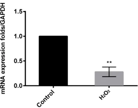
a sig-nificant decrease (p < 0.01) in the expression level of miR-181a (Fig. 2).
Fig. 1. TM cell viability was significantly suppressed by H2O2
in a dose-dependent manner, and 200 μM was selected as a lethal concentration of 50% used in the following analyses
* p < 0.05; ** p < 0.01.
Fig. 2. After H2O2 intervention, the miR-181a levels in TM cells were
Fig. 4. The overexpression of miR-181a enhanced TM cell viability, while the knockdown of miR-181a inhibited cell viability
Transfection efficiency of TM cells
TM cells were transfected and then divided into mature miR-181a mimic, si-miR-181a and negative control (siNC) which were then evaluated by qRT-PCR. The results showed a significant decrease (p < 0.01) in the mRNA expression level of si-miR-181a compared to the control. Furthermore, they showed a significantly increased (p < 0.01) mRNA ex-pression level of miR-181a mimic compared to the control. This suggested that transfection efficiency was observed when miR-181a was overexpressed or suppressed in TM cells (Fig. 3).
H
2O
2treatment and overexpression
of miR-181a increased TM cell viability
Cell viability was measured in the TM cells after treat-ment with H2O2, and the effect of miR-181a on cell
viabil-ity was measured after transfection. The results showed that cell viability was promoted with increasing time in the miR-181a mimic group compared to si-miR-181a and to control groups. These results suggest that the over-expression of miR-181a enhanced TM cell viability, while the knockdown of miR-181a inhibited cell viability (Fig. 4).
H
2O
2treatment and overexpression
of miR-181a decreased TM cell apoptosis
Cell apoptosis was measured in the TM cells after treatment with H2O2, and the effect of miR-181a on cell
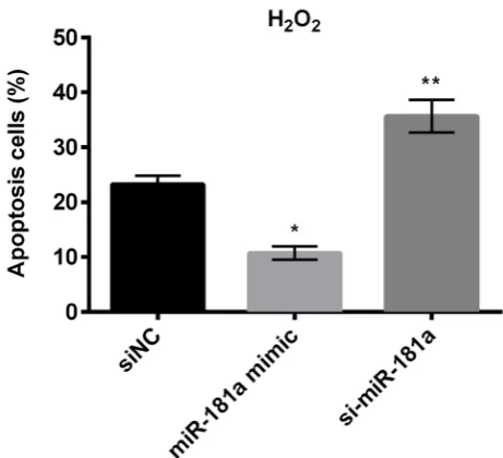
apoptosis was measured after transfection. The re-sults showed an increased percentage of cell apoptosis (p < 0.01) in the si-miR-181a group (mean: 35.7) compared to control groups. Treatment with H2O2 caused nuclear
condensation, which is an indicator of apoptosis. Also, the miR-181a mimic group (mean: 10.7) showed a sig-nificant reduction on cell apoptosis rate (p < 0.05), but not as significant as si-miR-181a. These results suggested that the overexpression of miR-181a suppressed TM cell apoptosis, while the knockdown of miR-181a induced apoptosis (Fig. 5).
miR-181a suppressed TM cell apoptosis via
blocking the NF-κB and JNK pathways
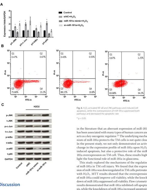
Generally, H2O2 activates the NF-κB and JNK pathways
and induces cell apoptosis. The results showed that the si-miR-181a group has a decreased expression of miR-181a and treatment with H2O2 suppresses
the expres-sion of miR-181a (p < 0.05). Thus, in the si-miR-181a + H2O2 group, the expression of miR-181a is low and
it can-not block the NF-κB and JNK pathways – it will rather produce just the opposite effect (Fig. 6). This suggest-ed that the overexpression of miR-181a could suppress both the pathways, thereby decreasing the rate of cell apoptosis.
Fig. 3. Overexpressed or suppressed miR-181a in TM cells showed transfection efficiency
** p < 0.01.
Fig. 5. The overexpression of miR-181a suppressed TM cell apoptosis, while the knockdown of miR-181a induced apoptosis
Discussion
Oxidative stress has been established as an important contributing factor in TM cell degeneration.5
The contrib-utory role of H2O2 in TM cell degeneration is also known.23
In the present study, we induced TM cell apoptosis through H2O2 and found that miR-181a was downregulated by H2O2
in a dose-dependent manner. It has been commonly shown
in the literature that an aberrant expression of miR-181a has been associated with many types of human cancers and acts as a key oncogenic regulator.23 The underlying
mecha-nism of miR-181a protects the TM cells is not quite clear. In the present study, we not only demonstrated an active change in the expression profile of miR-181a upon H2O2
-induced apoptosis, but also a protective role of the miR-181a overexpression on TM cell. Thus, these results high-light the functional role of miR-181a in glaucoma.
This study explored the mechanisms of the regulation of miR-181a in TM cell injury. We found that the expres-sion of miR-181a was downregulated in TM cells pretreated with H2O2. MTT results showed that the overexpression
of miR-181a could improve cell viability, while the knock-down of miR-181a suppressed cell viability. Flow cytometry results demonstrated that miR-181a inhibited cell apopto-sis, while the knockdown of miR-181a increased apoptosis. Western blotting assay showed a suppression of miR-181a cell apoptosis via blocking the NF-κB and JNK signaling pathways.
In this study, we also showed that during H2O2-induced
apoptosis downregulating miR-181a blocks the NF-κB and JNK signaling pathways. Our results were similar to one
Fig. 6. H2O2 activated NF-κB and JNK pathways and induced cell
apoptosis, while the overexpression of miR-181a suppressed both pathways and decreased the apoptotic rate
of the studies conducted by Kozloski et al. in B-cell-like diffuse large B-cell lymphoma.22 Thus, it is likely
that miR-181a upregulation may serve a protective role in glaucoma by exerting a protective effect on H2O2-induced apoptosis
in TM cells. This hypothesis was further supported by our results, which showed that miR-181a overexpression pro-moted TM cell apoptosis, which possibly induced the trans-differentiation of mesenchymal-like cells in the aqueous humor of TM cells.20 NF-кB is considered to be
an impor-tant nuclear transcription factor that helps in mediating the inflammatory response caused by oxidative stress.24
In endothelial cells, NF-кB is strongly activated by treatment with H2O2 for 4 h.25 However, to elucidate the exact
molecu-lar interaction between miR-181a and the NF-κB and JNK pathways in protecting trabecular meshwork cells, future experiments that require blocking of these pathways or its upstream pathways upon the inhibition of miR-18a would provide solid evidence about the direct targeting of miR-181a on anti-apoptotic pathways in TM cells.
Many miRNAs are involved in the molecular mechanisms of glaucoma; some miRNAs were up-regulated in cells, while others were down-regulated.12 These miRNAs facilitate
the evaluation of specific pathophysiology and offer diag-nosis. Further in-depth understanding will help in diagnos-ing the subtypes of glaucoma, the pathophysiological stages, and the prescription of appropriate drugs.12 miR-183 was
involved in the alteration of integrin-β1 expression, thereby effecting the physiology of TM, resulting in glaucoma.26
The miR-29 family regulates ECM synthesis in the TM.27
The changes associated with ECM synthesis result in al-terations in the IOP. miR-29b negatively regulates the ECM proteins, and its regulators – under chronic oxidative stress conditions – affect ECM homeostasis in human TM cells. The expression of miR-29b decreased under chronic oxida-tive stress conditions, resulting in ECM gene upregulation. Similarly, miR-24 plays an important role in the flow path-ways.28 One study identified the involvement of microRNAs
in glaucoma withhsa-let-7b-3p as the most prevalent factor in the aqueous humor.13
In conclusion, this study indicated that miR-181a could improve the survival rate of TM cells after H2O2
treat-ment by blocking the NF-κB and JNK signaling pathways. These findings might provide novel therapeutic oportuni-ties in the treatment of glaucoma.
References
1. Tham YC, Li X, Wong TY, Quigley HA, Aung T, Cheng CY. Global prev-alence of glaucoma and projections of glaucoma burden through 2040: A systematic review and meta-analysis: Ophthalmology.
2014:121:2081–2090.
2. Cook C, Foster P. Epidemiology of glaucoma: What’s new? Can J Oph-thalmol. 2012:47:223–226.
3. Li A, Chi TL, Peterson-Yantorno K, Stamer WD, Mitchell CH, Civan MM. Mechanisms of ATP release by human trabecular meshwork cells, the enabling step in purinergic regulation of aqueous humor out-flow. J Cell Physiol. 2012;227:172–182.
4. Luna C, Li G, Huang J, et al. Regulation of trabecular meshwork cell con-traction and intraocular pressure by miR-200c. Plos One. 2012;7:e51688.
5. Awaikasaoka N, Inoue T, Kameda T, Fujimoto T, Inouemochita M, Tanihara H. Oxidative stress response signaling pathways in tra-becular meshwork cells and their effects on cell viability. Mol Vis.
2013;19:1332–1340.
6. Pillai RS. MicroRNA function: Multiple mechanisms for a tiny RNA?
RNA. 2005;11:1753–1761.
7. Ali NM, Boo L, Yeap SK, et al. Probable impact of age and hypoxia on proliferation and microRNA expression profile of bone marrow-derived human mesenchymal stem cells. Peer J. 2016;4:e1536 8. Di LG, Croce CM. miRNA profiling of cancer. Curr Opin Genet Dev.
2013;23:3–11.
9. Nie Y, Han BM, Liu XB, et al. Identification of microRNAs involved in hypoxia- and serum deprivation-induced apoptosis in mesen-chymal stem cells. Int J Biol Sci. 2010;7:762–768.
10. Tomé M, López-Romero P, Albo C, et al. miR-335 orchestrates cell proliferation, migration and differentiation in human mesenchymal stem cells. Cell Death Differ. 2011;18:985–995.
11. Kong N, Lu X, Li B. Downregulation of microRNA-100 protects apop-tosis and promotes neuronal growth in retinal ganglion cells. BMC Mol Biol. 2014;15:25–25.
12. Raghunath A, Perumal E. Micro-RNAs and their roles in eye disor-ders. Ophthal Res. 2015;53:169–186.
13. Tanaka Y, Tsuda S, Kunikata H, et al. Profiles of extracellular miRNAs in the aqueous humor of glaucoma patients assessed with a micro-array system. Sci Rep. 2014;4:633–633.
14. Dismuke WM, Challa P, Navarro I, Stamer WD, Liu Y. Human aqueous humor exosomes. Exp Eye Res. 2015;132:73–77.
15. He S, Zeng S, Zhou ZW, He ZX, Zhou SF. Hsa-microRNA-181a is a regulator of a number of cancer genes and a biomarker for endometrial carcinoma in patients: A bioinformatic and clin-ical study and the therapeutic implication. Drug Des Devel Ther.
2015;9:1103–1175.
16. Dragu DL, Necula LG, Bieotu C, et al. Therapies targeting cancer stem cells: Current trends and future challenges. World J Stem Cells.
2015;7:1185–1201.
17. Nakamura A, Rampersand YR, Sharma A, et al. Identification of microRNA-181a-5p and microRNA-4454 as mediators of facet car-tilage degeneration. JCI Insight. 2016;1:e86820.
18. Brodie C, Buchris E, Lee HK. miRNA Expression and Functions in Gli-oma and Gliin Gli-oma Stem Cells. MicroRNA Targeted Cancer Therapy. 2014;112:225–231.
19. Thalyana Smith-Vikos FJS. MicroRNAs and their roles in aging. J Cell Sci. 2012;125:7–17.
20. Parikh A, Lee C, Joseph P, et al. MicroRNA-181a has a critical role in ovarian cancer progression through the regulation of the epithe-lial-mesenchymal transition. Nat Commun. 2014;5:149–168. 21. Takahashi E, Inoue T, Fujimoto T, Kojima S, Tanihara H. Epithelial
mes-enchymal transition-like phenomenon in trabecular meshwork cells. Exp Eye Res. 2014;118:72–79.
22. Kozloski GA, Jiang X, Bhatt S, et al. MiR-181a negatively regulates NF-κB signaling and affects activated B-cell like diffuse large B-cell lymphoma pathogenesis. Blood. 2016;127(23):2856–2866. 23. Guo LJ, Zhang QY. Decreased serum miR-181a is a potential new tool
for breast cancer screening. Int J Mol Med. 2012;30:680–687. 24. Bowie A, O’Neill LA. Oxidative stress and nuclear factor-κB
activa-tion A reassessment of the evidence in the light of recent discover-ies. Biochem Pharmacol. 2000;59:13–23.
25. Bowie AG, Moynagh PN, O’Neill LA. Lipid peroxidation is involved in the activation of NF-kappaB by tumor necrosis factor but not interleukin-1 in the human endothelial cell line ECV304. Lack of involvement of H2O2 in NF-kappaB activation by either cyto-kine in both primary and transformed endothelial cells. J Biol Chem. 1997;272:25941–25950.
26. Li G, Luna C, Qiu J, Epstein DL, Gonzalez P. Targeting of integrin β1 and kinesin 2α by MicroRNA 183. J Biol Chem. 2010;285:5461–5471. 27. Luna C, Li G, Qiu J, Epstein DL, Gonzalez P. Role of miR-29b
on the reg-ulation of the extracellular matrix in human trabecular meshwork cells under chronic oxidative stress. Mol Vis. 2009;15:2488–2497. 28. Luna C, Guorong LI, Qiu J, Epstein DL, Gonzalez P. MicroRNA-24