Barbara Iwańczak
A, C, D–F, Krzysztof Matusiewicz
B–D, F,
Franciszek Iwańczak
A, B, D–FClinical Picture of Classical, Atypical
and Silent Celiac Disease in Children and Adolescents
Obraz kliniczny choroby trzewnej klasycznej, atypowej i utajonej
u dzieci i młodzieży
Department of Pediatrics, Gastroenterology and Nutrition, Wroclaw Medical University, Poland
A – research concept and design; B – collection and/or assembly of data; C – data analysis and interpretation;
D – writing the article; E – critical revision of the article; F – final approval of article; G – other
Abstract
Background. Celiac disease is a frequent disease of the alimentary tract in children. Clinical presentation of the disease is variable and depends on type of the disease.
Objectives. The aim of the study was an analysis of clinical findings, selected laboratory features and coexisting diseases in children and adolescents with celiac disease.
Material and Methods. Material of the study comprised a series of 78 children aged 8 months – 13 years. Celiac disease was diagnosed basing on clinical symptoms, histological studies of intestinal specimens and positive sero-logic tests (EmA, TG2).
Results. Classical celiac disease was diagnosed in 40 children (51.3%), atypical celiac disease in 26 children (33.3%) and silent celiac disease in 12 children (15.4%). The most frequent clinical symptoms of classical form of celiac disease were chronic diarrhea (90.0%), recurrent abdominal pain (70.0%), development retardation (65%), hypo-cholesterolemia (35.0%) and IgA deficiency (22.5%). In atypical form of the disease dominated the following symptoms: recurrent abdominal pain (76.9%), failure to thrive (38.4%), short stature (42.3%), anemia (15.3%), hypertransaminasemia (11.5%), food allergy (19.2%) and thyroid diseases (11.5%). In silent celiac disease hyper-cholesterolemia was present in 33.3%, hypertriglycerydemia in 16.6%, type 1 diabetes in 50%, and celiac disease in parents or siblings in 33.3%.
Conclusions. Classical celiac disease is the most frequently diagnosed clinical form of the disease. Silent celiac disease occurs frequently in children with type 1 diabetes mellitus whose parents or siblings have celiac disease. Frequent diagnosis of atypical and silent forms of celiac disease is an indication to serological examination in
chil-dren with unclear clinical picture and genetic predisposition (Adv Clin Exp Med 2013, 22, 5, 667–673).
Key words: celiac disease: classical, atypical, silent, children.
Streszczenie
Wprowadzenie. Choroba trzewna jest częstą chorobą przewodu pokarmowego u dzieci. Obraz kliniczny choroby jest zróżnicowany i zależny od postaci klinicznej.
Cel pracy. Ocena objawów klinicznych, wybranych parametrów biochemicznych oraz chorób współistniejących.
Materiał i metody. Badaniem objęto 78 dzieci w wieku od 8 miesięcy do 13 lat. Choroba trzewna była rozpoznana na podstawie objawów klinicznych, oceny histopatologicznej kosmków jelitowych i obecności przeciwciał w suro-wicy (EmA, tTG).
Wyniki. Klasyczną chorobę trzewną rozpoznano u 40 dzieci (51,3%), atypową u 26 (33,3%), a postać utajoną u 12 (15,4%). Choroba trzewna klasyczna charakteryzowała się przede wszystkim: przewlekłą biegunką (90,0%), nawra-cającym bólem brzucha (75%), upośledzonym rozwojem fizycznym (65,0%), powiększonym obwodem brzucha (42,5%), hipertransaminazemią (40,0%), hipocholesterolemią (35,0%) oraz niedoborem IgA (22,5%). W choro-bie trzewnej atypowej dominowały bóle: brzucha (76,9%), upośledzenie rozwoju fizycznego (38,4%), niski wzrost
Adv Clin Exp Med 2013, 22, 5, 667–673 ISSN 1899–5276
OrIGINAl PAPErS
Celiac disease is an immune-mediated enter-opathy caused by permanent sensitivity to gluten and related prolamines, affecting genetically sus-ceptible individuals. Genetic, environmental and immunological factors play a role in pathogenesis, damage of small intestinal mucosa and develop-ment of enteropathy [1, 2]. The prevalence of ce-liac disease in children aged 2.5 to 15 years is 3 to 13 per 1000 [2–4]. Celiac disease presents variable clinical picture. Typical classical presentation oc-curs mainly in the first two years of life. After the introduction of gluten into the child’s diet, a set of clinical symptoms of malabsorption appears, such as chronic diarrhea, failure to thrive, abdominal distension and pain, growth delay and iron defi-ciency anemia. Atypical presentation is character-ized by scarce clinical symptoms. Frequently, only a single symptom is observed, such as lack of body mass increase and growth retardation, anemia, dental enamel hypoplasia, osteoporosis, or puber-tal delay. Silent presentation of celiac disease is di-agnosed in asymptomatic patients with increased risk of the disease. Among diseases and conditions predisposing someone to celiac disease are: type 1 diabetes mellitus (6%), first-degree relatives (10%). Down syndrome (5.5%), Turner syndrome (6%), Williams syndrome (9.5%), autoimmune diseases of the liver (13.5%) and of the thyroid gland (3%), juvenile arthritis (1.5%), and selective IgA defi-ciency (3%) [1–3]. Frequencies of celiac disease in those conditions are given in brackets. The aim of the study was an analysis of clinical and laborato-ry features in children and adolescents with diag-nosed classical, atypical and silent celiac disease.
Material and Methods
Material of the study comprised a series of 78 patients aged 8 months to 13 years, hospitalized in The Department of Children Gastroenterology of Wroclaw Medial University from January 2003 to December 2010. In all cases, celiac disease was diagnosed based on European and North Ameri-can Societies for Pediatric Gastroenterology, Hep-atology and Nutrition criteria [2, 5]. In all children, celiac disease was diagnosed based on a histological
study of intestinal specimens obtained during en-doscopic examination and assessed according to the Marsh scale (grades II and III a, b, c) and on elevated titers of antibodies against endomysium of smooth muscles and/or tissue transglutaminase type 2 [2, 3, 6–9]. According to clinical symptoms, the children were classified into three groups. The first group consisted of 40 children with gastroin-testinal symptoms such as chronic diarrhea and malabsorption, which was regarded as classical ce-liac disease. The second group included 26 chil-dren with recurrent abdominal pain, short stat-ure and anemia. These children were classified as having atypical celiac disease. Silent celiac disease was diagnosed in 12 children and adolescents with type 1 diabetes mellitus and first degree relatives of celiac patients (Table 1). For the assessment of physical development (body mass and stature) per-centile scale was used according to Palczewska and Niedźwiedzka [10]. The data was statistically ana-lyzed by chi-square test of proportions using Med-Cacl Software v.12.6.1, Ostend, Belgium. Values of p < 0.05 were regarded as statistically significant. Bioethical Committee agreement for the study had been obtained.
Results
Number, age and sex of the studied children are presented in Table 1. Classical celiac disease was diagnosed in 40 children (51.3%) at the age of 8 months to 6 years (average age 17.5 months). In 80% of children classical celiac disease was diag-nosed when they were under two years of age and in the remaining 20% at the age between three and six years. Girls outnumbered boys (75% vs. 25%, p < 0.05). In these children gluten was introduced into the diet between 3rd and 18th month of life (average 7.5 months). Atypical celiac disease was diagnosed in 26 children (33.3%) at the age from 2.5 to 12 years and in about 80% of them aged be-tween 5th and 13th year of life. Also in this group there were more girls than boys (61.5% vs. 38.4%); however, the difference was not statistically signif-icant. In the group with this clinical picture of the disease, gluten was introduced to the diet between
(42,3%) oraz u części dzieci niedokrwistość (15,3%), hipertransaminazemia (11,5%), alergia pokarmowa (19,2%), choroby tarczycy (11,5%). W chorobie trzewnej utajonej stwierdzono: hipercholesterolemię (33,3%), hipertrójglice-rydemię (16,6%), cukrzycę typu 1 (50%) i chorobę trzewną u rodziców lub rodzeństwa (33,3%).
Wnioski. Choroba trzewna klasyczna jest najczęściej rozpoznawaną postacią. Choroba trzewna utajona występuje często u dzieci chorych na cukrzycę typu 1 oraz u dzieci rodziców lub rodzeństwa chorującego na celiakię. Częste rozpoznawanie postaci atypowej i utajonej przemawia za prowadzeniem badań serologicznych w kierunku choroby
trzewnej u dzieci z niejasnym obrazem choroby oraz u dzieci predysponowanych genetycznie (Adv Clin Exp Med
2013, 22, 5, 667–673).
8th and 12th month of life, average 9.2 months. Silent celiac disease was diagnosed in 12 children (15.4%) at the age between 7 and 13 years. There were more boys than girls (58.1% vs. 41.6%) but the difference was not statistically significant. In this group gluten was introduced to the diet between 8th an 14th month of life, average 11.3 months.
The most frequent clinical symptoms of clas-sical form of celiac disease were: chronic diar-rhea (90.0%), frequently greasy, recurrent abdom-inal pain (70.0%), abdomen distention (40%) and
development retardation (65%) (Table 2). In chil-dren with atypical form of the disease recurrent ab-dominal pain (76.9%), failure to thrive 38.4%), short stature (42.3%) and abdominal distention (15.3%) were most frequent. In children with silent celiac disease there were no characteristic symptoms. On-ly in single cases, abdominal pain, delayed increase of stature and constipation were observed.
There was a statistically significant difference in diarrhea frequency between the group with clas-sical celiac disease and the groups with atypical or
Table 1. Clinical types of celiac disease in children Tabela 1. Postacie kliniczne choroby trzewnej u dzieci
Groupe
(Grupa) Celiac disease (Choroba trzewna)
Numer of children (liczba dzieci) n = 78
Age of diagnosis – years
(Wiek rozpozna-nia – lata)
Sex
(Płeć) Statistical significance (Istotność statystyczna) (p)
Gluten introduction into the diet – months (Wprowadzenie glutenu do diety – miesiące) female male
n % x from–to n % n % x from–to I Classical
celiac disease
40 51.3 1.75 8 months
–6 years 30 75.0 10 25.0 0.0001 7.5 3–18
II Atypical celiac disease
26 33.3 7.1 2.5–13 16 61.5 10 38.4 ns. 9.2 8–12
III Silent celiac disease
12 15.4 8.75 4–13 5 41.6 7 58.3 ns. 11.3 8–14
x – mean (średnia arytmetyczna).
ns. – not significant (nieistotne statystycznie).
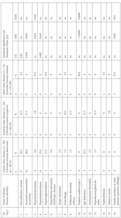
Table 2. Symptoms in patients with celiac disease according to type of the disease Tabela 2. Ocena objawów choroby trzewnej w zależności od postaci choroby
No
(lp.) Symptoms(Objawy) Classic celiac disease – I (n = 40) (Choroba trzew-na klasycztrzew-na – I (n = 40))
Atypical celiac disease – II (n = 26) (Choroba trzew-na nietypowa – II (n = 26))
Silent celiac disease – III (n = 12) (Choroba trzew-na utajotrzew-na – III (n = 12))
Statistical significance (p) (Istotność statystyczna (p))
n % n % n % I/II I/III II/III
1. Chronic diarrhea 36 90.0 0 0 0 0 < 0.0001 < 0.0001 ns. 2. Abdominal pain 30 75.0 20 76.9 2 16.6 ns. 0.0009 0.0017 3. Failure to thrive 26 65.0 10 38.4 0 0 ns. 0.0003 0.0354 4. Abdominal distension 16 40.0 4 15.3 0 0 ns. 0.02 ns. 5. Constipation 0 0 1 3.8 2 16.6 ns. ns. ns.
6. Vomiting 4 10.0 1 3.8 0 0 ns. ns. ns.
7. Short stature 5 12.5 11 42.3 1 8.3 0.01 ns. ns. 8. Peripheral odema 4 10.0 0 0 0 0 ns. ns. ns.
n – numer (liczba).
silent forms of the disease (p < 0.0001). Moreover, in the group with classical picture of the disease, the occurrence of recurrent abdominal pain, dis-turbed development and abdominal distention were more frequent than in children with silent form of the disease. In children with atypical celi-ac disease, growth retardation was observed more often than in the group with classic picture of the disease (p < 0.01) and in comparison with silent disease, abdominal pain and development retarda-tion were more frequent (p < 0.001 and p < 0.03, respectively).
The most frequent diseases coexisting with celiac disease depending of the clinical picture of the disease are presented in Table 3. In children with classical celiac disease iron deficiency anemia (42.5%), increased activity of transaminases (40%) and decreased serum cholesterol (35%) were fre-quently observed. In 22.5% of the children selec-tive IgA deficiency was observed. In single cases, Dühring disease, atopic dermatitis, food allergy and thyroid diseases were present.
In children with diagnosed atypical celiac dis-ease with untypical disdis-ease, the following problems occurred the most often: iron deficiency anemia (15%), increased activity of transaminases (11.5%), food allergy (19.2%), thyroid gland diseases and pollinosis in 11.5%. In children with silent picture of celiac disease, hypercholesterolemia was pres-ent in 33.3% and hypertriglicerydemia in 16.6%. Type 1 diabetes was diagnosed in 50% of these children and celiac disease in parents or siblings in 33.3%. Statistical significance of the difference be-tween frequencies of clinical symptoms in various types of celiac disease is presented in Table 3.
Discussion
Celiac disease belongs to one of the most fre-quently diagnosed diseases of the alimentary tract. The frequency of celiac disease in developed coun-tries is about 1% [11]. Among predisposing factors are genetic predispositions determined by HlA markers of DQ2 and DQ8 types, which are present in more than 95% of celiacs [11]. It should be em-phasized that in spite of the frequent occurrence of these markers in the general population (30–40%), only 1 to 4% of these subjects develop celiac dis-ease [12]. It is thought that about 20 other genes are the risk factors of celiac disease. It has been ob-served that celiac disease is present in about 10% of children whose parents have this disease [12]. En-vironmental factors are also of great importance. The most distinguished factor is the timing of in-troduction of gluten into the diet of a child. Glu-ten introduced before the 3rd month of life causes
Table 3. Occurrence of coexisting diseases in celiac disease Tabela 3. Choroby współistniejące z chorobą trzewną
No (lp.)
European Societies for Pediatric Gastroenterology, Hepatology and Nutrition (ESPGHAN), recom-mendations in breast fed infents gluten should be introduced to the diet between 4th and 6th month of life. This allows the organism to establish tole-rantion against gluten and to reduce the risk of ce-liac disease [22].
Among clinical symptoms of classical celiac disease the following dominated: chronic diarrhea, abdominal pain, distention of abdomen and retar-dation of development. In atypical celiac disease – abdominal pain, short stature and retardation of development. These symptoms were not observed in silent celiac disease.
laboratory tests and analysis of coexisting dis-eases demonstrated more frequent increase in ami-notransferases activity, hypocholesterolemia, iron deficiency anemia and IgA deficiency in children with classical celiac disease in comparison with atypical form. Thyroid gland diseases dominat-ed in atypical celiac disease. In silent celiac dis-ease significantly more frequent incrdis-ease of serum cholesterol and coexistence of type 1 diabetes was observed. Szaflarska-Popławska observed that in 40.85% of children with celiac disease activity of AlT or/and AST was elevated. According to this author, all patients with celiac disease should un-dergo assessment of liver function at the time of diagnosis of the disease and after 6–12 months of gluten-free diet. lack of normalization after such a time may indicate lack of adherence to the diet or coexistence of autoimmune liver diseases [21]. From our studies it can be seen that the frequency of hyperatransaminasemia depends on celiac dis-ease clinical form and on the age of patients. Oth-er studies confirm this obsOth-ervation [19, 23, 25]. Iwańczak et al. in earlier studies compared the fre-quency of hypertansaminasemia and iron deficien-cy anemia in children and adults with celiac dis-ease [23]. The age of the 59 children included in that study was 7 to 18 years (mean 12.5) and that of 52 adults from 19 to 75 years (mean (32.0). In chil-dren, the clinical picture of the disease was not an-alyzed. The study demonstrated significantly more frequent hypertransaminasemia in adults than in
children (p < 0.001) as well as more frequent ane-mia in adults (80.7% vs. 28.8%, p < 0.001). Dis-eases of thyroid gland were also more frequent in adults than in children with celiac disease (15.4% vs. 5.1%). In our present study from among coex-isting diseases, type 1 diabetes was the most fre-quent disease in children with silent form of celiac disease. According to numerous studies, type 1 di-abetes is the most frequent disease in patients with celiac disease [19, 24, 25]. Iwanicka et al. conduct-ed a study on frequency of celiac disease among patients with type 1 diabetes in a group of 83 chil-dren aged 3 to 18 years. Celiac disease was diag-nosed based on positive antiendomysial antibod-ies (IgAEmA) in the serum and villi atrophy in duodenum mucosa specimens. In 6% of the stud-ied children celiac disease was diagnosed. Titer of IgAEmA in those children was 160 to 2560. In one female patient concomitant celiac disease and Graves-Basedov disease were diagnosed. Accord-ing to the authors of the cited work, the risk of au-toimmune diseases is greater in patients with celiac disease than in the general population. Grzędo-Adamek et al., in a screening study conducted on a group of 109 children with autoimmune disease of the thyroid gland, found only one case of celiac disease without any symptoms [26].
In summary, it should be recapitulated that the most frequently diagnosed clinical form of ce-liac disease is classical cece-liac disease. Silent cece-liac disease occurred most frequently in children with type 1 diabetes whose parents or siblings had celiac disease. In classical celiac diseases dominated diar-rhea, abdominal pain, disturbance of physical de-velopment, iron deficiency anemia, hypertransam-inasemia, hypocholesterolemia and IgA deficiency. In silent celiac disease, hypercholesterolemia was observed the most frequently. High frequency of celiac disease in the general population and vari-able clinical picture is an indication of serolog-ical examination, which will allow detection and treatment of the disease, Early detection of celi-ac disease will enable the child to develop proper-ly and will prevent frequent and dangerous health complications.
References
[1] Husby S, Koletzko S, Korponay-Szabo IR, Marin ML, Phillips A, Shamir R, Troncone R, Gersiepen K, Branki D, Catassi C, Lelgeman M, Mäki M, Ribes-Koninckx C, Ventura A, Zimmer KP: ESPGHAN guidelines for the diag-nosis of coeliac disease in children and adolescents. An evidence – based approach. J Pediatr Gastroenterol Nutr 2012, 54, 136–160.
[2] Hill ID, Dirks MH, Liptath GS: Guideline for the diagnosis and treatment of celiac disease in children: recom-mendations of the North American Society for Pediatric Gastroenterology, Hepatology and Nutrition. J Pediatr Gatroenterol Nutr 2005, 40, 1–19.
[3] AGA Institute Medical Position Statment on the Diagnosis and Managament of celiac Disease. Gastroenterology 2006, 131, 1977–1980.
[5] revised criteria for doagnosis of coeliac disease. report of Working Group of European Society of Pediatric Gastroenterology and Nutrition. Arch Dis Child 1990, 65, 909–911.
[6] Hill ID, Dirks MH, Liptath GS: Guideline for the diagnosis and treatment of celiac disease in children. recommendations of the North American Society for Pediatric Gastroenterology, Hepatology and Nutrition. J Ped Gastroent Nut 2005, 40, 1–19.
[7] Marsh MN, Crowe PT: Morphology of the mucosal lesion in gluten sensitivity. Bailleres Clin Gastroenterol 1995, 9, 273–293.
[8] Oberhuber G, Granditsch G, Vogelsang H: The histopatology of coeliac disease: time for a standardized report scheme for pathologistis. Eur J Gastroenterol Hepatol 1999, 11, 1185–1194.
[9] Matusiewicz K, Iwańczak B, Iwańczak F: Anti-tissue transglutaminase antibodies in dependence on villous atro-phy in children with celiac disease. Gastroenterol Pol 2002, 9, 109–115.
[10] Palczewska I, Niedźwiecka Z: Percentile scale of somatic development of children and younrsters. Instytut Matki i Dziecka, Warsaw, 1999.
[11] Catassi C, Fasano A: Pediatrics and celiac disease. Gastroenterol Hepatol 2008, 77–80.
[12] Frangou CH: Environmental factors examined in celiac. Gastroenterol Endoscopy News 2001, 62, 11, 1–7.
[13] Norris JM, Barriga K, Hoffenberg EJ, Taki I, Miao D, Haas JE, Emery LM, Sokol RJ, Erlich HA, Eisenbarth GS, Rewers M: risk of celiac disease autoimmunity and timing of gluten introduction in the diet of infants at increased risk of disease. JAMA 2005, 293, 2343–2351.
[14] Akobeng AK, Ramanan AV, Buchan I, Heller RF: Effect of breast feeding on risk of coeliac disease: a systematic review and meta-analysis of observational studies. Arch Dis Child 2006, 91, 39–43.
[15] Decker E, Engelmann G, Findeisen A, Gerner P, Laass M, Ney D, Posovszky C, Hoy L, Hornef MW: Cesarean delivery is associated with celiac disease but not inflammatory bowel disease in children. Pediatrics 2010, 125, 1433–1440.
[16] Mårild K, Stephansson O, Montgomery S, Murray JA, Ludvigsson JF: Pregnancy outcome and risk of celiac disease in offspring: a nationwide case-control study. Gastroenterology 2012, 142, 39–45.
[17] Decker E, Hornef M, Stockinger S: Cesarean delivery is associated with celiac disease but not inflammatory bowel disease in children. Gut Microbes 2011, 2, 91–98.
[18] Stene LC, Honeyman MC, Hoffenberg EJ, Haas JE, Sokol RJ, Emery L, Taki I, Norris JM, Erlich HA, Eisenbarth GS, Rewers M: rotavirus infection frequency and risk of celiac disease autoimmunity in early child-hood: a longitudinal study. Am J Gastroenterol 2006, 101, 2333–2340.
[19] Kuloglu Z, Kirsachoglu CT, Kansu A, Eusari A, Girgin N: Celiac disease: presentation of 109 children. Yonsei Med J 2009, 50, 617–623.
[20] Grzybowska-Chlebowczyk U, Woś H, Więcek KS, Kajar M, Szymańska M, Staszewska-Kwak A, Piątkowska M, Gałka M: The contemporary clinical picture of the celiac disease in children. Pol Merk lek 2005, 103, 49–53.
[21] Szaflarska-Popławska A: liver injury in coeliac disease – own study and review of the literature. Przegl Gastroenterol 2011, 6, 4, 259–266.
[22] Agostoni C, Decsi T, Fewtrell M, Goulet O, Kolacek S, Koletzko B, Michaelsen KF, Moreno L, Puntis J, Rigo J, Shamir R, Szajewska H, Turck D, van Goudoever J, ESPGHAN Committee on Nutrition: Complementary feed-ing: a commentary by the ESPGHAN Committee on Nutrition. J Pediatr Gastroenterol Nutr 2008, 46, 1, 99–110.
[23] Iwańczak B, Mowszet K, Waszczuk E, Szlachcic A, Paradowski L: Clinical differences of Celiac Disease in Schoolchildren and Adults. Adv Clin Ex Med 2009, 18, 2, 153–158.
[24] Iwanicka Z, Iwańczak F, Wąsikowa R, Iwańczak B, Salmonowicz B: The incidence of coeliac disease in children with type 1 diabetes mellitus. Diabetol Pol 2001, 8, 278–281.
[25] Kaniewska M, Rydzewska G: Celiac disease in adults – pathogenesis, clinical manifestations, coexistens with inflammatory bowel disease and other diseases with immunological background. Przegl Gastroenterol 2009, 4, 173–177.
[26] Grzęda-Adamek Z, Kalicka-Kasperczyk A, Starzyk J, Fyderek K: Is sereening test for celiac disease in children with autoimmune thyroid disease justified? Pediatr Współcz Gastroenterol Hepatol Żyw Dziec 2008, 10, 125–128.
Adress for correspondence:
Barbara Iwańczak
Department of Pediatrics, Gastroenterology and Nutrition Wroclaw Medical University
M. Curie-Skłodowskiej 50/52 50-369 Wrocław
Tel./Fax: 71 770 30 45, 770 30 46 E-mail: [email protected]
Conflict of interest: None declared