O R I G I N A L A R T I C L E
Open Access
Laser microdissection-based gene
expression analysis in the aleurone layer
and starchy endosperm of developing rice
caryopses in the early storage phase
Tsutomu Ishimaru
1,2,7, Masashi Ida
1,8, Sakiko Hirose
1,9, Satoshi Shimamura
1,10, Takehiro Masumura
3,
Naoko K. Nishizawa
4,5, Mikio Nakazono
4,6and Motohiko Kondo
1*Abstract
Background:Rice endosperm is composed of aleurone cells in the outermost layers and starchy endosperm cells in the inner part. The aleurone layer accumulates lipids, whereas starchy endosperm mainly accumulates starch. During the ripening stage, the starch accumulation rate is known to be asynchronous, depending on the position of the starchy endosperm. Different physiological and molecular mechanisms are hypothesized to underlie the qualitative and quantitative differences in storage products among developing rice endosperm tissues.
Results:Target cells in aleurone layers and starchy endosperm were isolated by laser microdissection (LM), and RNAs were extracted from each endosperm tissue in the early storage phase. Genes important for carbohydrate metabolism in developing endosperm were analyzed using qRT-PCR, and some of the genes showed specific localization in either tissue of the endosperm. Aleurone layer-specific gene expression of a sucrose transporter,OsSUT1, suggested that the gene functions in sucrose uptake into aleurone cells. The expression levels of ADP-glucose pyrophosphorylase (AGPL2andAGPS2b) in each endosperm tissue spatially corresponded to the distribution of starch granules differentially observed among endosperm tissues. By contrast, expressions of genes for sucrose cleavage—hexokinase, UDP-glucose pyrophosphorylase, and phosphoglucomutase—were observed in all endosperm tissues tested. Aleurone cells predominantly expressed mRNAs for the TCA cycle and oxidative phosphorylation. This finding was supported by the presence of oxygen (8 % concentration) and large numbers of mitochondria in the aleurone layers. In contrast, oxygen was absent and only a few mitochondria were observed in the starchy endosperm. Genes for carbon fixation and the GS/GOGAT cycle were expressed highly in aleurone cells compared to starchy endosperm. Conclusions:The transcript level ofAGPL2andAGPS2bencoding ADP-glucose pyrophosphorylase appears to regulate the asynchronous development of starch granules in developing caryopses. Aleurone cells appear to generate, at least partially, ATP via aerobic respiration as observed from specific expression of identified genes and large numbers of mitochondria. The LM-based expression analysis and physiological experiments provide insight into the molecular basis of the spatial and nutritional differences between rice aleurone cells and starchy endosperm cells.
Keywords:Aleurone cells; Early storage phase; Laser microdissection;Oryza sativaL; Rice; Starchy endosperm cells
* Correspondence:[email protected] 1
NARO Institute of Crop Science, NARO, Kannondai, Tsukuba, Ibaraki 305-8518, Japan
Full list of author information is available at the end of the article
Background
Endosperm accumulates large amounts of nutrients, including starch, proteins, lipids, and minerals, during the ripening stage. Rice (Oryza sativa L.) is the staple food of nearly half of the world’s population (Carriger and Vallee 2007). Rice endosperm is used not only as a source of carbohydrate (mainly starch) energy in the form of steamed rice, but also as a source of oils.
Rice endosperm consists of aleurone cells and starchy endosperm. Lipid is accumulated in aleurone cells, whereas starch is accumulated in starchy endosperm, starting at 5 days after flowering (DAF) (Hoshikawa 1967). Temporal changes in expression of genes for carbohydrate-metabolizing enzymes are closely associated with seed development and sugar status. At the pre-storage phase in rice, the ratio of hexose to sucrose is high, but it starts decreasing with the onset of the storage phase (Ishimaru et al. 2005). Sucrose is the dominant sugar transported into endosperm in the storage phase. Concomitantly, genes important in starch accumulation begin to be expressed at 5 DAF with the onset of starch accumulation. Sucrose is apoplastically unloaded into endosperm from maternal tissues by sucrose transporters. The rice sucrose transporter gene family comprises five genes, OsSUT1-5 (Aoki et al. 2003). Among these five OsSUT genes, OsSUT1 is expressed after 5 DAF (Hirose et al. 1997; Hirose et al. 2002) in aleurone cells in developing endosperm (Furbank et al. 2001; Ishimaru et al. 2007) and plays a critical role in starch accumulation in endosperm (Scofield et al. 2002). After uptake by a sucrose transporter, sucrose is metabolized by sucrose-cleavage enzymes including cell wall invertase and sucrose synthase. These enzymes are crucial for development, growth, and carbon partitioning to sink organs in plants (Sturm and Tang 1999). In rice, eight genes encoding cell wall invertase have been identified, with only OsCIN2 expressed in developing endosperm (Cho et al. 2005). Functional analysis of a rice grain incomplete filling 1 (GIF1) mutant has revealed that the cause is a single mutation inOsCIN2(Wang et al. 2008). For sucrose synthase, six genes have been identified and spatio-temporal expression in organs has been well char-acterized.SUS3andSUS4are expressed predominantly in developing grains, indicating potential roles in carbon allocation in filling grains (Hirose et al. 2008). Hexokinase (HXK), UDP-glucose pyrophosphorylase, and phosphoglu-comutase (PGM) act at an intermediate metabolic step between sucrose cleavage and starch biosynthesis. Ten HXK genes have been cloned, of which five,HXK2,HXK4,
HXK5,HXK6,andHXK8, are expressed at a higher level in endosperm than in pericarp, suggesting their role in endo-sperm (Cho et al. 2006). Two isoforms of UDP-glucose pyrophosphorylase,OsUgp1andOsUgp2, have been cloned in rice (Chen et al. 2007). Inactivation of OsUgp1 causes
grain chalkiness in addition to genic male sterility (Koh et al. 1999; Woo et al. 2008). All of the genes associ-ated with starch biosynthesis, including ADP-glucose pyrophosphorylase (Ohdan et al. 2005), plastid translo-cator (Toyota et al. 2006), starch synthase (Hirose and Terao 2004), branching enzyme, starch debranching enzyme, phosphorylase, and disproportionating enzyme (Ohdan et al. 2005) have been cloned, and some of them are expressed concomitantly with the onset of endosperm starch accumulation at 5 DAF (Hirose and Terao 2004; Ohdan et al. 2005; Toyota et al. 2006). Genetic ana-lyses using mutants and gene manipulation of starch biosynthesis-related genes have revealed the critical role(s) of some genes in grain phenotypes and starch properties in rice endosperm (Lee et al. 2007; Fujita et al. 2006; Fujita 2014; Umemoto et al. 2004; Fujita et al. 2007; Ryoo et al. 2007; Itoh et al. 2003; Satoh et al. 2003; Satoh et al. 2008; Nishi et al. 2001).
Histological studies have revealed the time course of development of storage product in rice developing endo-sperm. Aleurone cells begin their differentiation at 4–5 DAF in the outermost endosperm, whereas active starch accumulation starts in the center of the endosperm at 5 DAF (Hoshikawa 1968; Ishimaru et al. 2003). Starch accumulation proceeds asynchronously depending on the region. During the early storage phase, starch rapidly accumulates around the center of the endosperm, whereas in the peripheral starchy endosperm, starch accumulation proceeds at a slower rate until the late storage phase (Hoshikawa 1968). Nitrogen and mineral contents are higher in peripheral endosperm layers corresponding to aleurone cells (Itani et al. 2002). Thus, storage products are quantitatively and qualitatively different depending on their position in the rice endosperm. In barley and maize, histochemical studies have revealed that the oxygen gradient in the endosperm tissues is associated with energy status and the accumulation of storage products. In barley kernels, oxygen-rich regions in the lateral and peripheral endosperm begin starch accumula-tion first in endosperm tissues under high-ATP condi-tions, whereas the hypoxic region in the inner endosperm accumulates starch at the later stage (Rolletschek et al. 2004). In developing maize kernels, the oil-storing embryo is in a high-O2 state with high levels of metabolites of
development of starch among rice endosperm tissues at molecular level. In addition, the oxygen gradient in the developing endosperm is not yet known in rice.
To advance our understanding at the molecular level of positional differences in storage products (lipid and starch) and the starch-accumulating rate in developing rice endosperm, expression analysis by dissection of tar-geted endosperm tissues is desirable. However, manual dissection of specific tissues is difficult, because aleurone cells and starchy endosperm are structurally connected and very soft in the early storage phase. Laser microdissection (LM) is a powerful tool for isolating targeted individual cells from heterogeneous tissue viewed under a microscope, using an intense laser beam (Emmert-Buck et al. 1996). With LM, we previously succeeded in developing a method for obtaining high-quality RNA from developing rice endosperm, facilitating precise expression analysis (Ishimaru et al. 2007).
In the present study, LM was applied to dissect endo-sperm tissues at the early storage phase, 7 DAF, when the differentiation of aleurone layers and starchy endosperm is already distinct (Ishimaru et al. 2003) and the degree of starch accumulation varies with endosperm region (Hoshikawa 1968). Using RNAs extracted from each endosperm tissue, qRT-PCR analysis was performed to quantify the expression levels of genes for carbohydrate-metabolizing enzymes. In addition, oxygen concentrations were measured in the developing rice endosperm to deter-mine whether O2 gradients are coupled with different
metabolic pathways among endosperm tissues.
Results
Microscopic observation of starch granules and lipids in the endosperm
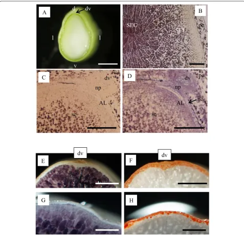
Under stereomicroscopic observation, the endosperm showed a uniform milky-white color (Fig. 1a), but morph-ology was qualitatively and quantitatively different among tissues observed with higher magnification. At 7 DAF, the starchy endosperm in the central region (SEC) accumulated much more starch than the starchy endosperm in the lateral regions (SEL) (Fig. 1b). In mature grain, iodine staining was lighter in SEL than in SEC (Fig. 1g). The surrounding outermost cell layer(s) were not stained with iodine solution (Fig. 1c, d, e, g). These cell layer(s) were identified as aleurone cells that were stained with Sudan IV with the accumulation of lipids at maturity (Fig. 1f, h). We defined the dorsal side of aleurone cells as AL (Fig. 1d).
Transmission electron microscopic observation of endosperm cells
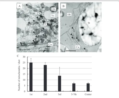
The development of organelles in endosperm cells at 7DAF was observed by transmission electron microscopy (TEM). Typical differences were observed in mitochondria
in AL and in starch granules in endosperm cells (Fig. 2a and b). The mitochondria in the cells of developing endosperm were quantified along a dorso-ventral axis (the lateral regions of the endosperm are not included in this axis). In the first and second cell layers of the AL, approximately 25 mitochondria were observed per image (310.2 μm2; Fig. 2a, c), but mitochondrial density decreased by half in the third cell layer of the AL (Fig. 2c) and dropped to one fourth in the fifth cell layer of the starchy endosperm compared with the outermost AL cells (Fig. 2b, c). A similar number of mito-chondria were observed below cell layers 5 to 7 (Fig. 2).
Oxygen concentration of developing endosperm along a dorso-ventral axis
In view of the differential distribution of mitochondria in the endosperm cell layers, oxygen concentration profiles along the dorso-ventral axis (SEL is not located on the x axis in Fig. 3) in developing rice caryopses at 7 DAF were investigated using a Clark-type O2
microelec-trode according to the method of Shimamura et al. (2010). We found that the oxygen concentration decreased steeply as the microelectrode was being inserted deeper into the starchy endosperm. In particular, 8 % oxygen was observed at 200 μm from the surface of the caryopsis (the pericarp), a region corresponding to the dorsal side of AL (Fig. 3). When the electrode was inserted to a depth greater than 300μm, where the microelectrode had already passed through the AL region and reached the inner starchy endosperm, oxygen concentration was lower than 2 % (Fig. 3). At 1200μm, a region corresponding to the SEC, oxygen was absent.
LM for endosperm cells at 7 DAF and identity confirmation of dissected tissues by qRT-PCR
because of the specific localization of starch granules and lipids in the endosperm (Fig. 1). Transcripts of
oleosin, 16 kDa isoform R16, were detectable only in AL, whereas those of SDBE were at almost negligible levels in AL (Additional file 3: Figure S2A). The tran-script level of SDBE was higher in the SEC than in the SEL (Additional file 3: Figure S2B) consistent with the gradients shown by iodine staining (Fig. 1b). These results confirmed the precise dissection of targeted endosperm tissues.
qRT-PCR for genes associated with sucrose transport, sucrose cleavage, and starch biosynthesis
Transcription levels of genes associated with sucrose transport (Hirose et al. 1997), sucrose cleavage (cell wall invertase; Cho et al. 2005 and sucrose synthase; Hirose et al. 2008), hexokinase (Cho et al. 2006), UDP-glucose pyrophosphorylase (Chen et al. 2007), phospho-glucomutase (Akiyama et al. unpublished), and plastidis translocator (Toyota et al. 2006) and starch biosynthesis (Hirose and Terao 2004; Ohdan et al. 2005) during rice
Fig. 1Observation of endosperm cells at 7 DAF (a–d) and in mature grain (e–h).aMedian transversal section.bMicroscopic observation of starchy endosperm at the center (SEC) and lateral side (SEL) stained with iodine.c,dMicroscopic observation of endosperm at the dorsal side stained with iodine (c) and post-stained with toluindine blue-O (d). Matured grain stained with iodine (e,g) and Sudan IV (f,h).ALalurone cells,d
grain filling were evaluated. The selected genes are consid-ered to play dominant roles in the developing endosperm among gene families with high expression or critical roles in grain phenotype and starch properties, according to previous genetic studies (see review by Fujita 2014, Table 2, and Background).
The expression ofOsSUT1was specific to aleurone cells and undetectable in the SEL and SEC. The expression levels were contrasting among the genes for sucrose-cleavage enzymes. The highest expression levels for OsCIN2,
OsSUS3, andOsSUS4were observed in AL, SEL, and SEC, respectively (Fig. 5).
A major role of hexokinase (OsHXK) genes in rice developing endosperm has not yet been clarified by genetic approaches, but the genes OsHXK2, OsHXK4,
OsHXK5,OsHXK6, andOsHXK8were selected in view of their preferential expression in developing endosperm,
based on the report of Cho et al. (2006). OsHXK4 and
OsHXK5 were expressed predominantly in SEC and AL, respectively. The expression of OsHXK2 and OsHXK6
was observed in all tissues, but preferentially in AL and SEL, respectively. The expression of OsHXK8 was higher in SEL and SEC compared to AL. Expression of OsUgp1 and phosphoglucomutase was observed in all tissues.
The expression of genes for starch biosynthesis was variable. Expression was at low level (with values below 25) in AL for all genes exceptOsSSIIaand OsISA1. The expression ofOsAGPL2andOsAGPS2b,OsBT1-1,OsSSI,
OsSSIIIa, and OsPHOL was preferential in SEC. The expression of genes for OsSSIIa, OsBEI, and OsBEIIb
was relatively high (with values over 60) in the SEL and highest in the SEC. The expression of genes forOsGBSSI
and OsISA1 was highest in SEL among endosperm
tissues tested. We also investigated the expression of disproportionating enzyme (OsDPE1; Ohdan et al. 2005), which plays a critical role in building the amylopectin structure in Chlamydomonas reinhardtii (Colleoni et al. 1999). The expression ofOsDPE1was undetectable in any tissues at 7DAF (data not shown), possibly because the transcript level abruptly decreases after 5DAF in the developing caryopsis (Ohdan et al. 2005).
qRT-PCR of genes associated with CO2fixation, the TCA
cycle, oxidative phosphorylation, and the GS/GOGAT cycle
The presence of oxygen (8 %) in the AL (Fig. 3), sug-gested the high expression of genes associated with aerobic respiration. In addition to the expression of genes for TCA cycle and oxidative phosphorylation, the expression of genes associated with CO2 fixation
and the GS/GOGAT cycle, a metabolic step is close to the TCA cycle and oxidative phosphorylation, was evaluated (Table 3; Fig. 6). Some of the selected genes have not yet been characterized, but they appear in NCBI (http://www.ncbi.nlm.nih.gov/nuccore/) and RAP-DB (http://rapdb.dna.affrc.go.jp/) as our target genes based on their sequence similarity to genes in other plant species (Table 3).
All the genes analyzed showed highest expression in AL, and their expression levels decreased in the order SEL > SEC (Fig. 6). Some of the genes were localized pre-dominantly in the AL (Osppc1, Acetyl-CoA carboxylase, genes for the TCA cycle,NADH-ubiquinone oxidoreductase subunit PSST,GS1;1,OsGDH1.2).
Discussion
Transcripts associated with metabolic steps from sucrose transport to starch biosynthesis were differentially distributed in developing endosperm
Genetic analyses using mutants and gene manipulation have revealed the critical role(s) of genes for carbohydrate-metabolizing enzymes in kernel phenotypes and starch properties (Table 2). In this study, LM was applied to the AL, SEL and SEC (Fig. 4) to quantify the distribution of transcripts at metabolic steps from sucrose trans-port to starch biosynthesis, relative to their spatial distribution in starch granules among tissues in the early storage phase (Fig. 1).
The transcript of sucrose transporter 1 (OsSUT1; Hirose et al. 1997) was specific to AL, as we reported previously for AL and SEC (Ishimaru et al. 2007). This study showed negligible expression in SEL (Fig. 5). An anti-sense transformant of OsSUT1 showed impaired grain filling (Scofield et al. 2002). The specific localization ofOsSUT1in AL (Fig. 5) suggests the critical function of aleurone cells for uptake of sucrose into developing endosperm tissues for starch accumulation. Our LM-based expression analysis revealed the specific localization of
OsCIN2in AL among endosperm tissues as that ofOsSUT1
(Fig. 5). A mutation of OsCIN2 (gif1) causes slower grain filling and chalky phenotype of the kernel with aberrant amyloplast formation, indicating that hexoses catalyzed byOsCIN2function as an important carbon energy source for grain development in rice (Wang et al. 2008). The specific distribution ofOsCIN2in AL (Fig. 5) was spatially
inconsistent with starch accumulation in the starchy endosperm, such as in SEL and SEC. OsCIN2 in AL is expected to contribute to the partitioning of hexoses to the inner endosperm tissues for starch accumulation in SEL and SEC. The organ expression analysis of OsSUS3
and OsSUS4 revealed their predominant localization in the developing caryopsis, especially in the endo-sperm, after the onset of starch accumulation at 5 DAF (Hirose et al. 2008). Our LM-based expression
analysis found OsSUS3 and OsSUS4 to be localized mainly in the SEL and SEC, respectively (Fig. 5). The expression profile of the sucrose-cleavage genes OsCIN2,
OsSUS3, andOsSUS4 was spatially complemented in the developing endosperm. The cleavage of sucrose in the developing endosperm is necessary for generating a sucrose gradient to maintain sink strength (Sturm and Tang 1999). The results suggest thatOsCIN2,OsSUS3, and
OsSUS4contribute to sink strength by cleaving sucrose in
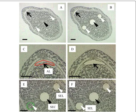
Fig. 4Microdissection of endosperm tissues.aMedian transverse section.bMicrodissection of aleurone cells from the dorsal side (AL;black arrow, magnified incandd), starchy endosperm from the center region (SEC;black arrowhead, magnified ineandf) and lateral regions (SEL;
white arrowheads, magnified ineandf).Bar: 200μm
Table 1Internal control and marker genes for aleurone cells and starchy endosperm
Category NCBI accession number Description Reference
Internal control X00755 18S rRNA Kim et al.2003
Marker for aleurone cells AF022148 16 kDa oleosin isoform R16 NCBIa; Medina and Quatrano, unpublished.
Marker for starchy endosperm D50602 Starch debranching enzyme Pullulanase; Nakamura et al.1996, OsPUL; Ohdan et al.2005
a
different locations in developing endosperm tissues. With respect to the expression profile for hexokinase,OsHXK4
andOsHXK5showed predominant expression in SEC and AL, respectively. For OsHXK2, a gradient in expression
level was observed from AL to SEC in descending order.
OsHXK6and OsHXK8showed highest expression level in SEL and SEC, respectively, and relatively high expression was observed remaining two tissues. Transcripts of
Table 2Information on the genes related to sucrose transport, sucrose cleavage, intermediate metabolic steps and starch biosynthesis
Category Accession No. Description Reference Major changes in grain appearance and
starch properties with the mutant and genetic manipulation of gene
Sucrose transport and cleavage
D87819 Sucrose transporter (SUT1) OsSUT1; Hirose et al.1997 Impaired grain filling (Scofield et al.2002)
AK072276 Cell wall invertase 2 OsCIN2; Cho et al.2005 Chalky phenotype with abnormal amyloplast (gif1; Wang et al.2008)
AK100306 Sucrose synthase 3 SUS3; Hirose et al.2008
-AK102158 Sucrose synthase 4 SUS4; Hirose et al.2008
-Metabolic step between sucrose cleavage and starch biosynthesis
DQ116384 Hexokinase 2 OsHXK2; Cho et al.2006
-DQ116386 Hexokinase 4 OsHXK4; Cho et al.2006
-DQ116387 Hexokinase 5 OsHXK5; Cho et al.2006
-DQ116388 Hexokinase 6 OsHXK6; Cho et al.2006
-DQ116390 Hexokinase 8 OsHXK8; Cho et al.2006
-AB062606 UDP-glucose pyrophosphorylase
UGPase; Abe et al.2002, OsUgp1; Chen et al.2007, UGPase1; Woo et al.2008
Chalky phenotype (Koh et al.1999)
AF455812 Phosphoglucomutase NCBIa; Akiyama, unpublished.
-Starch biosynthesis U66041 ADP-glucose pyrophosphorylase large subunit
OsAGPL2; Ohdan et al.2005; Lee et al.2007
shrunken phenotype (Lee et al.2007)
AK103906 ADP-glucose pyrophosphorylase small subunit
OsAGPS2b; Ohdan et al.2005; Lee et al.2007
shrunken phenotype (Lee et al.2007)
AK107368 ADP-glucose transporter OsBT1-1; Toyota et al.2006 brittle phenotype (maize; Shannon et al.
1998, barley; Patron et al.2004)
D16202 Soluble starch synthase 1 SSS; Baba et al.1993, SSI; Hirose and Terao2004, OsSSI; Ohdan et al.2005
Altered fine structure of amylopectin (Fujita et al.2006)
AF419099 Soluble starch synthase II-3 SSII-3; Hirose and Terao2004, SSIIa; Ohdan et al.2005
Altered fine structure of amylopectin (alk; Umemoto et al.2004)
AY100469 Soluble starch synthase III-2 SSIII-2; Hirose and Terao2004, OsSSIII-2; Dian et al.2005, OsSSIIIa; Ohdan et al.2005
White-cored chalky phenotype and altered fine structure of amylopectin (Fujita et al.2007, flo5; Ryoo et al.2007)
X62134 Granule-bound starch synthase I Okagaki1992, GBSSI; Hirose and Terao2004, OsGBSSI; Ohdan et al.2005
waxy phenotype with the absence of amylose (Itoh et al.2003)
D11082 Starch branching enzyme I RBEI; Mizuno et al.1992, OsBE1; Ohdan et al.2005
Altered fine structure of amylopectin (sbeI; Satoh et al.2003)
D16201 Starch branching enzyme IIb RBEIII; Mizuno et al.1993, OsBEIIb; Ohdan et al.2005
Chalky phenotype and altered fine structure of amylopectin (amylose-extender; Nishi et al.2001)
AB093426 Starch debranching enzyme: Isoamylase I
OsISA1; Ohdan et al.2005 sugary phenotype (sugary-1; Kubo et al.1999a)
AK063766 Plastidial phosphorylase OsPHOL; Ohdan et al.2005 shrunken to pseudonormal phenotypes (pho1; Satoh et al.2008), protein phosphorylation in amyloplast (wheat; Tetlow et al.2004) a
OsUgp1and phosphoglucomutasewere highest in AL and SEC, respectively, and relatively high expression was observed in the remaining two tissues. Thus, the expres-sion of each gene appeared to be functional in a different space in hexokinase.OsUgp1andphosphoglucomutaseare assumed to generate metabolites for subsequent starch biosynthesis in all three regions: AL, SEL, and SEC.
With respect to genes for starch biosynthesis, expres-sion was low in AL, and the highest values, except for
OsGBSSI and OsISA1, were observed in SEC (Fig. 5). Expression ofOsAGPL2,OsAGPS2b,OsBT1-1,OsSSI, and
SSIIIa was low in SEL, whereas expression of OsSSIIa,
OsGBSSI, OsBEI, OsBEIIb, and OsPHOL was relatively high (with values over 60) in SEL. T-DNA insertion into either OsAGPL2 or OsAGPS2b resulted in a shrunken phenotype of rice kernels with impaired grain filling (Lee et al. 2007). Mutant plants of brittle-1 in maize (Shannon et al. 1998) andlys5in barley (Patron et al. 2004) show drastically decreased kernel dry weight, owing to the absence of a plastidial ADP-glucose transporter. The gradient of transcript abundance for OsAGPL2,
OsAGPS2b,andOsBT1-1was consistent with the gradient of starch accumulation among AL, SEL, and SEC (Fig. 1), suggesting the in vivo regulation of starch accumulation by these genes at the transcriptional level. For starch synthase (both soluble and granule-bound type), starch branching enzymes, and starch debranching enzyme, greater attention has been paid to the effects of genes on starch properties such as amylopectin structure and amylose formation. Relatively high levels of expression of OsSSIIa, OsGBSSI, OsBEI, OsBEIIb, OsISA1, and
OsPHOL in SEL suggest that these genes are responsible for the production of amylopectin and amylose in SEL at the transcriptional level. The subcellular location of
OsAGPL2 andOsAGPS2b is cytosolic (Sikka et al. 2001), whereas that of OsBT1-1, a gene for a starch synthase branching enzyme, and starch debranching enzyme is plastidial. In addition, the metabolic step of ADP-glucose pyrophosphorylase is located upstream from that of starch synthase, starch branching enzyme, and starch debranching enzyme. In SEL, the substrate of ADP-glucose may be deficient in plastids, owing to the very low level of cytosolicOsAGPL2andOsAGPS2b. Starch biosynthesis in the cereal endosperm is a complex process engaged with many genes (Fig. 5). Overall results of expression of genes associated with metabolic steps from sucrose transport to starch biosynthesis revealed the clear differences in the distribution of transcripts in the de-veloping endosperm. The LM-based expression analysis conducted in this study provided the novel finding that the clear gradient of transcripts of OsAGPL2 and
OsAGPS2bmay be responsible for the large difference in starch accumulation among AL, SEL, and SEC in the early storage phase.
Aleurone cells are inferred to generate ATP by aerobic respiration during the early storage phase
Aleurone cells do not contain starch, but contain lipids (Fig. 1e and f ). Our LM-based expression analysis showed the specific localization of oleosin, 16 kDa isoform R16, in AL (Additional file 3: Figure S2), and the low level of transcripts for starch biosynthesis in AL (Fig. 5). The expression analysis is consistent with the large amount of lipid and the absence of starch in AL (Fig. 1c). In developing maize, oxygen concentration is maintained at the high level in the oil-storing embryo, corresponding to the steady-state levels of glycolytic intermediates and those of the TCA cycle, as well as free amino acids (Rolletschek et al. 2005). In the present study, we investigated the spatial distribution of oxygen (Fig. 3) and transcripts of genes associated with carbon fixation, the TCA cycle, oxidative phosphorylation, and
the GS/GOGAT cycle (Fig. 6). Oxygen was detectable in the outermost endosperm cells corresponding to the aleurone layers at 8 % (Fig. 3). All the genes exam-ined in Table 3 were expressed dominantly in AL (Fig. 6), supporting the findings in maize embryo (Rolletschek et al. 2005) with respect to oxygen con-centration and metabolites in oil-storage tissue. In the present study, we identified clear gradients in the number of mitochondria from the AL to the SEC in descending order (Fig. 2). Oparka et al. (1981) reported the presence of mitochondria in the aleurone and sub-aleurone layers with TEM observation. The present quantitative investigation showed that the profile of oxygen distribution agrees well with the gradient in numbers of mitochondria in rice endosperm in the early storage phase. Xu et al. (2008) reported that accumulation of proteins associated with the TCA cycle increased during 6–10 DAF
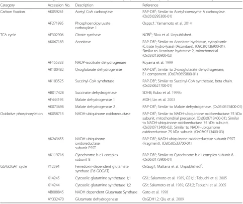
Table 3Information on the genes related to carbon fixation, TCA cycle, oxidative phosphorylation and GS/GOGAT cycle
Category Accession No. Description Reference
Carbon fixation AK059261 Acetyl CoA carboxylase RAP-DBa; Similar to Acetyl-coenzyme A carboxylase. (Os05t0295300-01)
AF271995 Phosphoenolpyruvate carboxylase 1
Osppc1; Yamamoto et al.2014
TCA cycle AF302906 Citrate synthase NCBIb; Silva et al. Unpublished.
AK067183 Aconitase RAP-DBa; Similar to Aconitate hydratase, cytoplasmic (Citrate hydro-lyase) (Aconitase). (Os03t0136900-01). Similar to Aconitate hydratase 2, mitochondrial. (Os03t0136900-02)
AF155333 NADP-isocitrate dehydrogenase Koyama et al.1999
AK100482 Oxoglutarate dehydrogenase RAP-DBa; Similar to 2-oxoglutarate dehydrogenase, E1 component. (Os07t0695800-01)
AK103525 Succinyl-CoA synthetase RAP-DBa; Similar to Succinyl-CoA synthetase, beta chain. (Os02t0621700-01)
AB017428 Succinate dehydrogenase SDHB; Kubo et al.1999b
AF444195 Malate dehydrogenase 1 MDH; Lin et al.2003
AK073698 Malate dehydrogenase 2 RAP-DBa; Similar to Malate dehydrogenase. (Os05t0574400-01)
Oxidative phosphorylation AK058713 NADH-ubiquinone oxidoreductase RAP-DBa; Similar to NADH-ubiquinone oxidoreductase 75 kDa subunit, mitochondrial precursor. (Os03t0713400-01); Similar to NADH-ubiquinone oxidoreductase 75 kDa subunit. (Os03t0713400-02); Similar to NADH-ubiquinone oxidoreductase 75 kDa subunit. (Os03t0713400-03)
AK243655 NADH-ubiquinone oxidoreductase subunit PSST
RAP-DBa; NADH-ubiquinone oxidoreductase subunit PSST (Fragment). (Os05t0533700-01)
AK119716 Cytochrome b-c1 complex subunit 8
RAP-DBa; Similar to Cytochrome b-c1 complex subunit 8. (Os06t0175900-01)
GS/GOGAT cycle Y12594 Ferredoxin-dependent glutamate synthase (Fd-GOGAT)
OsGog1; Mattana et al. Unpublishedb.
X14245 Cytosolic glutamine synthetase 1;1 GS1; Sakamoto et al.1989, GS1;1; Tabuchi et al.2005
X14244 Cytosolic glutamine synthetase 1;2 GSr; Sakamoto et al.1989, GS1;2; Tabuchi et al.2005
AB008845 NADH dependent Glutamate Synthase Goto et al.1998
AY332470 Glutamate dehydrogenase OsGDH1.2; Qiu et al.2009
a
The Rice Annotation Project Database
b
(in the early storage phase), based on proteomic analysis of developing rice kernels. Thus, mitochondria localized in the oxygen-rich cells of AL are expected to contribute to ATP generation through aerobic respiration, thereby assisting the initial formation and accumulation of lipids via expression of genes listed in Table 3. In barley, starch accumulation is initiated in the lateral region, where the tissues retain a high level of oxygen sup-plied by photosynthesis in the surrounding pericarp (Rolleschek et al. 2004). In rice, starch accumulation is ini-tiated in the center of the endosperm (Fig. 1b), where the tissue is most distant from the pericarp and in a condition
of hypoxia (Fig. 3). Energy production for starch accumu-lation may be different between barley and rice, given the differences in oxygen availability in the starch-storing tis-sue in the early storage phase. SEC is assumed to produce ATP via anaerobic respiration to supply energy for starch development in the absence of oxygen (Fig. 3). Whether gradients in oxygen concentration are coupled with the differences in storage product between endosperm tissues (i.e. lipids in aleurone cells and starch in starchy endo-sperm) through the different process of energy production is still elusive. Further evidence are required to reveal the relationship between the energy production and biological
process of storage product accumulation in the different positions of developing endosperm. Knockout mutants of cytosolic glutamine synthetase 1;1 showed a signifi-cant reduction in rice kernel weight (Tabuchi et al. 2005), indicating the involvement of this gene in car-bon partitioning through nitrogen metabolism in the AL. The function of cytosolic glutamine synthetase 1;1 in the developing endosperm is still unclear, but there may be physiological linkages between nitrogen metabolism in aleurone cells and starch synthesis in starchy endosperm.
Conclusions
We revealed the expression pattern of carbohydrate-metabolizing genes in the different positions of developing endosperm with an assistance of LM. The expression of
OsSUT1 was specific to the AL, and the expression of sucrose-cleavage enzymes such as OsCIN1, OsSUS3, and
OsSUS4was preferential in AL, SEL and SEC, respectively. The gradients of transcript abundance for OsAGPL2
and OsAGPS2b were assumed to be associated with the differential spatial distribution of starch granules among endosperm tissues in the early storage phase. These results in carbohydrate-metabolizing genes suggested the roles of each gene in carbon partitioning and starch synthesis in the different positions of developing endo-sperm. The presence of oxygen and large number of mitochondria in AL were consistent with the predominant expression of genes involved in TCA cycles and oxidative phosphorylation, inferring the energy production via aerobic respiration at least in part in AL. The LM-based expression analysis conducted in this study expanded molecular and physiological knowledge on the pos-itional differences in starch accumulation and energy production in the developing rice endosperm in the early storage phase.
Methods Plant materials
Oryza sativa cv. Koshihikari (a Japonica rice variety) was used. Seeds were sown in a nursery box filled with soil, and 4 weeks-old seedlings were transplanted into 0.02 m2pots. As a basal dressing, 0.5, 2.3, and 2.2 g each of N, P2O5, and K2O was applied, and 0.4 g of nitrogen
per pot was applied as a top dressing approximately two weeks before heading. At the booting stage, plants were transferred into a naturally illuminated temperature-controlled chamber. Day (13 h) and night (11 h) air temperatures were maintained at 26 and 20 °C, respectively until maturity. Spikelets were marked on the flowering day. Caryopses at 7 DAF located on the four primary rachis branches counted from the top of the panicles were used in all experiments.
Laser microdissection (LM)
Four developing rice caryopses were collected from one panicle. Three biological replicates (panicles) were prepared from three rice plants. The preparation of specimens for LM followed Ishimaru et al. (2007). Briefly, the developing caryopses were immediately fixed with an ice-cold mixture of 3:1 ethanol: acetic acid, and embedded with 2 % carboxy-methylcellulose. Transverse sections (8μm thickness) were made at the median part of the developing caryopses with a cryomicrotome (Leica, CM1850). Aleurone cells at the dorsal side (AL) and starchy endosperm in the center (SEC) and the lateral regions (SEL) were microdissected with an AS LMD system (Leica Microsystems, Wetzlar, Germany).
RNA extraction, quantification, and quality check
Total RNA was extracted with a Picopure RNA isolation kit (Molecular Devices, Sunnyvale, CA) using DNase I. Quantification of total RNA was determined by the fluorescence based method, using a RiboGreen RNA Quantification kit (Molecular Probes, Eugene, OR). The integrity of RNA from aleurone cells and starchy endosperm was assessed using a 2100 Bioanalyzer (Agilent technologies, Santa Clara, CA). The average RNA integrity number (RIN) with clear rRNA peaks from each tissue exceeded 7.0 for the three biological replications (Additional file 1: Table S1).
cDNA synthesis and quantitative RT-PCR
Quantitative RT-PCR was performed following Ishimaru et al. (2009) with three biological replications. Total RNA (10 ng) was amplified and cDNA was synthesized with a WT-OvationTM RNA Amplification System (NuGEN technologies Inc., San Carlos, CA) according to the manu-facturer’s instructions. For quantitative RT-PCR, SYBR Premix Ex Taq (TaKaRa Bio Inc, Shiga, Japan) was used with a real time RT-PCR system (7500 Real Time PCR System, Applied Biosystems, Foster, CA). Gene-specific primer pairs were designed with Primer 3 (version 0.4.0., http://frodo.wi.mit.edu/primer3/) or taken from previous reports (Additional file 4: Table S2). Dissociation curves confirmed the presence of a single amplicon in each PCR. cDNA of each gene was further amplified with Taq polymerase (ExTaq; TaKaRa Bio Inc), and PCR products were sequenced to confirm that the fragments were the targeted gene. For each gene, the highest value in a given tissue was adjusted to 100 after normalization with the transcript level of 18S rRNA (Kim et al. 2003; Additional file 2: Figure S1), and relative values less than 100 were determined for two other tissues.
Oxygen concentration in developing caryopses
the glume, the caryopsis was placed in an aluminum block and fixed with dental silicone impression material (Provil novo light, Heraeus Kulzer GmbH, Germany). The base of the rachilla was covered with dental silicone impression material to prevent an inflow of external air after detachment. The impression material hardened within 2 min. A Clark-type O2 microelectrode (OX-25,
Unisence A/S, Denmark) with a guard cathode and a tip diameter of 25 μm was inserted into a developing caryopsis at 50-μm intervals along a dorso-ventral axis. The microelectrode was connected to a pA meter (PA2000, Unisense A/S) and output was logged at 5-s intervals on a computer using an analog-to digital converter (ADC-16, Pico Technology, UK). The electrode was calibrated in air before measurement and in O2-free
N2. The experiment was conducted at a temperature of
25 °C and 12 μmol m−2 s−1 photon flux density. Three developing caryopses sampled from different panicles were used. O2 concentrations obtained from three developing
caryopses were averaged.
Microscopic observation
Stereo microscope
Median transverse sections (1.0–1.5 mm thickness) of developing caryopses and matured grain were manually cut with a sharp razor. Sectioned mature kernels were immersed into a solution of 2 % KI and 0.4 % I2or 2 %
Sudan IV (w/v) in 70 % ethanol for staining starch or lipid, respectively. After staining, specimens were viewed under a stereomicroscope (SZX12, Olympus, Japan), and immediately photographed.
Light microscope
Developing caryopses were immersed in FAA (formalin: acetic acid: 70 % ethanol = 1: 1: 18), dehydrated in an ethanol series, and embedded in Technovit 7100 (Heraeus Kulzer GmbH, Hanau, Germany). Sections 3 μm thick were cut with a microtome (HM335E, Leica Microsystems, Germany). Sections were stained with a solution of 2 % KI and 0.4 % I2to observe starch granules and the same
speci-mens were then stained again with 0.1 % toluidine blue-O to observe median transverse endosperm.
Transmission electron microscope (TEM)
Developing caryopses at 7 DAF were hand-cut into 1.0 mm-thick sections at the median with a sharp razor and immediately fixed with ice-cold 4.0 % paraformalde-hyde and 2.0 % glutaraldeparaformalde-hyde for 3 h. Specimens were fixed again overnight at 4 °C. After washing with 100 mM phosphate buffer (pH 7.2), the specimens were post-fixed with 1 % osmium tetroxide overnight at 4 °C. They were then washed with distilled water, dehydrated in an ethanol series, and embedded in Spurr’s resin (Spurr Low Viscosity Embedding kit, Polyscience Inc., Warrington, PA). Sections
200 nm thick were cut with glass knives with an ultrami-crotome (MT2-B, Sorvall, Newtown, CT) at the median part of the specimen. Sections were stained with TI blue (Nissin EM, Tokyo, Japan) for 2 h and with lead citrate for 7 min, and viewed under an H-7100 transmission electron microscope (Hitachi, Tokyo, Japan) at 75 kV. Mitochondria were counted from the images at ×3500 magnification on 76 × 50 mm films (310.2 μm2). Mean values were calculated with 13–29 independent images (cells) from two biological replications.
Additional files
Additional file 1: Table S1.RNA integrity number (RIN) used for qRT-PCR analysis.
Additional file 2: Figure S1.qRT-PCR for18S rRNA.The gene accession numbers and primer pairs are shown in Table 1. Values are the means of three biological replications. The value in each tissue was used for the normalization.
Additional file 3: Figure S2.qRT-PCR for 16 kDaoleosin(A) andstarch debranching enzyme(B;SDBE). The gene accession numbers and primer pairs are shown in Table 1. Values are the means of three biological replications.
Additional file 4: Table S2.Gene-specific primers used in this study.
Competing interests
The authors declare that they have no competing interests.
Authors’contributions
TI, TM, MN, and MK designed the experiments. TI, MI, SH, and SS conducted the experiments. TI, SS, TM, NKN, MN, and MK wrote the manuscript. All authors read and approved the submitted version of manuscript.
Acknowledgements
T. I. gratefully acknowledges Prof. N. Tsutsumi (The University of Tokyo) for technical advice on the measurement of mitochondria. T. I. also thanks Dr. Taniguchi (NICS), Dr. Ishikawa (NICS), Ms. Ito, and Ms. Hiroki for technical assistance. This work was supported by research grants from the Ministry of Education, Culture, Sports, Science and Technology of Japan (No.20780013 to T. I.).
Author details
1NARO Institute of Crop Science, NARO, Kannondai, Tsukuba, Ibaraki 305-8518, Japan.2Japan International Research Center for Agricultural Sciences, Ohwashi, Tsukuba, Ibaraki 305-8686, Japan.3Graduate School of Life and Environmental Science Kyoto Prefectural University, Shimogamo, Sakyo-ku, Kyoto 606-8522, Japan.4Graduate School of Agricultural and Life Sciences, University of Tokyo, Yayoi, Bunkyo, Tokyo 113-8657, Japan. 5Research Institute for Bioresources and Biotechnology, Ishikawa Prefectural University, 1-38 Suematsu, Nonoichi, Ishikawa 921-8836, Japan.6Graduate School of Bioagricultural Sciences, Nagoya University, Furo, Chikusa, Nagoya 464-8601, Japan.7International Rice Research Institute (IRRI), DAPO Box 7777, Metro Manila, Philippines.8Life Science Research Institute, Kumiai Chemical Industry Co., Ltd., Shizuoka 439-0031, Japan.9National Institute of Agrobiological Sciences, Kannondai, Tsukuba, Ibaraki 305-8602, Japan. 10NARO Tohoku Agricultural Research Center (TARC), NARO, Kari-wano, Daisen, Akita 019-2112, Japan.
Received: 19 March 2015 Accepted: 25 June 2015
References
Aoki N, Hirose T, Scofield GN, Whitfeld PR, Furbank RT (2003) The sucrose transporter gene family in rice. Plant Cell Physiol 44:223–232
Baba T, Nishihara M, Mizuno K, Kawasaki T, Shimada H, Kobayashi E, Ohnishi S, Tanaka K, Arai Y (1993) Identification, cDNA cloning, and gene expression of soluble starch synthase in rice (Oryza sativaL.) immature seeds. Plant Physiol 103:565–573
Carriger S, Vallee D (2007) More crop per drop. Rice Today 6:10–13 Chen R, Zhao X, Shao Z, Zhu L, He G (2007) Multiple isoforms of UDP-glucose
pyrophosphorylase in rice. Physiol Planta 129:725–736
Cho JI, Lee SK, Ko S, Kim HK, Jun SH, Lee YH, Bhoo SH, Lee KW, An G, Hahn TR, Jeon JS (2005) Molecular cloning and expression analysis of the cell-wall invertase gene family in rice (Oryza sativaL.). Plant Cell Rep 24:225–236 Cho JI, Ryoo N, Ko S, Lee SK, Lee J, Jung KH, Lee YH, Bhoo SH, Winderickx J, An
G, Hahn TR, Jeon JS (2006) Structure, expression, and functional analysis of the hexokinase gene family in rice (Oryza sativaL.). Planta 224:598–611 Colleoni C, Dauvillée D, Mouille G, Morell M, Samuel M, Slomiany MC, Liénard L,
Wattebled F, d’Hulst C, Ball S (1999) Biochemical characterization of the Chlamydomonas reinhardtiiα-1,4 glucanotransferase supports a direct function in amylopectin biosynthesis. Plant Physiol 120:1005–1014 Dian W, Jiang H, Wu P (2005) Evolution and expression analysis of starch
synthase III and IV in rice. J Exp Bot 56:623–632
Emmert-Buck MR, Bonner RF, Smith PD, Chuaqui RF, Zhuang Z, Goldstein SR, Weiss RA, Liotta LA (1996) Laser capture microdissection. Science 274:998–1001
Fujita N (2014) Starch biosynthesis in rice endosperm. AGri-Biosci Monographs 4:1–18
Fujita N, Yoshida M, Asakura N, Ohdan T, Miyao A, Hirochika H, Nakamura Y (2006) Function and characterization of starch synthase I using mutants in rice. Plant Physiol 140:1070–1084
Fujita N, Yoshida M, Kondo T, Saito K, Utsumi Y, Tokunaga T, Nishi A, Satoh H, Park JH, Jane JL, Miyao A, Hirochika H, Nakamura Y (2007) Characterization of SSIIIa-deficient mutants of rice: The function of SSIIIa and pleiotropic effects by SSIIIa deficiency in the rice endosperm. Plant Physiol 144:2009–2023 Furbank RT, Scofield GN, Hirose T, Wang XD, Patrick JW, Offler CE (2001) Cellular
localization and function of a sucrose transporter OsSUT1 in developing rice grains. Aust J Plant Physiol 28:1187–1196
Goto S, Akagawa T, Kojima S, Hayakawa T, Yamaya T (1998) Organization and structure of NADH-dependent glutamate synthase gene from rice plants. Biochim Biophys Acta 1387:298–308
Hirose T, Terao T (2004) A comprehensive expression analysis of the starch synthase gene family in rice (Oryza sativa L.). Planta 220:9–16
Hirose T, Imaizumi N, Scofield GN, Furbank RT, Ohsugi R (1997) cDNA cloning and tissue specific expression of a gene for sucrose transporter from rice (Oryza sativaL.). Plant Cell Physiol 38:1389–1396
Hirose T, Takano M, Terao T (2002) Cell wall invertase in developing rice caryopsis: molecular cloning of OsCIN1 and analysis of its expression in relation to its role in grain filling. Plant Cell Physiol 43:452–459
Hirose T, Scofield GN, Terao T (2008) An expression analysis profile for the entire sucrose synthase gene family in rice. Plant Sci 174:534–543
Hoshikawa K (1967) Studies on the development of endosperm in rice: 1. Process of endosperm tissue formation. Proc Crop Sci Soc Jpn 36:151–161 Hoshikawa K (1968) Studies on the development of endosperm in rice: 11.
Development of amyloplasts in endosperm tissue. Proc Crop Sci Soc Jpn 37:207–216
Ishimaru T, Matsuda T, Ohsugi R, Yamagishi T (2003) Morphological development of rice caryopses located at the different positions in a panicle from early to middle stage of grain filling. Funct Plant Biol 30:1139–1149
Ishimaru T, Hirose T, Matsuda T, Goto A, Takahashi K, Sasaki H, Terao T, Ishii R, Ohsugi R, Yamagishi T (2005) Expression patterns of genes encoding carbohydrate-metabolizing enzymes and their relationship to grain filling in rice (Oryza sativa L.): comparison of caryopses located at different positions in a panicle. Plant Cell Physiol 46:620–628
Ishimaru T, Nakazono M, Masumura T, Abiko M, San-oh Y, Nishizawa NK, Kondo M (2007) A method for obtaining high integrity RNA from developing aleurone cells and starchy endosperm in rice (Oryza sativa L.) by laser microdissection. Plant Sci 173:321–326
Ishimaru T, Horigane AK, Ida M, Iwasawa N, San-oh YA, Nakazono M, Nishizawa NK, Masumura T, Kondo M, Yoshida M (2009) Formation of grain chalkiness and changes in water distribution in developing rice caryopses grown under high-temperature stress. J Cereal Sci 50:166–174
Itani T, Tamaki M, Arai E, Horino T (2002) Distribution of amylase, nitrogen, and minerals in rice kernels with various characters. J Agri Food Chem 50:5326–5332 Itoh K, Ozaki H, Okada K, Hori H, Takeda Y, Mitsui T (2003) Introduction ofWx
transgenic into ricewxmutants leads to both high- and low-amylose in rice. Plant Cell Physiol 44:473–480
Kim BR, Nam HY, Kim SU, Kim SI, Chang YJ (2003) Normalization of reverse transcription quantitative-PCR with housekeeping genes in rice. Biotech Let 25:1869–1872
Koh HJ, Son HY, Heu MH, Lee HS, McCouch SR (1999) Molecular mapping of a new genic male-sterility gene causing chalky endosperm in rice (Oryza sativaL.). Euphytica 106:57–62
Koyama H, Kihara T, Takita E, Hara T (1999) Isolation of Two cDNA Clones Encoding NADP-Specific Isocitrate Dehydrogenase from Rice. Plant Physiol 120:1207 Kubo A, Fujita N, Harada K, Matsuda T, Satoh H, Nakamura Y (1999a) The
starch-debranching enzymes isoamylase and pullulanase are both involved in amylopectin biosynthesis in rice endosperm. Plant Physiol 121:399–409 Kubo N, Harada K, Hirai A, Kadowaki K (1999b) A single nuclear transcript
encoding mitochondria RPS14 and SDHB of rice is processed by alternative splicing: Common use of the same mitochondrial targeting signal for different proteins. PNAS 96:9207–9211
Lee SK, Hwang SK, Han M, Eom JS, Kang HG, Han Y, Choi SB, Cho MH, Bhoo SH, An G, Hahn TR, Okita TW, Jeon JS (2007) Identification of the ADP-glucose pyrophosphorylase isoforms essential for starch synthesis in the leaf and seed endosperm of rice (Oryza sativa L.). Plant Mol Biol 65:531–546
Lin CF, Jiang LZ, Zhang XN, Qian XY, Liang ZS, Yang JS (2003) Cloning and prokaryotic expression of a cDNA encoding a putative mitochondrial malate dehydrogenase inOryza sativa. DNA Seq 14:75–77
Mizuno K, Kimura K, Arai Y, Kawasaki T, Shimada H, Baba T (1992) Starch branching enzymes from immature rice seeds. J Biochem 112:643–651 Mizuno K, Kawasaki T, Shimada H, Satoh H, Kobayashi E, Okumura S, Arai Y, Baba
T (1993) Alternation of the structural properties of starch components by the lack of an isoform of starch branching enzyme in rice seeds. J Biol Chem 268:19084–19091
Nakamura Y, Umemoto T, Ogata N, Kuboki Y, Yano M, Sasaki T (1996) Starch debranching enzyme (R-enzyme or pullulanase) from developing endosperm: purification, cDNA and chromosomal localization of the gene. Planta 199:209–218
Nishi A, Nakamura Y, Tanaka N, Satoh H (2001) Biochemical and genetic analysis of the effects ofAmylose-Extendermutation in rice endosperm. Plant Physiol 127:459–472
Ohdan T, Francisco PB Jr, Sawada T, Hirose T, Terao T, Satoh H, Nakamura Y (2005) Expression profiling of genes involved in starch synthesis in sink and source organs of rice. J Exp Bot 56:3229–3244
Okagaki RJ (1992) Nucleotide sequence of a long cDNA from the ricewaxygene. Plant Mol Biol 19:513–516
Oparka KJ, Gates PJ, Boulter D (1981) Regularly aligned mitochondria in aleurone and sub-aleurone layers of developing rice endosperm. Plant Cell Environ 4:355–357
Patron NJ, Greber B, Fahy BF, Laurie DA, Parker ML, Denyer K (2004) The lys5 mutations of barley reveal the nature and importance of plastidial ADP-Glc transporters for starch synthesis in cereal endosperm. Plant Physiol 135:2088–2097
Qiu X, Xie W, Lian X, Zhang Q (2009) Molecular analyses of the riceglutamate dehydrogenasegene family and their response to nitrogen and phosphorus deprivation. Plant Cell Rep 28:1115–1126
Rolletschek H, Weschke W, Weber H, Wobus U, Borisjuk L (2004) Energy state and its control on seed development: starch accumulation is associated with high ATP and steep oxygen gradients within barley grains. J Exp Bot 55:1351–1359
Rolletschek H, Koch K, Wobus U, Borisjuk L (2005) Positional cues for the starch/lipid balance in maize kernels and resource partitioning to the embryo. Plant J 42:69–83
Ryoo N, Yu C, Park CS, Baik MY, Park IM, Cho MH, Bhoo SH, An G, Hahn TR, Jeon JS (2007) Knockout of a starch synthase geneOsSSIIIa/Flo5causes white-core floury endosperm in rice (Oryza sativaL.). Plant Cell Rep 26:1083–1095 Sakamoto A, Ogawa M, Masumura T, Shibata D, Takeba G, Tanaka K, Fujii S (1989)
Three cDNA sequences coding for glutamate synthetase polypeptide in
Oryza sativaL. Plant Mol Biol 13:611–614
Satoh H, Nishi A, Yamashita K, Takemoto Y, Tanaka Y, Hosaka Y, Sakurai A, Fujita N, Nakamura Y (2003) Starch-branching enzyme I-deficient mutation specifically affects the structure and properties of starch in rice endosperm. Plant Physiol 133:1111–1121
Scofield GN, Hirose T, Gaudron JA, Upadhyaya NM, Ohsugi R, Furbank RT (2002) Antisense suppression of the rice sucrose transporter gene,OsSUT1, leads to impaired grain filling and germination but does not affect photosynthesis. Funct Plant Biol 29:815–826
Shannon JC, Pien FM, Cao H, Liu KC (1998) Brittle-1, an adenylate translocator, facilitates transfer of extraplastidial synthesized ADP-glucose into amyloplasts of maize endosperms. Plant Physiol 117:1235–1252
Shimamura S, Yamamoto R, Nakamura T, Shimada S, Komatsu S (2010) Stem hypertrophic lenticels and secondary aerenchyma enable oxygen transport to roots of soybean in flooded soil. Ann Bot 106:277–284
Sikka VK, Choi SB, Kavakli H, Sakulsingharoj C, Gupta S, Ito H, Okita TW (2001) Subcellular compertmentation and allosteric regulation of the rice endosperm ADPglucose pyrophosphorylase. Plant Sci 161:461–468 Sturm A, Tang GQ (1999) The sucrose-cleaving enzymes of plants are crucial for
development, growth and carbon partitioning. Trends Plant Sci 4:401–407 Tabuchi M, Sugiyama K, Ishiyama K, Inoue E, Sato T, Takahashi H, Yamaya T (2005)
Severe reduction in growth rate and grain filling of rice mutants lacking OsGS1;1, a cytosolic glutamine synthetase 1;1. Plant J 42:641–651 Tetlow IJ, Wait R, Lu Z, Akkasaeng R, Bowsher CG, Esposito S, Kosar-Hashemi B,
Morell MK, Emes MJ (2004) Protein phosphorylation in amyloplasts regulates starch branching enzyme activity and protein-protein interactions. Plant Cell 16:694–708
Toyota K, Tamura M, Ohdan T, Nakamura Y (2006) Expression profiling of starch metabolism-related plastidic translocator genes in rice. Planta 223:248–257 Umemoto T, Aoki N, Lin HX, Nakamura Y, Inouchi N, Sato Y, Yano M, Hirabayashi H,
Maruyama S (2004) Natural variation in ricestarch synthase IIaaffects enzyme and starch properties. Funct Plant Biol 31:671–684
Wang E, Wang J, Zhu X, Hao W, Wang L, Li Q, Zhang L, He W, Lu B, Lin H, Ma H, Zhang G, He Z (2008) Control of rice grain-filling and yield by a gene with a potential signature of domestication. Nat Genet 40:1370–1374
Woo MO, Ham TH, Ji HS, Choi MS, Jiang W, Chu SH, Piao R, Chin JH, Kim JA, Park BS, Seo HS, Jwa NS, McCouch SR, Koh HJ (2008) Inactivation of theUGPase1
gene causes genic male sterility and endosperm chalkiness in rice (Oryza sativaL.). Plant J 54:190–204
Xu SB, Zhu TL, Deng Y, Chong K, Xue Y, Wang T (2008) Dynamic proteomic analysis reveals a switch between central carbon metabolism and alcoholic fermentation in rice filling grains. Plant Physiol 148:908–925
Yamamoto N, Kubota T, Masumura T, Shiraishi N, Tanaka K, Sugimoto T, Oji Y (2014) Molecular cloning, gene expression and functional expression of a phosphoenolpyruvate carboxylaseOsppc1in developing rice seeds: implication of involvement in nitrogen accumulation. Seed Sci Res 24:23–36
Submit your manuscript to a
journal and benefi t from:
7Convenient online submission
7Rigorous peer review
7Immediate publication on acceptance
7Open access: articles freely available online
7High visibility within the fi eld
7Retaining the copyright to your article