Original Research Article
Incidence and association of TCF7, TCF7L2 single nucleotide
polymorphisms in type 1 diabetes mellitus patients
of South Tamil Nadu, India
Kumaravel Velayutham, Balaji Ramanathan, Sivan Arul Selvan,
Rohini Gomathinayagam*
INTRODUCTION
Several linkage studies have revealed that the HLA class I, class II genes and several other loci harbor a significant number of alleles that incur genetic susceptibility to the incidence and onset of T1DM.1,2 Updated and recent studies also reveal that T1DM patients exhibit differences in the clinical, metabolic, immunological and genetic characteristics. Emerging evidences bring to light that such heterogeneity in the disease course could strongly be
associated with the genotypic variations occurring in the T1DM patients.3-5
The human TCF7 gene is located on chromosome 5q31, and initial gene deletion studies using mice models revealed the importance of the gene in T cell formation, function.6,7 It is predominantly expressed in the thymocytes, natural killer cells, and functionally related to the cytokine cluster and hence stands out with a strong linkage to T1DM.7,8 As a transcription factor, the TCF1
ABSTRACT
Background: The TCF family genes TCF7 (T cell specific transcription factor-7) and TCF7L2 (transcription factor 7 like 2) are increasingly recognized to play a pivotal role in the incidence, pathophysiology of type 1 diabetes mellitus (T1DM). However, the prevalence and the influence of these allelic variants in the Indian/south Indian T1DM population is completely obscure.
Methods: Genomic DNA was isolated from the peripheral blood samples of healthy controls, T1DM patients, and PCR (polymerase chain reaction), restriction fragment length polymorphism (RFLP), allele specific PCR (ASP), PCR product sequencing strategies were utilized to determine the prevalence of the TCF7 (exon 3, flanking intron 2, 3 regions) and TCF7L2 (intron 4) polymorphisms. Clinical investigations included assessment of the blood glucose/ estimated average glucose levels (EAG) and C-peptide levels.
Results: The results indicate that 34.9% and 3.17% of the T1DM patients harbored the TCF7L2 rs7903146 and the TCF7 rs386692598 polymorphisms, respectively. Assessment of biochemical parameters indicated that the rs7903146 positive T1DM patients exhibited significantly lower EAG levels (p<0.05), suggesting that these patients may exhibit phenotypic heterogeneity, a milder disease course. The study further demonstrates that PCR based strategies enable reliable molecular diagnosis of T1DM in small scale diagnostic units.
Conclusions: T1DM patients from south Tamil Nadu present TCF7, TCF7L2 genetic variations and screening for these polymorphisms will empower physicians to provide appropriate therapy and genetic counselling.
Keywords: Type 1 diabetes mellitus, TCF7, TCF7L2, Polymorphisms, C883A, rs7903146
Department of Molecular Genetics, Alpha Health Foundation, Mela Anuppanady, Madurai, Tamil Nadu, India
Received: 03 September 2019
Accepted: 20 September 2019
*Correspondence:
Dr. Rohini Gomathinayagam,
E-mail: [email protected]
Copyright: © the author(s), publisher and licensee Medip Academy. This is an open-access article distributed under the terms of the Creative Commons Attribution Non-Commercial License, which permits unrestricted non-commercial use, distribution, and reproduction in any medium, provided the original work is properly cited.
protein encoded by the TCF7 gene primarily regulates the expression of Th1/humoral immune responses, and coordinates with the Wnt signaling candidates, further governing important physiological functions.9 The exon 2 (NC_000005.9) / exon 3 (NC_000005.10, updated version) of the TCF7 gene has been identified to exhibit around 11 SNPs that include the C883A (rs5742913; Pro-Thr) non synonymous coding region polymorphism, and confer susceptibility to T1DM. However, till date, the incidence/relevance of the multi-ethnic, high risk polymorphism C883A and other reported SNPs in the exon 3 of TCF7 has not been assessed in the T1DM patients of India/ South India.
On parallel grounds, rs7903146, an intronic variant of TCF7L2, located in chromosome 10q25.2-q25.3 has also been strongly associated with T2DM risk and the healthy subjects carrying the homozygous or heterozygous version of the T allele present features pertaining to impaired insulin secretion, processing; defective glucagon suppression and incretin secretion.10,11 Further, the TT phenotype of the rs7903146 polymorphism in TCF7L2 is also connected with the presence of single islet cell autoantibody, mild islet cell auto immunity and heterogeneity in the T1DM disease course.12 Based on such strong literature pertaining to TCF7, TCF7L2 genetic variants, the present study focused to examine the presence, influence of the allelic imbalance in the regionally predominant T1DM study population.
METHODS
The prevalence and risk of disease incidence pertaining to the TCF7, TCF7L2 polymorphisms was aimed to be determined in a pilot population of T1DM patients from Alpha Hospital and Research Center/ Alpha Health Foundation, south Tamil Nadu, India. The study was conducted over a period of 11-12 months (February 2018 to January 2019) with the approval of the Institutional Ethical Committee (AHRC). Informed consent from
patients/parents/guardians, routine biochemical
parameters (blood glucose levels, C-peptide) were measured according to previously published protocols.13 The assessed T1DM patients were insulin dependent and presented a minimum of 2 years from the time of diagnosis. All participating T1DM patients were <30 years of age and did not have a medical history pertaining to other pancreatic conditions. Healthy volunteer samples without a history of T1DM served as negative controls. Genomic DNA was extracted from peripheral blood samples of 21 T1DM patients and 6 healthy normal controls, using QIAamp DNA blood mini kit (QIAGEN India Pvt. Ltd, New Delhi, India) according to the manufacturer’s protocol. The concentration of the DNA, the purity of DNA (260/280 nm absorbance ratio) was estimated using a NanoDrop, Thermo scientific, India.
Primers were designed to amplify a 563bp TCF7 gene product that would encompass the amplification of the exonic region containing the C883A and other reported
SNPs. The PCR conditions pertaining to the initial denaturation of 2 min at 96°C, followed by 35 cycles of 30 sec at 94°C, 30 sec at 60.1°C and 2 min at 72°C were utilized for the 563bp product amplification using a forward 5’-TGCCCTGACCTTTATAGGAGTAAAC-3’ and a reverse 5’-CGTCTATTTTGTTCCAGGCAGAA-3’ primer. Based on prior published literature the TCF7L2 polymorphism rs7903146 was also identified by
combining Allele Specific PCR (ASP), RFLP
(Restriction fragment length polymorphism), PCR
product sequencing strategies (PCR conditions
corresponded to an initial denaturation of 5 min at 95°C, followed by 35 cycles of 30 sec at 95°C, 30 sec at 55°C, 45 sec at 72°C and final extension of 5 min at 72°C).14 In brief, for the RFLP study, PCR was carried out using a 5'-ACAATTAGAGAGCTAAGCACTTTTTAGGTA-3' forward primer and 5'-GTGAAGTGCCCAAGCTTCTC-3' reverse primer, and a 188bp PCR product for the TCF7L2 gene was obtained. Restriction digestion with
RsaI resulted in a 158, 30bp cleavage of the wild type/ normal C allele, whereas the SNP T allele was not cleaved. Recognition of the T alleles were also carried out using ASP primers that corresponded to the forward primer
5′- GAACAATTAGAGAGCTAAGCACTTTTTAGAGAT-3′ (product size: 205bp) specific for the T allele and the
reverse primer 5’
AGATGAAATGTAGCAGTGAAGTGC 3’ (initial
denaturation of 3 min at 94°C, followed by 32 cycles of 1 min at 94°C, 1 min at 65°C, 40sec at 72°C and final extension of 5 min at 72°C). Direct sequencing was carried out in a 318bp product that was amplified by
using 5′-GGTAATGCAGATGTGATGAGATCT-3’
forward primer and
5'-AGATGAAATGTAGCAGTGAAGTGC-3’ reverse
primer with PCR conditions that corresponded to an initial denaturation of 3 min at 94°C, followed by 32 cycles of 1 min at 94°C, 1 min at 58°C, 40 sec at 72°C and final extension of 5 min at 72°C. The PCR products were visualized after electrophoresis using a UV gel documentation system (Medicare, Chennai, India) and were gel extracted, purified and sequence verified (Scigenome or Agrigenome, Kochi, Kerala). Statistical analysis of the obtained data was assessed using GraphPad Prism version 7.04 for Windows, GraphPad Software, La Jolla California USA.
RESULTS
present study aimed in assessing the incidence of C883A SNP and other reported polymorphisms in the regional population by means of PCR amplification of a 563 bp region of TCF7 (Figure 1A), and direct sequencing the PCR product. While it can be evidenced from the
sequencing data presented in Figure 1B that the SNP rs5742913/C883A is non-incident in the assessed population, the data revealed the presence of rs386692598, a prior reported intronic variant incident in the study group.
Figure 1: Determination of the incidence of the TCF7 SNPs in the TIDM population: (A) Lane 1-100bp ladder; L2-L3: Non-TIDM controls, L4-L9:T1DM patients. PCR amplification for a 563bp product was carried out, was
electrophoretically separated and visualized in a 1% agarose gel; (B) representative Images for the direct sequencing results for the PCR amplified 563bp product of TCF7 of control sample. The forward and reverse sequences were aligned and the wild type alleles are shown in the aligned sequence; (C) representative Images for the direct sequencing results for the PCR amplified 563bp product of TCF7 of rs386692598 positive patient sample.
The forward and reverse were aligned and the change in nucleotide from GC to TT in forward and CG to CA in reverse sequences is shown in the aligned sequence.
We further examined the influence of the TCF7L2 rs7903146 polymorphism that has recently been associated with phenotypic heterogeneity, late onset in T1DM as the first of its kind, in the present study population. In order to determine the presence of the SNP in the regional T1DM population, we amplified a 188bp SNP associated region and performed RFLP with RsaI enzyme. As indicated in Figure 3, while carriers of the SNP presented with an undigested 188bp product, the wild type carriers presented with an enzymatically cleaved 158bp and 30bp based product. To further substantiate the data the study included direct sequencing of a larger 318bp region (Figure 4) and revalidated the
Table 1: Percentage of allelic expression of rs7903146 in the TIDM patients of south Tamil Nadu, India.
rs7903146 genotype patients
Allelic expression in (%)
Wild type Heterozygous Homozygous
CC CT TT
61.90 28.57 9.52
Genotyping for the rs7903146 group identified that 28.57% of the patients were heterozygous positive and 9.52% of the patients were homozygous positive in the assessed TIDM group.
Figure 2: Percentage of prevalence of TCF7/TCF7L2 polymorphisms in TIDM patients: the incidence and prevalence of the TCF7 563 base pair PCR product that is inclusive of the exon 3 (NC_000005.10) and the 318 bp
TCF7L2 intronic variant (NC_000010.11) was assessed in the TIDM patients of south Tamil Nadu, India; about 3.17% of the patients were positive for the TCF7 intronic variant rs386692598 and 34.90% were positive for the
rs7903146 TCF7L2 polymorphism.
Figure 3: Determination of the incidence of the TCF7L2 rs7903146 polymorphism in the TIDM population: (A) PCR amplified for a 188bp product that would enable the identification of the intron 4-rs7903146; (B) representative image of the RFLP analysis of the 188bp product with RsaI that results in a 158 and 30bp digested
product for patient samples without the SNP. Lane 5, 6, 7: rs7903146 positive patient samples; (C) allele specific PCR was carried out to detect the C to T transition using allele specific primers; patients presenting the T allele
Table 2: Assessment of disease onset age, BMI and C-peptide levels in T1DM patients of south Tamil Nadu, India.
T1DM patients with rs7903146 polymorphism
T1DM patients with rs386692598 polymorphism
Age onset (years) 9.13±2.3 14
BMI (kg/m2) 18.4±1.33 23.2
C-peptide (ng/ml) 0.42±0.17 1.3
Tabular presentation of the mean±SEM values of the assessed parameters in the T1DM patients with TCF7L2 rs7903146 and the TCF7 rs386692598 SNPs.
Figure 4: Determination of TCF7L2 rs7903146 SNP by direct sequencing: (A) M-100bp ladder; L1- L3: Non-TIDM controls, L4-L7:T1DM patients. PCR amplification for a 318bp product was carried out, was electrophoretically
separated and visualized in a 1% agarose gel; (B) representative Images for the direct sequencing results for the PCR amplified 318bp product of TCF7L2 of control sample. The forward and reverse sequences were aligned and the wild type alleles are shown in the aligned sequence; (C) representative Images for the direct sequencing results for the PCR amplified 318bp product of TCF7L2 of homozygous positive TIDM patient sample. The forward and reverse were aligned and the change in nucleotide from C to T in both forward and reverse sequences is shown in
the aligned sequence.
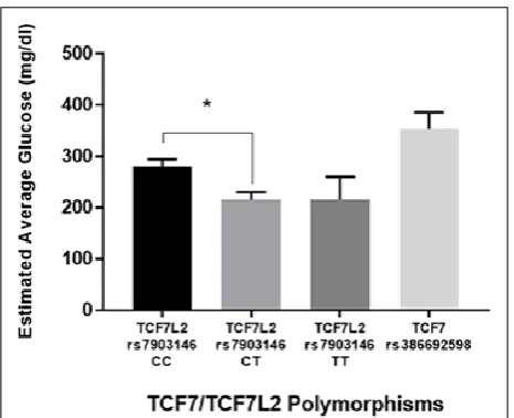
Assessment of the mean age, BMI in the study group T1DM patients who are carriers of the TCF7L2 rs7903146 polymorphism (Table 2) indicates that the mean age group of the carriers is 9.13±2.3, a mean range of BMI as 18.4±1.3 and C-peptide levels in the range of 0.42±0.17. Interestingly and as presented in Figure 5, the mean values of the estimated average glucose was significantly lower in the TCF7L2 rs7903146 carriers
(heterozygous CT, p<0.05) when compared to the non-carriers.
Figure 5: Estimated average glucose levels (EAG) of TIDM patients: TIDM patients without TCF7 gene
polymorphisms/wild type allele and the TIDM patients carrying the TCF7L2 rs7903146, the TCF7 rs386692598 SNP were assessed for differences in the EAG levels; a significant difference in the EAG levels between T1DM patients without SNPs and T1DM patients with TCF7L2 rs7903146 was observed and
the significance is indicated by*. P<0.05 was considered significant and statistical significance was assessed using Mann-Whitney test. All values for EAG
are the mean±SEM for triplicate data sets.
DISCUSSION
An increase in the incidence of T1DM in India has been observed in the recent years, and hence successful management of the disease requires understanding the etiology of the condition.15 Shared genetic variants in T1DM, T2DM and their influence in the incidence and progression of diabetes mellitus has also been explored intensely in the recent years. Of the several candidate genetic variants associated with diabetes, the HMG group of TCF or LEF family genes, TCF7, and TCF7L2, are identified to be significantly associated with the incidence and disease progression of both T1DM and T2DM.16 Primarily, as a candidate gene, TCF7 is being increasingly recognized for its association with the disease incidence and onset of T1DM in multi ethnic populations.9 While the major regions of HLA I, HLA II that comprise of the genetic variations are well examined and established to play a pivotal role in the susceptibility to T1DM in India, cohort studies pertaining to candidate genes from other loci are still being investigated. Very exciting reports had also brought to light that the other member of the TCF family, the TCF7L2 and its genetic
variants (rs7903146, rs4506565, rs7901695) are
associated with a distinct phenotype in T1DM pediatric patients that resemble a T2DM pathological course and is characterized by milder immunological, metabolic phenotype, a late age onset (≥12 years). In corollary, our present study aimed to understand the significance and
incidence of the polymorphisms incident in the exon 3 of the TCF7 gene that would include the globally studied C883A, and decipher the incidence/prevalence of the TCF7L2 polymorphism rs7903146 in the regional population of India or southern India, our efforts have been the first of the kind, till date.
The exon 3 of the TCF7 gene is a hub for several reported polymorphisms that render the risk of incidence of T1DM, and in particular encompasses the C883A (rs5742913; Pro-Thr) non synonymous coding region polymorphism that stands out as a non-DR3/DR4, high risk variant.6 Earlier research investigations have revealed that the TCF7 gene, and its product TCF-1 play an essential role in β-cell survival, apoptosis, responses to the cytokine/pro-inflammatory molecules, insulin secretion, receptor-hormone functions.9 Additively, recent reports have also demonstrated that the TCF7-TCF1 axis regulates the incretin hormone (Glucose dependent insulinotropic peptide, GIP) mediated insulin responses. While our efforts to determine the presence of C883A and other associated SNPs in the regional population indicated that only 4% of the study group presented with a reported intronic variant rs386692598, and none presented the C883A polymorphism, further large scale studies would bring to light the absence of association of C883A polymorphisms in the incidence of T1DM of the regional population. The limited number of samples assessed in the present pilot study could also attribute for the observed absence in the incidence of C883A. Further detailed analysis of the presence of the intronic rs386692598 variant and the associated pathogenicity may bring to light its prevalence, role of such intronic variants in the regional T1DM disease course.
Another recent 2018 study in T1DM patients had also reported that TCF7L2 genetic variants exhibited phenotypic heterogeneity, differences in the disease course progression, the mean glucose values, C-peptide values and the effective age of the disease (≤12 years).19 In sequence, clinical assessment of the influence of the TCF7L2 genetic variations that include (Table 2, Figure 5) age of disease onset, BMI, C-peptide levels, EAG glucose levels, indicated in the present study that the patients positive for the TCF7L2 rs7903146 presented a significantly lower estimated average glucose levels (EAG, p<0.05). However, the present study could not bring to light significant associations with age onset and C-peptide values, most likely owing to the sample size limitation.
Taken together, the present pilot study data for the first time demonstrates that the incidence of TCF7 gene variants in exon 3 and the flanking upstream, downstream intronic variants are sparse in the south Indian/Indian population. However, the TCF7L2 SNP rs7903146 is observed to be present in a relatively higher percentage of the regional T1DM population and that larger scale studies would further bring out the influence of the SNP in late onset, milder disease course.
CONCLUSION
Our study results have brought to light for the first time that the TCF7L2 SNP rs7903146 is of relatively higher incidence in the T1DM population of south India/India and could possibly be associated with a milder disease course. Based on the results it can further be recommended that the incorporation of molecular diagnosis in routine clinical T1DM diagnosis in India, would greatly aid patients and their family to understand the incidence and address the disease course of T1DM.
Funding: No funding sources Conflict of interest: None declared
Ethical approval: The study was approved by the Institutional Ethics Committee
REFERENCES
1. Concannon P, Erlich H A, Julier C, Morahan G, Nerup J, Pociot F, et al. Type 1 Diabetes Genetics C: Type 1 diabetes, Evidence for susceptibility loci from four genome-wide linkage scans in 1,435 multiplex families. Diabetes. 2005;54(10):2995. 2. Rich SS, Concannon P, Erlich H, Julier C, Morahan
G, Nerup J, et al. The Type 1 Diabetes Genetics Consortium. Ann New York Acad Sci. 2006;1079:1. 3. Qu HQ, Polychronakos C. The TCF7L2 locus and
type 1 diabetes. BMC Med Genetics. 2007;8:51.
4. Grant SF, Thorleifsson G, Reynisdottir I,
Benediktsson R, Manolescu A, Sainz J, et al. Variant of transcription factor 7-like 2 (TCF7L2) gene confers risk of type 2 diabetes. Nature Genetics. 2006;38(3):320.
5. Kimber CH, Doney AS, Pearson ER, McCarthy MI,
Hattersley AT, Leese GP, et al. TCF7L2 in the Go-DARTS study: evidence for a gene dose effect on both diabetes susceptibility and control of glucose levels. Diabetologia. 2007;50(6):1186.
6. Verbeek S, Izon D, Hofhuis F, Robanus-Maandag E, te Riele H, van de Wetering M, et al. An HMG-box-containing T-cell factor required for thymocyte differentiation. Nature. 1995;374(6517):70.
7. van de Wetering M, Oosterwegel M, Holstege F, Dooyes D, Suijkerbuijk R, Geurts van Kessel A, et al. The human T cell transcription factor-1 gene.
Structure, localization, and promoter
characterization. J Biol Chem. 1992;267(12):8530. 8. Noble JA, White AM, Lazzeroni LC, Valdes AM,
Mirel DB, Reynolds R, Grupe A, et al. A polymorphism in the TCF7 gene, C883A, is associated with type 1 diabetes. Diabetes. 2003;52(6):1579.
9. Campbell JE, Ussher JR, Mulvihill EE, Kolic J, Baggio LL, Cao X, Liu Y, et al.TCF1 links GIPR signaling to the control of beta cell function and survival. Nature Med. 2016;22(1):84.
10. Zimdahl H, Ittrich C, Graefe-Mody U, Boehm BO, Mark M, Woerle HJ, et al. Influence of TCF7L2 gene variants on the therapeutic response to the
dipeptidylpeptidase-4 inhibitor linagliptin.
Diabetologia. 2014;57(9):1869.
11. Tkac I, Gotthardova I. Pharmacogenetic aspects of the treatment of Type 2 diabetes with the incretin
effect enhancers, Pharmacogenomics.
2016;17(7):795.
12. Redondo MJ, Muniz J, Rodriguez LM, Iyer D, Vaziri-Sani F, Haymond MW, et al. Association of TCF7L2 variation with single islet autoantibody expression in children with type 1 diabetes. BMJ Open Diabetes Res Care. 2014;2(1):e000008. 13. Unnikrishnan AG, Bhatia E, Bhatia V, Bhadada SK,
Sahay RK, Kannan A, Kumaravel V, et al. Type 1 diabetes versus type 2 diabetes with onset in persons younger than 20 years of age. Ann New York Acad Sci. 2008;1150:239.
14. Dutra LA, Costa PG, Velasco LF, Amato AA, Barra GB. Allele-specific PCR assay to genotype SNP rs7903146 in TCF7L2 gene for rapid screening of diabetes susceptibility. Arquivos Brasileiros De Endocrinologia e Metabologia. 2008;52(8):1362. 15. Philip B, Isabel W. Association of cytotoxic T
lymphocyte-associated antigen 4 gene single nucleotide polymorphism with type 1 diabetes mellitus in Madurai population of Southern India, Indian J Human Genetics, 2011;17(2):85.
16. Redondo MJ, Geyer S, Steck AK, Sosenko J, Anderson M, Antinozzi P, et al. TCF7L2 Genetic Variants Contribute to Phenotypic Heterogeneity of Type 1 Diabetes. Diabetes care. 2018;41(2):311. 17. Lukacs K, Hosszufalusi N, Dinya E, Bakacs M,
the gene effect is modified by obesity: a meta-analysis and an individual study. Diabetologia. 2012;55(3):689.
18. Frohnert BI, Ide L, Dong F, Baron AE, Steck AK, Norris JM et al. Late-onset islet autoimmunity in childhood: the Diabetes Autoimmunity Study in the Young (DAISY). Diabetologia. 2017;60(6):998. 19. Redondo MJ, Grant SF, Davis A, Greenbaum C,
Biobank TDE. Dissecting heterogeneity in
paediatric Type 1 diabetes: association of TCF7L2
rs7903146 TT and low-risk human leukocyte antigen (HLA) genotypes. Diabetic Med: J Br Diabetic Assoc. 2017;34(2):286.