Nervous System Changes in Patients
with Multiple Sclerosis
Hormoz Ayromlou1,2, Hadi Mohammad-KhAnli1, Mohammad Yazdchi-mArAndi1,2, Reza riKhtegAr1,2, Sina ZArrintAn3, Samad ej golZAri4,5, Kamyar ghAbili6
1 Department of Neurology, Imam Reza Hospital, Tabriz University of Medical Sciences, Tabriz 51666-14756, Iran
2 Neurosciences Research Center, Tabriz University of Medical Sciences, Tabriz 51666-14756, Iran
3 Department of General & Vascular Surgery, Imam Reza Hospital, Tabriz University of Medical Sciences, Tabriz 51656-65811, Iran
4 Cardiovascular Research Center, Tabriz University of Medical Sciences, Tabriz 51666-15573, Iran
5 Students’ Research Committee, Tabriz University of Medical Sciences, Tabriz 51656-65811, Iran
6 Physical Medicine and Rehabilitation Research Center, Tabriz University of Medical Sciences, Tabriz 51656-65811, Iran
Submitted:22 Jan 2013
Accepted:21 Apr 2013
Abstract
Background: There is supportive evidence that multiple sclerosis (MS) could potentially affect the peripheral nervous system. We assessed peripheral sensory and motor nerve involvement in patients with MS by a nerve conduction velocity test.
Methods: We studied 75 patients who had a relapsing-remitting or secondary progressive pattern. We measured amplitude, latency, conduction velocity, Hoffmann reflex (H-Reflex), and F-Waves.
Results: The amplitude of the right tibial, right proneal, left tibial, left proneal, and left median motor nerves was less than the mean for the normal population. Right ulnar sensory conduction in the patients showed an amplitude that was less than that of the normal population; there was no significant change in the amplitude of other sensory nerves. Latencies of the right and left median and right proneal motor nerves and left ulnar sensory nerves were statistically less than that of the normal population. Mean motor conduction velocity and F-wave conduction did not differ significantly from the normal population. H-reflex latencies of the right and left lower limbs were significantly more prolonged than those of the normal population.
Conclusion: Our results suggest possible peripheral motor nerve abnormalities in MS patients, especially with the amplitude of the motor nerves; however, our results do not demonstrate any significant difference among the nerve conduction velocity parameters of sensory nerves between MS patients and the normal population.
Keywords: demyelination, latency, multiple sclerosis, nerve conduction velocity, peripheral neuropathy
Introduction
Multiple sclerosis (MS) is the most common demyelinating disorder of the central nervous system (CNS). The main pathophysiology of MS is immune-mediated destruction of myelin in the CNS and subsequent plaque formation in the
suggestive of PNS involvement in MS raise the possibility of central and peripheral demyelination due to a same mechanism (3–6).
PNS involvement in MS patients is more than clinically evident peripheral neurologic symptoms, and subtle PNS axonal demyelination
may be present in a significant number of MS
patients (7). However, peripheral motor and sensory axonal degeneration in MS still remains a matter of controversy, and frequency and degree of peripheral involvement necessitates further research and evaluation.
Electrodiagnostic assessment of peripheral nerve involvement in MS may indicate peripheral
demyelination with respect to significant changes
in nerve conduction velocity (NCV) parameters. According to Misawa et al. (7), all MS patients may not necessarily develop peripheral axonal degeneration, but approximately 5% of MS patients show NCV changes suggestive of peripheral involvement. This association could result from a common pathogenesis, possibly due to epitope spreading during the long course of MS.
In the present study, we assess peripheral sensory and motor nerve involvement in patients with relapsing-remitting (RR) or secondary progressive (SP) MS by electrodiagnostic examination. We measured standard NCV parameters to show peripheral neuropathy and compared study subjects with the normal population.
Materials and Methods
This study was designed as a cross-sectional study. We studied 75 MS patients who had an RR or SP pattern. The patients were chosen randomly from a list of MS patients in Eastern Azerbaijan province, Iran, who were registered with the Eastern Azerbaijan Multiple Sclerosis Association. Diagnosis of MS in the study patients
was confirmed by using the 2005 McDonald
Criteria (8). We used systematic random sampling to select our patients by selecting registration numbers ending in the number 3.
Seventy-five patients were chosen from 752 registered
individuals with MS. The exclusion criteria for the study subjects were a documented history of diabetes mellitus, any history of traumatic injuries to the limbs, patients with myasthenia gravis, any history of myopathy, and any history of metabolic, toxic, or drug-induced neuropathies (9). An informed consent was obtained from all patients prior to their admittance to the study.
For the purpose of evaluating PNS changes
in patients with MS, we measured amplitude, latency, and conduction velocity of peripheral nerves in the upper and lower limbs. We assessed the sensory and motor components of median and ulnar nerves in the left and right upper limbs, the motor components of tibial and proneal nerves, and the sensory components of sural nerves in the left and right lower limbs (10–12). In addition,
we calculated the Hoffmann reflex (H-Reflex)
in lower limbs and F-wave in upper and lower peripheral nerves. The measured electrodiagnostic parameters were calculated using TOENNIES electromyograph and NeuroScreen plus software (Erich Jaeger GmbH, Hoechberg, Germany).
Demographic variables were age, sex, pattern of MS (i.e., SP or RR), duration of MS, number of brain and spinal plaques in magnetic resonance imaging (MRI), and Expanded Disability Status Score (EDSS). These variables were analysed by descriptive statistics and were expressed by the mean (standard deviation-SD) or frequency. The mean (SD) of the analysed PNS electrodiagnostic parameters was recorded for all parameters and compared with the mean NCV parameters of the normal population (13) by a one-sample t test; a P value less than 0.05 was considered
to be statistically significant. We used Statistical
Package of Social Science (SPSS Inc., Chicago, IL) for Windows version 18.0 for all statistical analyses.
Results
Nineteen (25.3%) patients in our study were male and 56 (74.7%) were female. Patients had a mean age of 32.3 years (SD 11.3); for male patients the mean age was 35.7 years (SD 10.1), while for female patients, this was 32.5 years (SD 11.7). The SP pattern was present in 20 patients (26.7%), and the RR pattern was present in 55 patients (73.3%). The mean duration of disease was 4.2 years (4.5); 3.8 years (SD 3.3) for males and 4.3 years (SD 4.9) for females. The mean EDSS was 3.8 (SD 1.9) with males having a mean score of 3.8 (SD 3.3) and females 4.3 (SD 4.9). The mean number of plaques in the brain and spine in MRI scans was 16.0 (SD 8.0) and 0.8 (SD 1.1), respectively.
The amplitude of the right tibial, right proneal, left tibial, left proneal, and left median motor nerves were less than the mean for the normal population (Table 1). The amplitude of motor conductance in the median, ulnar, tibial,
and proneal nerves decreased in one (1.33%), five
Table 1: Amplitude of sensory and motor components of peripheral nervous system in patients with multiple sclerosis compared to normal population
Peripheral nerve Mean (SD)* Mean of normal
population* 95% Confidence Interval P value**
Motor
Right median 13.6 (5.6) 14.6 (12.3, 14.9) 0.133
Right ulnar 11.6 (3.6) 11.5 (10.8, 12.4) 0.857
Left median 13.0 (4.7) 14.6 (11.9, 14.1) 0.004
Left ulnar 11.8 (3.5) 11.5 (11.0, 12.6) 0.421
Right tibial 12.2 (9.2) 19.1 (10.1, 14.3) < 0.001
Right proneal 3.9 (2.4) 10.1 (03.4, 04.4) < 0.001
Left tibial 11.6 (5.8) 19.1 (10.3, 12.9) < 0.001
Left proneal 3.5 (2.1) 10.1 (03.0, 04.0) < 0.001
Sensory
Right median 45.2 (20.6) 41.6 (40.5, 49.9) 0.136
Right ulnar 44.0 (22.2) 50.0 (39.0, 49.0) 0.023
Left median 22.2 (22.1) 41.6 (17.2, 27.2) 0.284
Left ulnar 48.8 (25.4) 50.0 (43.1, 54.6) 0.688
Right sural 19.0 (11.9) 18.1 (16.3, 21.7) 0.547
Left sural 17.5 (10.1) 18.1 (15.2, 19.8) 0.594
*The numbers are in µV and mV for sensory and motor nerves respectively.
**One sample t test; a P value less than 0.05 has been considered to be statistically significant.
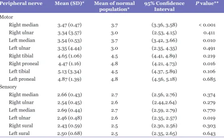
Table 2: Latency of sensory and motor components of peripheral nervous system in patients with multiple sclerosis compared to normal population
Peripheral nerve Mean (SD)* Mean of normal
population* 95% Confidence Interval P value**
Motor
Right median 3.47 (0.47) 3.7 (3.36, 3.58) < 0.001
Right ulnar 3.34 (3.57) 3.0 (2.53, 4.15) 0.411
Left median 3.54 (0.53) 3.7 (3.42, 3.66) 0.010
Left ulnar 3.35 (4.44) 3.0 (2.35, 4.35) 0.491
Right tibial 4.65 (1.06) 4.5 (4.41, 4.89) 0.219
Right proneal 4.47 (1.16) 4.8 (4.21, 4.73) 0.016
Left tibial 5.13 (3.34) 4.5 (4.37, 5.89) 0.106
Left proneal 4.87 (1.39) 4.8 (4.56, 5.18) 0.685
Sensory
Right median 2.66 (0.43) 2.7 (2.56, 2.76) 0.374
Right ulnar 2.54 (0.45) 2.6 (2.44,2.64) 0.279
Left median 2.69 (0.44) 2.7 (2.59, 2.79) 0.770
Left ulnar 2.46 (0.48) 2.6 (2.35, 2.57) 0.019
Right sural 2.43 (0.59) 2.5 (2.30, 2.56) 0.303
Left sural 2.50 (0.68) 2.5 (2.35, 2.65) 0.643
*The numbers are in millisecond (ms).
Table 3: Motor conduction velocity in patients with multiple sclerosis compared to normal population
Peripheral nerve Mean (SD)* Mean of normal
population* 95% Confidence Interval P value**
Right median 56.7 (5.5) 57 (55.5, 57.9) 0.628
Right ulnar 60.3 (7.6) 61 (58.6, 62.0) 0.454
Left median 56.0 (5.6) 57 (54.7, 57.3) 0.121
Left ulnar 59.6 (6.4) 61 (58.2, 61.1) 0.071
Right tibial 46.1 (4.6) 47 (45.1, 47.1) 0.117
Right proneal 46.0 (8.7) 47 (44.0, 48.0) 0.319
Left tibial 46.5 (5.2) 47 (45.3, 47.7) 0.443
Left proneal 45.8 (7.2) 47 (44.2, 47.4) 0.195
*The numbers are in meter per second (m/s).
**One sample t test; a P value less than 0.05 has been considered to be statistically significant.
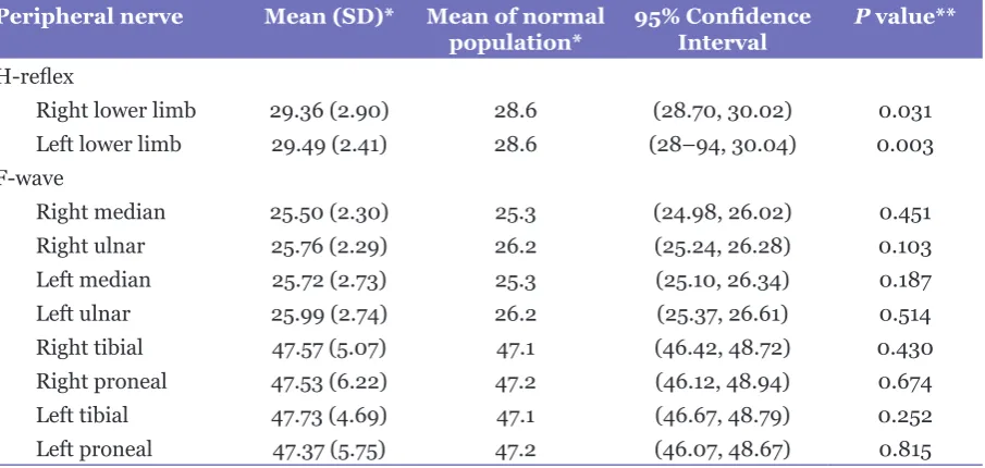
Table 4: H-reflex and F-wave in peripheral nervous system in patients with multiple sclerosis compared
to normal population
Peripheral nerve Mean (SD)* Mean of normal
population* 95% Confidence Interval P value**
H-reflex
Right lower limb 29.36 (2.90) 28.6 (28.70, 30.02) 0.031 Left lower limb 29.49 (2.41) 28.6 (28–94, 30.04) 0.003 F-wave
Right median 25.50 (2.30) 25.3 (24.98, 26.02) 0.451
Right ulnar 25.76 (2.29) 26.2 (25.24, 26.28) 0.103
Left median 25.72 (2.73) 25.3 (25.10, 26.34) 0.187
Left ulnar 25.99 (2.74) 26.2 (25.37, 26.61) 0.514
Right tibial 47.57 (5.07) 47.1 (46.42, 48.72) 0.430
Right proneal 47.53 (6.22) 47.2 (46.12, 48.94) 0.674
Left tibial 47.73 (4.69) 47.1 (46.67, 48.79) 0.252
Left proneal 47.37 (5.75) 47.2 (46.07, 48.67) 0.815
*The numbers are in m/s.
**One sample t test; a P value less than 0.05 has been considered to be statistically significant. compared to the normal population, while
other sensory amplitudes were not significantly
different. The latency of the right and left median and right proneal motor nerves and left ulnar sensory nerve were statistically less than that of the normal population (Table 2), but all were within the normal range because only prolongation of distal latency is pathologic. Mean motor conduction velocity and F-wave did not
differ significantly from the normal population (Table 3 and Table 4), but H-reflex latencies of
the right and left lower limbs were statically more prolonged in the study population compared to the normal population (Table 4).
Discussion
In the present study we found that the amplitude of peripheral motor nerves may diminish during the course of MS. The affected motor nerves in this study were the right and left tibial, right and left proneal, and left median nerves.
MS is a debilitating disorder of the CNS that potentially causes demyelination in central
axons, and subsequent neurologic deficits
central demyelination or originate from primary involvement of the peripheral neurons. There is a suggestion that peripheral neuropathy in MS results from the same pathogenesis that affects the CNS (7); this has been investigated by a number of researchers using electrodiagnostic tests.
Recent studies on the pathogenesis of MS have shown that in addition to white matter involvement in the CNS, a neurodegenerative process also begins in the gray matter in the early stages of disease; brain cortical atrophy is a good example. Magnetic resonance spectroscopy techniques used for chemical analysis of gray matter in these studies showed some lesions in the cortex, basal ganglia, and gray matter of the brainstem and spinal cord (14–17). The involvement of motor neurons in gray matter of the spinal cord can cause decreased compound muscle action potential (CMAP) of motor nerves in nerve conduction studies. In our study we also found decreased CMAP of the tibial, proneal, and left median nerves that support the neurodegenerative process in gray matter of the spinal cord.
Pogorzelski et al. (2), evaluated the subclinical lesions in the PNS of MS patients. They found electrophysiologic evidence that peripheral nerve lesions could be present at least in one peripheral nerve in 74.2% of patients. Sarova-Pinhas et al. (18), showed NCV parameter abnormalities in 14.7% of examined nerves and in 45.5% of MS patients. They concluded that neurologic symptoms may not necessarily correlate with electrodiagnostic abnormalities. Our study suggests possible peripheral motor nerve abnormalities in MS patients but does not
demonstrate any significant difference among
NCV parameters of sensory nerves of MS patients and the normal population.
Anlar et al. (3), stated that a high frequency of sensory motor electrophysiological nerve abnormalities may be present in MS patients. Gartzen et al. (19), also demonstrated that electrodiagnostic abnormalities may be present in MS patients with subtle neurologic symptoms. Our
study confirms primary peripheral neuropathy in
MS, but in contrast to several studies (3,18), only motor nerves had NCV changes.
NCV abnormalities may originate from primary pathophysiology of MS in peripheral nerves. Grana and Kraft (20), showed electrodiagnostic abnormalities in MS patients and evaluated and ruled out concurrent peripheral or entrapment neuropathies. Their evaluation suggests that peripheral neurologic symptoms in MS are of potential clinical interest and may
include peripheral axonopathy and demyelination, indicative of primary peripheral neuropathy, as part of the disease process. Results of NCV electrodiagnostic testing in our study suggest a motor peripheral neuropathy, supporting the hypothesis that MS primarily affects peripheral myelinated neurons.
A number of other studies have evaluated peripheral neuropathy (21–23) and electromyographic changes (24) in MS. The outcomes are indicative of peripheral nervous system involvement in the studied patients (3– 8,13,18–23). Petajan (24), used electromyography in 29 patients with MS and reported neurogenic atrophy in six patients of whom two patients had peripheral atrophy from peripheral neuropathy.
Sharma et al. (25), described chronic inflammatory
demyelinating disease (CIDP) in association with MS. In their study, MS patients developed peripheral involvements suggestive of CIDP after 4–22 years. Sharma and colleagues suggested that CIDP may be a common pathogenesis that affects central and peripheral axons during the course of autoimmunity.
The present study demonstrated that peripheral neuropathy in MS mainly affects motor neurons. Peripheral neurologic signs and symptoms are present in the course of MS in a number of patients (1). Evidence indicates that the autoimmune process in MS is initiated by CD4+ TH1 and TH17 T-cells that react against
self-myelin antigens (26). Peripheral deficits in the
course of MS may potentially result from central demyelination. It is also possible that similar autoimmune mechanisms cause peripheral demyelination and that peripheral neurologic signs and symptoms develop because of primary peripheral demyelination.
We compared our NCV parameter findings
with the mean of the normal population that was based on previous studies (13). This is a major limitation of our study because we could not compare our results based on gender and age characteristics of the studied sample. Future studies with larger samples and matched control groups are proposed.
Conclusion
In our study, decreased amplitude of motor nerves (CMAP) in tibial, proneal, and left median nerves is supportive of a new theory on the neurodegenerative process in gray matter of the spinal cord in early stages of MS. In addition,
prolonged H-reflexes in our study suggest nerve
neurologic involvement could be present in MS. Central demyelination may create peripheral
neurologic deficits, but primary peripheral
demyelination can, in turn, lead to peripheral symptoms. NCV parameters may indicate motor neuropathy; thus, electromyographic studies may also be of potential clinical interest for further assessment of motor neurons.
Acknowledgments
The authors would like to acknowledge Neurosciences Research Center of Tabriz University of Medical Sciences, Tabriz, Iran for their kind support of this research. We also thank the Eastern Azerbaijan Multiple Sclerosis Association (EAMSA), Tabriz, Eastern Azerbaijan, Iran for providing facilities and access to MS patients. In addition, we thank Dr Hossein Jabbari Khamnei and Dr Mohammad Zakaria Pezeshki for their useful comments regarding the statistical analysis of the presented data.
Conflict of Interest
None.
Fund(s)
This work was supported by the Neurosciences Research Center of Tabriz University of Medical Sciences, Tabriz, Iran.
Authors’ Contributions
Conception and design: MY
Final approval of the article: MY, HA, HMK, RR, SZ, SEG, KG
Critical revision of the article for the important intellectual content: HA, SEG, KG
Analysis and interpretation of the data and statistical expertise: SZ
Drafting of the article: RR, SEG, KG
Correspondence
Dr Mohammad Yazdchi-Marandi
MD (Tabriz University of Medical Sciences) Department of Neurology
Imam Reza Hospital
Tabriz University of Medical Sciences Golgasht St.
Tabriz 51666-14756, Iran Tel: +98 (411) 334 2889 Fax: +98 (411) 334 2889
E-mail: [email protected]
References
1. Lublin FD, Miller AE. Multiple sclerosis and other
inflammatory demyelinating diseases of the central
nervous system. In: Bradley WG, Daroff RB, Fenichel GM, Jankovic J, editors. Neurology in clinical practice. 5th ed. Philadelphia (PA): Elsevier; 2008. p. 1584–1612. doi: 10.1016/B978-0-7506-7525-3.50002-9.
2. Khoei NS, Atashpaz S, Ghabili K, Khoei NS, Omidi Y. Melittin and hyaluronidase compound derived from bee venom for the treatment of multiple sclerosis. Iran J Med Hypotheses Ideas. 2009;39(4): 1139–1142.
3. Pogorzelski R, Baniukiewicz E, Drozdowski W. Subclinical lesions of peripheral nervous system in multiple sclerosis patients. Neurol Neurochir Pol. 2004;38(4):257–264.
4. Anlar O, Tombul T, Kisli M. Peripheral sensory and motor abnormalities in patients with multiple sclerosis. Electromyogr Clin Neurophysiol. 2003;43(6):349–351.
5. Eisen A, Paty D, Hoirch M. Altered supernormality in multiple sclerosis peripheral nerve. Muscle Nerve. 1982;5(5):411–414. doi: 10.1002/mus.880050513.
6. Shefner JM, Mackin GA, Dawson DM. Lower motor neuron dysfunction in patients with multiple sclerosis. Muscle Nerve. 1992;15(11):1265–1270. doi: 10.1002/ mus.880151108.
7. Misawa S, Kuwabara S, Mori M, Hayakawa S, Sawai S, Hattori T. Peripheral nerve demyelination in multiple sclerosis. Clin Neurophysiol. 2008;119(8):1829. doi: 10.1016/j.clinph.2008.04.010.
8. McDonald WI, Compston A, Edan G, Goodkin D, Hartung HP, Lublin FD, et al. Recommended diagnostic criteria for multiple sclerosis: guidelines from the International Panel on the Diagnosis of Multiple Sclerosis. Ann Neurol. 2001;50(1):121–127. doi: 10.1002/ana.1032.
9. Etemadi J, Shoja MM, Ghabili K, Talebi M, Namdar H, Mirnour R. Multiple etiologies of axonal sensory motor polyneuropathy in a renal transplant recipient: a case report. J Med Case Rep. 2011;5(1):530. doi: 10.1186/1752-1947-5-530.
10. Farhoudi M, Ayromlou H, Bazzazi AM, Shadi FB, Golzari SE, Ghabili K, et al. Time frequency of Guillain-Barre syndrome in northwest of Iran. Life Sci J. 2013;10(1):223–225.
11. Ayromlou H, Tarzamni MK, Daghighi MH, Pezeshki MZ, Yazdchi M, Sadeghi-Hokmabadi E, et al. Diagnostic value of ultrasonography and magnetic resonance imaging in ulnar neuropathy at the elbow. ISRN Neurol. 2012;2012:491892. doi: 10.5402/2012/491892.
12. Yazdchi M, Tarzemani MK, Mikaeili H, Ayromlu
H, Ebadi H. Sensitivity and specificity of median
13. Ropper AH, Samuels MA. Adams and Victor’s Principles of Neurology. 9th ed. New York (NY): McGraw Hill; 2009. p. 1293. doi: 10.1001/archneurol. 2009.258.
14. Lisak RP. Neurodegeneration in multiple sclerosis:
defining the problem. Neurology. 2007;68(22
Suppl 3):S5–12.
15. Charil A, Filippi M. Inflammatory demyelination
and neurodegeneration in early multiple sclerosis. J Neurol Sci. 2007;259(1–2):7–15. doi: 10.1016/j. jns.2006.08.017.
16. Lassmann H. Multiple sclerosis: is there
neurodegeneration independent from inflammation?
J Neurol Sci. 2007;259(1–2):3–6. doi: 10.1016/j. jns.2006.08.016.
17. Fisniku LK, Chard DT, Jackson JS, Anderson VM, Altmann DR, Miszkiel KA, et al. Gray matter atrophy is related to long-term disability in multiple sclerosis. Ann Neurol. 2008;64(3):247–254. doi: 10.1002/ ana.21423.
18. Sarova-Pinhas I, Achiron A, Gilad R, Lampl Y. Peripheral neuropathy in multiple sclerosis: a clinical and electrophysiologic study. Acta Neurol Scand. 1995;91(4):234–238. doi: 10.1111/j.1600– 0404.1995.tb06996.x.
19. Gartzen K, Katzarava Z, Diener HC, Putzki N. Peripheral nervous system involvement in multiple sclerosis. Eur J Neurol. 2011;18(5):789–791. doi: 10.1111/j.1468-1331.2010.03149.x.
20. Grana EA, Kraft GH. Electrodiagnostic abnormalities in patients with multiple sclerosis. Arch Phys Med Rehabil. 1994;75(7):778–782.
21. Koszewicz M, Martynów R. Electrophysiological analysis of changes in peripheral nervous system in multiple sclerosis. Neurol Neurochir Pol. 1999;33(1):53–62.
22. Dainese R, Brazzo F, Hanau R. Motor conduction velocities and distal latencies in multiple sclerosis. Riv Patol Nerv Ment. 1982;102(5):201–204.
23. Argyriou AA, Karanasios P, Makridou A, Makris N. F-wave characteristics as surrogate markers of spasticity in patients with secondary progressive multiple sclerosis. J Clin Neurophysiol. 2010;27(2):120–125. doi: 10.1097/ WNP.0b013e3181d64c94.
24. Petajan JH. Electromyographic findings in multiple
sclerosis: remitting signs of denervation. Muscle Nerve. 1982;5(9 Suppl):S157–160.
25. Sharma KR, Saadia D, Facca AG, Bhatia R, Ayyar DR,
Sheremata W. Chronic inflammatory demyelinating
polyradiculoneuropathy associated with multiple sclerosis. J Clin Neuromuscul Dis. 2008;9(4):385– 396. doi: 10.1097/CND.0b013e31816f18e3.