University of Pennsylvania
ScholarlyCommons
Publicly Accessible Penn Dissertations
Spring 5-17-2010
Deconvoluting the Engineering and Assembly
Instructions for Complex Iii Activity
Sarah C. Hokanson
University of Pennsylvania, [email protected]
Follow this and additional works at:http://repository.upenn.edu/edissertations Part of theBiochemistry, Biophysics, and Structural Biology Commons
This paper is posted at ScholarlyCommons.http://repository.upenn.edu/edissertations/105 For more information, please [email protected].
Recommended Citation
Hokanson, Sarah C., "Deconvoluting the Engineering and Assembly Instructions for Complex Iii Activity" (2010).Publicly Accessible Penn Dissertations. 105.
Deconvoluting the Engineering and Assembly Instructions for Complex Iii
Activity
Abstract
In respiratory systems, membrane-bound Complex III catalyzes the oxidation of ubiquinone and the reduction of a soluble cytochrome with the bioenergetic formation of a transmembrane proton gradient (∆μH+). Complex III turnover is initiated by a unique two electron oxidation of ubiquinone at the Qo site; one electron is delivered to a high potential chain containing an iron-sulfur cluster, cytochrome c1 and cytochrome c2, and a second electron is transferred to a low potential chain that terminates at the Qi site. All Complex III electron tunneling reactions are reversible, and a critical part of Complex III maintaining productive turnover is its suppression of energy-wasting reverse electron transfer reactions. The key to uncovering the controversial mechanism of Qo oxidation is determining how Complex III is regulated such that productive electron-transfer steps overwhelm unproductive steps.
This thesis focuses on understanding the structural and biochemical tolerances of the redox cofactors in Complex III and applying that knowledge towards the design of a simple, but robust, amphiphilic maquette that is capable of transmembrane proton and electron transfer. In chapter two, kinetic studies of heme c1 mutants reveal that R. sphaeroides Complex III is engineered to withstand large changes in heme c1 active site residues while still preserving heme c1 midpoint potential and enzyme turnover. In chapter three, the
maquette approach was applied toward developing a simple model protein (AP6) that retained the minimum engineering requirements for Complex III electron and proton transfer reactions but lacked the complexity found in the natural system. The AP6 peptide assembles as a four-α-helix bundle protein and can potentially bind up to six hemes tightly across a membrane interface. Chapter four demonstrates that AP6 successfully performs quinol-cytochrome c oxidoreductase activity in hundreds of milliseconds. AP6 is the first example of a synthetic enzyme capable of near-natural turnover rates. Chapter five focuses on defining the
thermodynamic limit for maquette activity. This work supports the further development of simple model proteins to study aspects of Complex III mechanism.
Degree Type Dissertation
Degree Name
Doctor of Philosophy (PhD)
Graduate Group
Biochemistry & Molecular Biophysics
First Advisor P. Leslie Dutton
Keywords
Subject Categories
DECONVOLUTING THE ENGINEERING AND ASSEMBLY INSTRUCTIONS FOR COMPLEX III ACTIVITY
Sarah Chobot Hokanson A DISSERTATION
in
Biochemistry and Molecular Biophysics
Presented to the Faculties of the University of Pennsylvania in
Partial Fulfillment of the Requirements for the Degree of Doctor of Philosophy
2010
Supervisor of Dissertation:
________________________________ P.Leslie Dutton, Ph.D., FRS
Eldridge Reeves Johnson Professor of Biochemistry and Biophysics and Director of the Johnson Foundation for Molecular Biophysics
Graduate Group Chair:
________________________________ Kathryn Ferguson, Ph.D.
Associate Professor of Physiology and Chair, Biochemistry and Molecular Biophysics Graduate Group
Dissertation committee:
Jane M. Vanderkooi, Ph.D., Professor of Biochemistry and Biophysics Feng Gai, Ph.D., Associate Professor of Chemistry
S. Walter Englander Ph.D., Jacob Gershon-Cohen Professor of Medical Science and Professor of Biochemistry and Biophysics
Cecilia Tommos, Ph.D., Research Assistant Professor of Biochemistry and Biophysics Tomoko Ohnishi, Ph.D., Professor of Biochemistry and Biophysics
Deconvoluting the engineering and assembly requirements for Complex III assembly
COPYRIGHT
2010
Sarah Chobot Hokanson
To Les, for believing in second chances and in me
and
To Chris, the patron saint of patience
r\' L
4F
Acknowledgements:
I came to Penn from the very supportive research environment of the Elliott laboratory at Boston University, and I must thank Sean (HP) for believing in my abilities as a science major when countless others (including me) had their doubts. Sean, I would not be writing this thesis without your influence and support. But most importantly, I must thank you for making me apply to Penn, and more importantly, for not letting me apply to Clemson. I will finally admit that perhaps you knew what you were doing all along when you pushed me to believe in my own potential, and I certainly appreciate the impact your mentoring has had on my development as a scientist.
I have always said that alongside every successful Les Dutton, there must be a Chris Moser. I am so glad this principle continues to be true. Chris, without your patience and willingness to share your (seemingly unlimited) breadth of knowledge, this thesis would just be a string of “Friday afternoon experiments” that seemed interesting to me rather than the complete story that it has become. I have enjoyed our time together, and I will sincerely miss working and talking with you on a daily basis.
I also had the pleasure of working with collaborators at universities near and far, and those discussions were crucial to the development of my thesis project, as well as my personal development as a scientist. Much of my electron transfer calculations were done in conjunction with ongoing experiments in the Weiner and Cecchini laboratories, and their perspective as EPR spectroscopists taught me a whole new way of thinking about electron transfer reactions in proteins. I also had several wonderful discussions with Artur Osyczka, Fevzi Daldal, and Colin Wraight as I developed my Complex III projects, and their input really helped me appreciate many aspects of the large field of Complex III bioenergetics how my work contributed to it.
I really appreciate the support and guidance of my thesis committee members: Dr. Jane Vanderkooi, Dr. S. Walter Englander, Dr. Feng Gai, Dr. Cecilia Tommos, Dr. Tomoko Ohnishi, and Dr. Marilyn Gunner. My committee members have always provided excellent feedback during our sessions together, and I consistently looked forward to meeting with them. I would also like to thank Dr. Gunner for serving as my external reviewer during the final stages of my dissertation. It has been a pleasure to interact with you thus far and I am so grateful that you took time out of your busy schedule to be a part of my thesis defense.
my parents, my sister, and my extended family means so much to me, and I am so lucky that they are such a big part of my life. I love all of you very much. My three best friends, Kristen, Elliot, and Victoria, are the glue that holds me together in both good times and in stressful times. I always feel close to all of you, even though we are cities apart. And finally, I must thank the love of my life, my husband David. I never expected that the best thing to come out of graduate school would be our marriage, but then again, before I met you, I also wasn’t fully aware that life existed outside of the laboratory doors. Though you are a fantastic scientist yourself, your contribution to this thesis work is ironically that you were able to tear me away from it every now and again. (Though maybe when I am a post-doc, I will let you teach me more about molecular biology.) You continually make me a better and more complete person, and I am so happy that we found one another.
ABSTRACT
DECONVOLUTING THE ENGINEERING AND ASSEMBLY INSTRUCTIONS FOR COMPLEX III ACTIVITY
Sarah Chobot Hokanson
P. Leslie Dutton, Ph.D., FRS
In respiratory systems, membrane-bound Complex III catalyzes the oxidation of ubiquinone and the reduction of a soluble cytochrome with the bioenergetic formation of a transmembrane proton gradient (∆µH+). Complex III turnover is initiated by a unique
two electron oxidation of ubiquinone at the Qo site; one electron is delivered to a high potential chain containing an iron-sulfur cluster, cytochrome c1 and cytochrome c2,and a
second electron is transferred to a low potential chain that terminates at the Qi site. All Complex III electron tunneling reactions are reversible, and a critical part of Complex III maintaining productive turnover is its suppression of energy-wasting reverse electron transfer reactions. The key to uncovering the controversial mechanism of Qo oxidation is determining how Complex III is regulated such that productive electron-transfer steps overwhelm unproductive steps.
This thesis focuses on understanding the structural and biochemical tolerances of the redox cofactors in Complex III and applying that knowledge towards the design of a simple, but robust, amphiphilic maquette that is capable of transmembrane proton and electron transfer. In chapter two, kinetic studies of heme c1 mutants reveal that R.
sphaeroides Complex III is engineered to withstand large changes in heme c1 active site
chapter three, the maquette approach was applied toward developing a simple model protein (AP6) that retained the minimum engineering requirements for Complex III electron and proton transfer reactions but lacked the complexity found in the natural system. The AP6 peptide assembles as a four-α-‐helix bundle protein and can potentially
Table of Contents
Dedication ... iii
Acknowledgments ...iv
Abstract ... viii
List of Illustrations ...xiv
List of Tables ... xvii
Chapter 1: Introduction ... 1
1.1 A summary of mitochondrial respiration...2
1.2 Redox-active cofactors in the mitochondrial respiratory chain ...3
1.1.1 Quinones...4
1.1.2 Hemes...6
1.1.3 NAD+ / NADP+...7
1.1.4 Flavin adenine dinucleotide ...7
1.1.5 FeS cluster...8
1.3 Complex III – what we know...8
1.3.1 Considering Qo movement in its active site...10
1.3.2 Previous models for Qo site mechanism...13
1.3.3 Possibilities for Qo site thermodynamics...14
1.3.4 Previous attempts to isolate SQo ...16
1.3.5 Complex III activity is unaffected by Qo binding site mutations...19
1.3.6 Redox properties of the quinone sites in Complex III...19
1.3.7 Redox properties of other Complex III cofactors...21
1.4 Developing a simple tunneling view of Complex III activity ... 22
1.4.1 A single turnover view of Complex III electron tunneling...22
1.5 Finding new ways to study an old problem... 25
Chapter 2: Mutations to heme c1 binding site residues reveal R.
sphaeroides Complex III activity is robust ... 30
2.1 Introduction... 31
2.1.1 Mutations at the heme c1 binding site...31
2.1.2 Inhibition of heme c1...32
2.2 Materials and Methods... 33
2.2.1 Bacterial strains and growth...33
2.2.2 Chromatophore membrane / protein purification...33
2.2.3 Flash-induced single turnover kinetics...34
2.2.4 Redox titrations of heme c1 mutants ...35
2.2.5 CO binding...36
2.2.6 Reduction of decylubiquinone (DBH) substrate...36
2.2.7 Steady-state measurement of Complex III activity...36
2.3 Results ... 37
2.3.1 Flash kinetics of cytochrome c ...37
2.3.2 Flash kinetics of heme bH...40
2.3.3 Heme c1 – CO ...41
2.4 Discussion ... 42
2.4.1 Heme c1 binding motifs are not required for midpoint potential stabilization...42
2.5 Conclusion ... 44
2.6 References ... 44
Chapter 3: Design and characterization of an amphiphilic
maquette, AP6 ... 46
3.1 Introduction... 47
3.1.1 Step 1: reproducing membrane protein function in HP maquettes...48
3.1.2 Step 2: modifying HP designs to create proteins that can assemble in detergent micelles or phospholipid bilayers...51
3.1.3 Step 3: integrating properties from the HP and LP proteins into an AP maquette...52
3.2 Materials and Methods... 54
3.2.1 Peptide synthesis and purification...54
3.2.2 Peptide solubilization...56
3.2.3 Heme binding...56
3.2.4 Analytical ultracentrifugation ...57
3.2.6 Oxygen binding determination...57
3.2.7 Midpoint potential determination...58
3.3 Results ... 59
3.3.1 AP6 assembly ...59
3.3.2 Heme binding in AP6...62
3.3.3 Redox potentials of hemes bound to AP6...65
3.3.4 Generation of the oxy-ferrous state in AP6 ...66
3.3.5 AP6 variants...68
3.4 Discussion ... 69
3.4.1 Design characteristics...69
3.4.2 AP6 may require additional structural constraints...71
3.4.3 Heme properties in AP6...72
3.4.4 Proton coupling to heme midpoint potentials...74
3.5 Conclusion ... 76
3.6 References ... 77
Chapter 4: Demonstrating quinol-cytochrome c oxidoreductase
kinetic activity by AP6 ... 80
4.1 Introduction... 81
4.2 Materials and Methods... 83
4.2.1 Complex III growth and purification...83
4.2.2 Peptide synthesis and purification...83
4.2.3 Peptide solubilization...84
4.2.4 Heme binding...84
4.2.5 Reduction of decylubiquinone (DBH) substrate ...84
4.2.6 Enzyme steady state activity measurements...85
4.2.7 Computational work...86
4.3 Results ... 86
4.3.1 Quinol-cytochrome c oxidoreductase activity of AP6 ...86
4.3.2 Testing AP6 activity with alternative quinone substrates...90
4.3.3 Observing changes in AP6 activity in the presence of oxygen...92
4.4 Discussion ... 93
4.4.1 Developing a model for AP6 heme reactivity...93
4.4.2 Thermodynamic contraints on AP6 activity...95
4.4.3 AP6 activity is not decylubiquinone specific...95
4.5 Conclusion ...100
4.6 References ...102
Chapter 5: Testing the thermodynamic limits of maquette quinol-
cytochrome c oxidoreductase activity ... 104
5.1 Introduction...105
5.2 Materials and Methods...107
5.2.1 Maquette preparation... 107
5.2.2 Heme stock preparation... 108
5.2.3 Heme binding... 108
5.2.4 Midpoint potential determination... 108
5.2.5 Enzyme steady state activity measurements ... 109
5.3 Results ...110
5.3.1 Determining the midpoint potential of alternative hemes bound to HP7 ... 110
5.3.2 Comparing AP6 activity to soluble maquette proteins... 111
5.4 Discussion ...114
5.4.1 Approaching and exceeding the thermodynamic limit for quinol to heme electron transfer... 114
5.4.2 Impact of thermodynamic threshold on future AP6 designs... 115
5.5 Conclusion ...116
5.6 References ...117
Chapter 6: Conclusions ... 119
6.1 Complex III assembly instructions...120
6.2 Developing a simpler model for Complex III...120
6.3 Impact of this work on future experiments ...124
6.3.1 Exploiting Complex III thermodynamics to access Qo states... 124
6.3.2 Maquettes as enzymes – the potential for future designs... 128
6.4 Conclusion ...131
6.5 References ...131
List of Illustrations
Chapter 1
1.1 A contemporary view of the mitochondrial respiratory chain 2
1.2 Quinone redox/protonation states 5
1.3 Schematic representation of the Q-cycle in Complex III 9
1.4 ket vs. Qo-FeS distance 11
1.5 Electron transfer reaction energy surfaces for Qo 15
1.6 Eight redox states of Qo 20
1.7 Eh vs. pH for Complex III cofactors 22
1.8 Simple tunneling-based view of Complex III turnover 23
Chapter 2
2.1 Difference spectra for Complex III and cysteine variants 34
2.2 Flash kinetics for wild-type and mutant C145A poised at 100 mV 39
2.3 Flash kinetics for wild-type and mutant C145A poised at 250 mV 40
2.4 CO binding determination 41
2.5 Redox titration of C145A in different buffer solutions 43
Chapter 3
3.1 Maquette family tree 50
3.2 Polar plot of AP6 sequence 53
3.3 Analytical ultracentrifugation of apo- and holo-AP6 60
3.5 Heme binding titration in AP6 63
3.6 ZnPPX binding titration in AP6 65
3.7 AP6 redox titration 65
3.8 Midpoint potential vs. pH relationship for AP6 66
3.9 Creation of the oxy-ferrous state in AP6 67
3.10 AP6 variant APab heme titration 68
3.11 Structure of Complex III b-subunit α-helices 71
Chapter 4
4.1 Cartoon representation of the assay 87
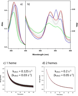
4.2 Demonstration of quinol-cytochrome c activity 88
4.3 Dependence of AP6 activity on number of hemes / bundle 89
4.4 Varying [cytochrome c] 90
4.5 Testing AP6 specificity for the quinone reductant 91
4.6 Measuring AP6 activity under aerobic conditions 92
4.7 Considering the energetics of AP6 activity 96
4.8 Modeling quinone stability constants and electron tunneling distances 98
4.9 Comparing AP6 turnover rates to other de novo enzymes and natural
Chapter 5
5.1 Structures and midpoint potentials of heme cofactors capable of binding
to HP7 105
5.2 Absorbance spectra for heme cofactors bound to HP7 106
5.3 Redox titration of HP7 with 1 equivalent of heme a bound 110
5.4 Redox titration of HP7 with 1 equivalent of iron(III)-
dicyanodeuteroporphryin bound 111
5.5 Quinol-cytochrome c oxidoreductase activity for soluble maquettes 112
5.6 Quinol-cytochrome c oxidoreductase activity for HP7 with
iron(III)-dicyanodeuteroporphryin bound 113
Chapter 6
Figures
6.1 A single chain view of Complex III electron and proton transfer 123
6.2 Manipulating Qo and FeS cluster midpoint potentials to prolong Qo
redox states 125
6.3 Relationship between FeS cluster midpoint potential and Kstab of Qo
6.4 Catalytic rates for natural and synthetic proteins 128
List of Tables
Chapter 2
2.1 Mutagenesis of non-heme c1 ligating cysteine residues in
Rb. sphaeroides Complex III 38
Chapter 3
3.1 Summary of the sequences and molecular weights of AP6 and AP6
variants 55
3.2 Heme binding constants for AP6 proteins 64
Chapter 4
4.1 Summary of the kinetics of Complex III and AP6 93
Chapter 5
5.1 Summary of midpoint potentials and kcat values at pH 8 110
Chapter 1: Introduction
1.1 A summary of mitochondrial respiration
We now mostly understand the basic electron tunneling reactions (Figure 1.1 blue arrows) that connect the complexes within the mitochondrial respiratory chain together. In Complexes I and II, flavin-containing nicotinamide adenine dinucleotide (NADH) and succinate dehydrogenase sites are connected via ubiquinone sites in the membrane domain. These sites collect single electrons of the tunneling chains into pairs for two electron and two proton quinone oxidation-reduction before exchanging with a membranous quinone pool. Complex III interacts with the quinone pool via two ubiquinone oxidation-reduction sites, Qo and Qi, Once reduced Qo is bound to Complex
Figure 1.1: A contemporary view of the mitochondrial respiratory chain, which contains (left to right) Complex I, Complex II, Complex III, cytochrome c, and Complex IV. Blue arrows indicate electron tunneling pathways, red arrows indicate the direction of proton translocation, black arrows indicate the arrival/placement of substrate molecules, and green arrows indicate the position and movement of quinone molecules. This figure was modified from a figure presented in Biochimica et
III, one electron reduces cytochrome c1 while the other electron equivalent passes through
two b-type hemes and reduces a quinone molecule at the Qi site. In the terminal Complex IV, cytochrome c electron transfer leads to dioxygen reduction.
Although the overall stoichiometries of redox-linked proton exchange (red arrows, Figure 1.1) are known, the basics these are coupled to the electron tunneling reactions of ubiquinone (Complexes I, II and III) or dioxygen (Complex IV) that generate the transmembrane electrochemical proton gradient (ΔµH+) remain undefined and
controversial. The most information is known about Complex IV, as the key intermediate redox states of the stepwise four-electron reduction of dioxygen have been resolved structurally [7, 8] and spectroscopically [9-11]. However, the molecular mechanism of its driven proton pump remains to be determined. Information about the operation of the Qs site in Complex II is limited [12]; most experiments have been focused on the redox chemistry of its FeS clusters [13, 14] and heme b [15-17] instead. Incomplete structural information available for Complex I has limited the resolution of its proton pumping mechanism, though several hypotheses exist in the literature at present [18-23]. One of the most controversial questions left unanswered about the operation of the mitochondrial respiratory chain is the molecular mechanism of ubiquinone oxidation-reduction, proton exchange and energy coupling at the Qo site in Complex III. In sharp contrast to Complex IV, the redox intermediate states of Qo have proven difficult to access, lack clear spectroscopic signatures, and remain highly uncertain.
1.2 Redox cofactors in the mitochondrial respiratory chain
space, creating an electrochemical proton gradient across the mitochondrial inner membrane (ΔµH+). This electrochemical proton gradient allows adenosine triphosphate
(ATP) synthase to use the flow of protons to generate ATP from adenosine diphosphate (ADP) and inorganic phosphate. In the absence of ADP, protons cannot flow back to the matrix, and the pH and electrical gradients are at maximum. As respiration with outward proton pumping proceeds, the free energy change for proton expulsion increases and approaches the magnitude of that for electron transfer. When the coupled reaction becomes non-spontaneous, respiration stops, and this state is called a static head. The formation of reactive oxygen species is supported by static head conditions in the mitochondrial respiratory chain [24].
There are many different types of redox-active cofactors responsible for proton-coupled electron transfer reactions within the mitochondrial respiratory chain. These cofactors span a wide range of midpoint potentials, and are linked together in long chains that extend over long distances within these proteins to link together remote catalytic sites.
1.2.1 Quinones. There are two main types of quinones found in biological
depicted in Figure 1.2, though only the states depicted in red have been observed in biology.
Quinones are regulated in membrane complexes through the stability of their semiquinone redox state, which is related to the redox potential difference between their
Figure 1.2: Thermodynamic box of nine possible ubiquinone redox/protonation states (ubiquinone is drawn here without its isoprenoid side chain). Shown in red are the quinone states that have been observed in biological systems, while shown in black are thermodynamically unfavored states. This figure was modified from a similar figure shown in the thesis of Bruce Lichtenstein [2].
QH2/SQ and SQ/Q couples. The stable equilibrium constant for the intermediate
semiquinone state at complete redox equilibrium is referred to as a semiquinone stability constant, or Kstab:
Equation 1.1 Log Kstab values greater than one mean that the semiquinone is a dominant species during a redox titration, while log Kstab < -4 means that the semiquinone will be barely
observable spectroscopically, if at all.
1.2.2 Hemes. Hemes consist of an iron ion coordinated to a large heterocyclic
organic ring called a porphyrin. The most common porphyrin scaffold in biology is protoporphyrin IX (PPIX), otherwise known as heme b. The heme b biosynthesis pathway is highly conserved across biological organisms, and is initiated in the mitochondrion by the synthesis of D-aminolevulinic acid (dALA) from glycine and succinyl-CoA.
In addition to heme b, there are two other main forms of hemes found in the mitochondrial respiratory chain. c-type hemes contain a modified FePPIX where the two vinyl side chains are uniquely covalently attached to cysteine thiol residues within the protein. In the mitochrondrial respiratory chain, heme c is most notably found in the cytochrome c1 subunit of Complex III and its redox partner, cytochrome c2. Heme a is
1.2.3 NAD+ / NADP+. Nicotinamide adenine dinucleotide (NAD+
) and its close analog nicotinamide adenine dinucleotide phosphate (NADP+
) consist of two nucleotides joined by a pair of bridging phosphate groups. The plus sign in the abbreviations for these cofactors does not indicate their net charge, but rather that the nicotinamide ring is in its oxidized form with a positive charge on the nitrogen atom. Both cofactors undergo reversible reduction of the nicotinamide ring. As a substrate molecule undergoes oxidation, the oxidized form of the nucleotide accepts a hydride ion (or the equivalent of a proton and two electrons) and is transformed into the reduced form (NADH or NADPH). More than 200 enzymes are known to catalyze reactions in which NAD+
(or NADP+
) accepts a hydride ion from a reduced substrate, or NADH (or NADPH) donates a hydride ion to an oxidized substrate. In Complex I, hydride transfer from NADH to a bound quinone cofactor initiates the proton pumping mechanism of the mitochondrial respiratory chain.
1.2.4 FAD / FADH˙ / FADH2. Flavin adenine dinucleotide (FAD) consists of a
1.2.5 FeS cluster. In FeS clusters, iron molecules are present in association with inorganic sulfur atoms or with the sulfur atoms of cysteine residues, or both. There are three main structural motifs of biological FeS clusters, though exceptions do exist in hydrogenase enzyme families. The simplest FeS cluster is the [2Fe-2S] cluster, which is comprised of two iron ions bridged by two sulfide ions and coordinated by four cysteine-based thiol ligands. [4Fe-4S] clusters feature four iron ions and four sulfide ions placed at the vertices of a cubane-like structure. In these FeS clusters, the iron ions are typically coordinated by cysteine thiol ligands. Finally, proteins also coordinate 3Fe-4S centers, which feature one less iron ion than the more common [4Fe-4S] form. Three sulfide ions bridge two iron ions each, while the fourth sulfide bridges three iron ions. Complexes I and II each contain combinations of these different types of FeS clusters. Complex III contains a variation of a [2Fe-2S] cluster, where two histidine residues, rather than two cysteine thiol groups, coordinate one of the two iron atoms. This type of [2Fe-2S] cluster is named for John Rieske, after the Complex III iron-sulfur protein (ISP) domain was isolated in 1964 [25, 26].
1.3 Complex III – what we know
One electron is accepted by the iron-sulfur cluster (FeS), and follows the c-chain via cytochrome c1 to external c-type cytochromes (Figure 1.3). The other electron passes
through two b-type hemes and reduces a quinone molecule at the Qi site. This unique
Figure 1.3: Each panel represents a step in the electron and/or proton transfer process of one complete Q-cycle (Complex III is represented here as a monomer for simplicity). a) One reduced quinol molecule is delivered to the Qo site from the Q-pool. b) The Qo site becomes oxidized, transferring two protons across the membrane and distributing two electrons to the high and low potential chains of bc1. c) A proton
and electron are delivered to the Qi site, forming a stable semiquinone intermediate. d) The Q-pool delivers another reduced quinol molecule to the Qo site to restart Complex III turnover. e) The Qo site turns over again, delivering a second set of electrons to the high potential c-chain and the low potential b-chain, as well as two more protons across the membrane. f) Upon receiving an additional proton and an electron from heme bH, the Qi semiquinone becomes fully reduced, and delivers a
reduced quinol back to the Qpool. The net result of this cycle is oxidation of one quinol, reduction of two cytochromes c, two electrogenic transmembrane electron transfers, uptake of two protons from one membrane face and delivery of 4 protons to the other. Reprinted with permission from SpringerLink and the Journal of Bioenergetics and Biomembranes [3].
electron split sets into motion a quasi-equilibrium state of Complex III that balances the redox potentials of the high potential c-chain and low potential b-chain evenly on either side of the redox potential of the quinone pool. This quasi-equilibrium behavior shows that Complex III catalysis is rapidly and readily reversible [6]. Collapse of this quasi-equilibrium into complete quasi-equilibrium, in which all chains and pools reach the same redox potential, takes place on a tens of seconds timescale and can be assisted by the short-circuiting action of redox mediator dyes.
Under normal forward electron transfer conditions, the efficiency of Complex III turnover is high, and deleterious short circuit or bypass reactions involving either Qo or heme bL are minimal [31]. Short circuit reactions are defined as any electron transfer
reaction between two Complex III cofactors that results in an unproductive loss of energy [6]. Alternatively, bypass reactions result when the electron transfer processes at the Qo site are intercepted by an extraneous reagent, such as oxygen, which steals the electron from Complex III and initiates harmful side reactions that produce reactive oxygen species [32, 33]. In order for the Qo site to be efficient, these reactions must be suppressed, even though the driving forces for these reactions are highly favorable. Understanding Qo site engineering means understanding how electron transfer is regulated such that productive electron-transfer steps overwhelm unproductive steps.
1.3.1 Considering Qo movement in its active site. Crystal structures have
electron tunneling distances between Qo-FeS and Qo-bL are, the distances in Complex III
in general are engineered to allow for millisecond Qo oxidation and avoid short-circuit reactions that would be a danger even to a concerted mechanistic model, such as direct electron transfer between FeS and bL.
Figure 1.4 (top panel): Crystal structure of FeS-Qo-bL active sites, drawn from PDB
file 1PPJ [5]. Heme bL is depicted in red, the FeS cluster is drawn in orange, and two
Qo site inhibitors myxothiazol (yellow) and stigmatellin (green) are shown bound in the Qo site. Distances between the cofactors were calculated using Pymol, where 23 Å represents the distance between the FeS cluster and bL , 7 and 11 Å represent
stigmatellin distances from the FeS cluster and bL respectively, and 13 and 6 Å
represent myxothaizol distances from the FeS cluster and bL respectively. (bottom
Shown in Figure 1.4 are two Qo inhibitor molecules, myxothiazol (yellow) and stigmatellin (green), bound at the Qo site, with their respective distances to the other cofactors measured using Pymol software [41]. Myxothiazol binds closer to bL in the Qo
niche than stigmatellin does, and could still allow some quinone to approach the Qo pocket distally. This could explain the observations in the literature that myxothiazol has no effect on the semiquinone signal attributed to Qo [42], and permits considerably more superoxide production by Complex III than stigmatellin [43, 44].
As described in the Appendix, the Dutton laboratory has developed an electron transfer rate expression that offers a simple way to examine parameters that directly relate to the oxidoreductase activity of a particular mitochondrial enzyme [1, 45, 46]. The Moser-Dutton equation defines the rate of electron transfer as a function of distance (R), midpoint potential (included in ΔG°), reorganization energy (λ), and protein packing density (ρ). Using this equation, a range of Qo-FeS distances can be considered to determine the effect changing the location of Qo in its active site has on the rate of bL
reduction. Figure 1.4 depicts the halftime of heme bL reduction vs. Qo-FeS cluster
distance and demonstrates that millisecond bL reduction rates are maintained as long as
the Qo-FeS distance falls between 6 and 11 Å at pH 7. Though it is well-established that the uphill electron tunneling step between reduced Qo and the FeS cluster is rate-limiting as long as Qo falls within this 6-11 Å range [47], should Qo favor binding more closely (within 4 Å) to the FeS cluster, Qo to bL electron tunneling becomes rate limiting and
slows bL reduction by two orders of magnitude. Likewise, should Qo bind closely to the bL heme, Qo oxidation rates are limited by Qo-FeS cluster electron tunneling. This
values, though the reaction rates for Qo oxidation increase as the pH becomes more alkaline, increasing the range of FeS-Qo distances that will maintain millisecond catalysis.
1.3.2 Previous models for Qo site mechanism. Without the direction of
structural information on Qo binding, a range of mechanistic models has emerged with no real consensus as to which one is the most successful. The work of Osyczka et al.
prompted revision of all contemporary Q-cycle models in order to accommodate suppression of unwanted short-circuits in a reversible, energy-coupling mechanism evident in Complex III [6, 48]. However, it was made clear that models that are truly concerted two-electron transfer reactions [6], with no detectable semiquinone intermediate state on more than a femtoseconds timescale, were not prone to the same kind of short circuit possibilities as sequential models that included a semiquinone intermediate state [47, 49]. Therefore, all sequential mechanisms that exploit the properties of a semiquinone intermediate [24, 48, 50-52], must be modified to include effective gating mechanisms, sensitive to different combinations of redox states of the FeS, Qo and heme bL redox partners, to promote productive catalysis and prevent
unproductive short-circuits.
quinone is forbidden from binding if both of its redox partners are not reduced. This can, in principle, be achieved by regulation of oxidized and reduced quinone binding by manipulation of the conformations of Qo hydrogen bonding partners by the redox and protonation states of heme bL and FeS (see also [6, 48].
Crofts et al. modified their sequential mechanism [53] to introduce more redox-state sensitive gating to avoid short circuits [24, 50]. They propose that when a semiquinone intermediate is formed, it can move closer to oxidized heme bL and
participate in productive electron transfer. However, when heme bL is reduced, there is a
Coulombic repulsion which keeps the semiquinone away from reduced heme bL, towards
the FeS end of the site, and inhibits the unproductive, and energetically favorable, short-circuit reduction of semiquinone by heme bL. While in principle Coulombic interactions
could provide a redox-state sensitive gate to inhibit a short-circuit reaction, more than one gate is needed. The Coloumbic push of semiquinone from reduced heme bL moves it
closer to FeS, which when oxidized, can accept an electron from the semiquinone in another type of short circuit. A second gate for this type of model requires the FeS and semiquinone to overcome their coulombic attraction and enter some sort of conformation to prevent energetically favorable, but wasteful electron transfer. In addition, the site must be designed to overcome a coulombic repulsion between semiquinone and reduced FeS. This would allow a favorable interaction between semiquinone and reduced FeS, fostering productive and rapid reverse reactions in which oxidized quinone is doubly reduced by FeS and heme bL
1.3.3 Possibilities for Qo site thermodynamics. The thermodynamics of
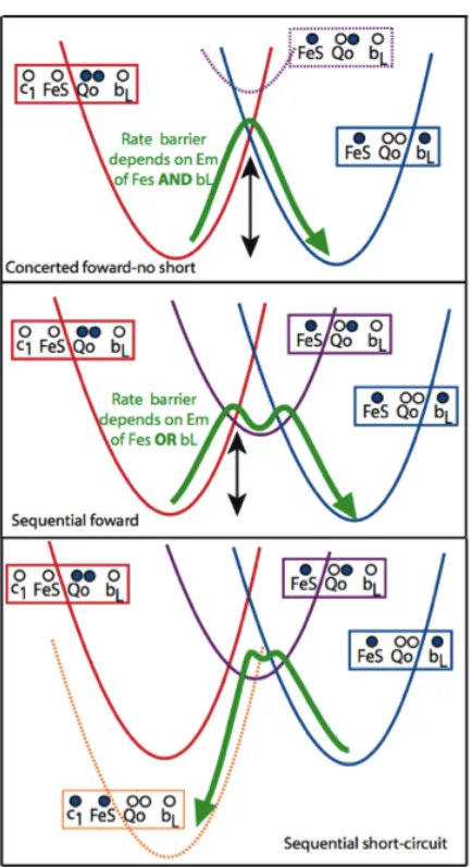
III. Figure 1.5 illustrates that the energetic landscape for a concerted mechanism at Qo
has an activation barrier that depends on both FeS and heme bL redox midpoint
potentials[3]. In this mechanism, the activation barrier for forming the semiquinone
Figure 1.5: Electron transfer reaction energy surfaces for Qo site operation by concerted and sequential mechanisms. Marcus-like parabolic potential surfaces are shown for each redox state. In a concerted reaction (top), electron transfer to form a semiquinone state (purple) takes more thermal energy than forming the two-electron transition state. In a sequential mechanism (middle), electron transfer to form a semiquinone intermediate state takes less energy than the simultaneous two electron transfers, but leaves Complex III potentially exposed to highly exergonic short-circuit reactions such as the one illustrated in the bottom panel. Reprinted with permission from SpringerLink and the Journal of Bioenergetics and Biomembranes [3].
intermediate (purple curve) is significantly higher; normally semiquinone formation
would be minimal, but not impossible. In contrast, the sequential mechanism will have a
reduced quinone oxidation rate that depends on either the midpoint potential of the FeS
or the heme bL. Experimentally, changing the midpoint potential of the FeS often
changes the rate; however, changing the heme bL midpoint potential has proven more
difficult and the changes that have been made are modest. Thus, how or if the heme bL
midpoint potential affects the rate is still unclear.
The bottom panel of Figure 1.5 illustrates the energetic picture of one of the
short-circuit reactions [3]. The rapid reversibility of the reactions at the Qo site means reverse
electron transfer is on roughly the same timescale as the forward reaction, and will
reform the same amount of semiquinone that is formed by the initial electron transfer
between Qo and the FeS cluster. However, the short-circuit reduction of reoxidized FeS
is highly favorable and energetically disastrous; if Complex III is operating by a
sequential mechanism, redox-state activated gates must be present to avoid unproductive
short-circuit reactions.
1.3.4 Previous attempts to isolate SQo. Direct observation of a Qo
also examined the redox-poised membranes for evidence of a Qo semiquinone by EPR spectroscopy, but did not detect a clear signal.
de Vries et al. announced the trapping of a Qo semiquinone in mitochondrial Complex III as a 2,3-dimercaptopropanol (BAL)-sensitive signal uncovered under non-equilibrium conditions [55]. BAL chelates heavy metals and is suggested to destroy the Rieske FeS center, allowing for a direct probe of the Qo site. Compared with the antimycin-sensitive semiquinone signal discovered at Qi (g = 2.005, 10 G wide) [56], the signal attributed to Qo in these experiments was narrower (8.8 G), had a slightly different g value (2.006), and required less power to saturate. Since this signal was obtained using non-equilibrium methods, the redox midpoint potential of the semiquinone couples could not be determined. However, Junemann et al. revisited these experiments and revealed that de Vries’s semiquinone signal was insensitive to a more modern collection of Qo site inhibitor compounds, such as myxothiazol, MOA-stilbene, and stigmatellin [57]. Therefore, based upon this evidence, it remains unlikely the data published by de Vries et al. was indicative of a Qo semiquinone state.
Rieske FeS cluster, raising questions about whether or not it is representative of a true Qo semiquinone intermediate.
The Dutton laboratory explored the second approach; their experiments used light-activated photosynthetic membranes with heme(s) bL and/or heme c1 knocked out,
deliberately driving the thermodynamics of Qo to maximize semiquinone production [42]. After a series of light flashes, the high potential c-chain became highly oxidized in this system, stimulating the oxidation of Qo by the quinone pool and loading the low potential b-chain with reducing equivalents. This experimental setup, where the high potential chain is very oxidized, proved to be very favorable for stripping an electron from a second quinol at Qo to form a semiquinone. The g-value of the signal observed under these conditions was determined to be 2.0040 and the line width was 11.7 G. Because the stoichiometry of the redox components involved in the pseudo-equilibrium between the b- and c-chains and the quinone pool was known, the split between the redox couples for this quinone species was estimated to be approximately 880 mV, with the QH2/SQ couple ~410 mV at pH 9. Therefore, from this data the effective stability
constant for Qo was estimated to be 10-15
. However, this data was obtained using chromatophore membranes with massive mutational changes (cofactor knockouts), causing many in the field to argue that this signal, even if it was attributable to a Qo semiquinone, was not indicative of a mechanistic state that could be observed in wild-type chromatophores.
experimental design. Therefore, it is still too soon to reject the possibility that the normal Qo site mechanism is concerted, with the transfer of both electrons occurring in a very short interval, leaving no time for an oxidized quinone to relax into any intermediate state.
1.3.5 Complex III activity is unaffected by Qo binding site mutations. The
Qo site mechanism is also surprisingly resilient to mutational modifications at the Qo site. Though the mostly conserved glutamate (E) of the Qo active site PEWY sequence is often given a carefully orchestrated role controlling the proton transfer reactions of QH2
oxidation at the Qo site [24, 50], replacement of the glutamate group with non-polar or even basic groups had almost no effect on any of the equilibrium properties of the Qo site or its redox partners [51]. As the Qo site appears to have residue redundancy that compensates for mutagenic changes easily, a specific network of protons within the active site may not be necessary for preserving productive Qo site electron tunneling. Instead, the tunneling network itself must be designed to raise barriers that suppress unwanted short circuit reactions.
1.3.6 Redox properties of the quinone sites in Complex III. The bifurcated
electron transfer that occurs within Complex III is completely dependent upon its ability to modify the redox environment of the ubiquinone pool at each of its quinone binding sites, Qo and Qi. The environment of the Qi binding site forces the quinone redox couples to be very similar. The redox couples and stability constant for Qi have been measured (log Kstab is -2.3 at neutral pH) using EPR spectroscopy, and under mildly
the first electron is removed from the reduced quinol at a relatively oxidizing midpoint potential to head down the high potential c-chain, while the second electron is removed at a relatively reducing midpoint potential and is transferred to the low potential b-chain. Therefore, unlike Qi, the redox couples at Qo have a very large difference in midpoint potential, and a very low Kstab value.
As shown in figure 1.6, there are eight possible combinations of redox states for Qo and its redox partners, heme bL and FeS. Half of these states are equilibrium states
readily achieved by simple redox poising (shown in black), and half are non-equilibrium states have yet to be experimentally characterized (shown in red and green). The green states are particularly important since they are the enzyme-substrate and enzyme-product
Figure 1.6: Eight possible redox state combinations for the Qo site and its electron transfer partners, FeS and heme bL. Equilibrium states are shown in black, productive
transient intermediates are shown in green, and non-physiological quasi-equilibrium states are shown in red. Solid lines indicate closed gates, and solid lines with a break in the middle indicate open gates for electron transfer. Dashed lines indicate that a gating mechanism is optional for this electron transfer. Reprinted with permission from SpringerLink and the Journal of Bioenergetics and Biomembranes [3].
states immediate to physiologically productive quinone oxidation- and reduction-coupled energy transduction at the Qo site, while the states shown in red are subject to short-circuit reactions that are high in driving force and physiologically unproductive. Assuming a sequential-gated Qo site mechanism in this model, the gates controlling semiquinone activity must be open so that catalysis can occur in the green states, while the gates are closed in the red states in order to protect Complex III from short circuiting.
1.3.7 Redox properties of other Complex III cofactors. Understanding the
engineering design of Complex III and the operating limits of failure of the Q-cycle requires an understanding of the redox properties of each redox-active component. The redox properties of the cofactors in Complex III are well-described by redox titrations performed across a wide range of pH values, as shown in Figure 1.7 [4]. The FeS cluster has a pKox near neutral pH in both R. capsulatus and R.sphaeroides. Cytochrome c1 has
pKox and pKred values close to one another near neutral pH, and a weak pH dependency (much less than 1 H+
/ 1 e
-). Previous measurements of the Qpool size and redox properties are included on the left graph as a green dashed line [54], and Qi redox properties were also previously measured under conditions where its semiquinone species is readily accessible by EPR measurements [58].
The Dutton [4] and Crofts [50] laboratories have measured redox prorperties of the b-hemes in recent years. Original redox titrations of the b-hemes revealed three redox active species [59], which are now resolved in Figure 1.7 as bL and two different forms
of bH - bH high
and bH low
. Heme bL displays a strong coupling to protons at acidic pH
redox behavior of bH is directly coupled to the redox state of Qi, and is also altered when
antimycin is bound to the Qi site. 80% of heme bH centers are in the low potential form
at acidic pH values, though this number is reduced to 55% at alkaline pH values [4].
1.4 Developing a simple tunneling view of Complex III activity
The redox midpoint potentials and electron tunneling distances of the cofactors in Complex III were inputted into the Moser-Dutton equation to calculate ket. These rates
are used to present an an elementary view of Complex III electron tunneling that is expanded upon experimentally in chapter two.
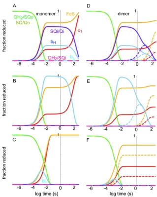
1.4.1 A single turnover view of Complex III electron tunneling. Figure 1.8
shows a single, two electron turnover of Complex III when all of its redox components are oxidized except for the Qo site, with its measured 10-15
Kstab constant [42] providing
Figure 1.7: Redox midpoint potentials of the components of Complex III in two different species of photosynthetic bacteria as a function of pH, as determined experimentally by Haibo Zhang. Reprinted with permission from SpringerLink and the Journal of Bioenergetics and Biomembranes [4].
for millisecond catalysis of quinone oxidation. FeS is modeled as moving between two sites, distal and proximal to the Qo site with a rate of about 105
s-1
[60]. Heme bL is only
transiently reduced as the electron comes to reside on heme bH and Qi. Also, when the
Figure 1.8: Electron tunneling calculations for Complex III. Shown on the left (A, C, E) are rate calculations that assume that Complex III is functional as a monomer, and shown on the right (B, D, F) are those for a functional dimer. Dashed lines in the right-hand side graphs represent the redox centers in the second half of the dimer introduced into the calculations. (A and D) All redox centers considered. (B and E) Calculating all centers except Qi, which has been effectively “inhibited” by extending the electron transfer distance to 50 Å. C and F) Considering site directed mutants where heme bH is not incorporated [6]. Figure modified from data presented in Biochimica et Biophysica Acta – Bioenergetics [1].
electron can get as far as Qi, short-circuit reactions through reverse reactions and a Qo semiquinone are slowed to a minutes timescale.
These calculations can be modified to include inhibitors or mutations that limit Complex III turnover. For example, the addition of antimycin at the Qi site or bH
knockout mutations can be considered in our system of rate equations. Heme bH
reduction is dominant in the presence of antimycin, with short-circuits still taking place relatively slowly on a tens of seconds timescale (under non-membrane energized conditions), representing what is observed under single turnover flash experiments. However, when heme bH is knocked out, the electron cannot escape from bL and is prone
to millisecond short-circuit reactions that are comparable to the rate of catalysis. Because rapid short-circuit reactions are not observed experimentally in these knockouts, the redox couples of Qo (and therefore, its stability constant), must be modulated in some way to allow catalysis in certain redox states while making the semiquinone (and therefore, short-circuit reactions) inaccessible in others.
Structurally, Complex III is a dimer, but whether or not it acts as a functional dimer remains controversial. We can compare Complex III as a functional monomer or dimer using the Moser-Dutton equation. With an edge-to-edge spacing of 14.7 Å between the two heme bL cofactors, functionally significant electron tunneling between
However, with the same fixed stability constant, of 10-15
, the heme b knockout allows the electron to linger on bL, with cross-dimer redox equilibration occurring on a millisecond
timescale. Therefore, the electron tunneling contact between both halves of the dimer is occurs on the same timescale as catalysis, indicating that Complex III has effectively increased the concentration of active Qo sites available when the complex is partially reduced.
1.5 Finding new ways to study an old problem
It has been decades since Peter Mitchell developed the Q-cycle mechanism with little resolved about the mechanism of Qo turnover in Complex III. In order to address these unanswered questions, the field of bioenergetics must develop a fresh perspective and new experimental techniques. In chapter two, R. sphaeroides Complex III proteins with modifications to their heme c1 are studied to understand how Complex III redox
reactions within the high and low potential chains are interconnected.
for Complex III that can be applied by the field of bioenergetics to study specific Qo redox states.
1.6 References
1. Moser, C.C., et al., Electron tunneling chains of mitochondria. Biochimica Et Biophysica Acta-‐Bioenergetics, 2006. 1757(9-‐10): p. 1096-‐1109.
2. Lichtenstein, B.R., Graduate Thesis. 2010.
3. Chobot, S.E., et al., Breaking the Q-cycle: finding new ways to study Qo through
thermodynamic manipulations. Journal of Bioenergetics and Biomembranes,
2008. 40(5): p. 501-‐507.
4. Zhang, H.B., et al., Quinone and non-quinone redox couples in Complex III. Journal of Bioenergetics and Biomembranes, 2008. 40(5): p. 493-‐499.
5. Berry, E.A. and L.S. Huang, Observations concerning the quinol oxidation site of
the cytochrome bc1 complex. FEBS Lett, 2003. 555(1): p. 13-‐20.
6. Osyczka, A., et al., Reversible redox energy coupling in electron transfer chains. Nature, 2004. 427(6975): p. 607-‐12.
7. Tsukihara, T., et al., The whole structure of the 13-subunit oxidized cytochrome c oxidase at 2.8 A. Science, 1996. 272(5265): p. 1136-‐44.
8. Yoshikawa, S., et al., Redox-coupled crystal structural changes in bovine heart
cytochrome c oxidase. Science, 1998. 280(5370): p. 1723-‐9.
9. Adelroth, P., P. Brzezinski, and B.G. Malmstrom, Internal electron transfer in
cytochrome c oxidase from Rhodobacter sphaeroides. Biochemistry, 1995.
34(9): p. 2844-‐9.
10. Karpefors, M., et al., Formation of the "peroxy" intermediate in cytochrome c
oxidase is associated with internal proton/hydrogen transfer. Biochemistry,
2000. 39(47): p. 14664-‐9.
11. Wikstrom, M., M.I. Verkhovsky, and G. Hummer, Water-gated mechanism of
proton translocation by cytochrome c oxidase. Biochim Biophys Acta, 2003.
1604(2): p. 61-‐5.
12. Cheng, V.W., et al., Alternative sites for proton entry from the cytoplasm to the
quinone binding site in Escherichia coli succinate dehydrogenase.
Biochemistry, 2008. 47(35): p. 9107-‐16.
13. Cheng, V.W., et al., The iron-sulfur clusters in Escherichia coli succinate
dehydrogenase direct electron flow. J Biol Chem, 2006. 281(37): p. 27662-‐8.
14. Hagerhall, C., et al., The trinuclear iron-sulfur cluster S3 in Bacillus subtilis succinate:menaquinone reductase; effects of a mutation in the putative cluster
ligation motif on enzyme activity and EPR properties. Biochim Biophys Acta,
1995. 1229(3): p. 356-‐62.
16. Tran, Q.M., et al., The quinone binding site in Escherichia coli succinate dehydrogenase is required for electron transfer to the heme b. J Biol Chem, 2006. 281(43): p. 32310-‐7.
17. Maklashina, E., et al., Retention of heme in axial ligand mutants of succinate-
ubiquinone xxidoreductase (complex II) from Escherichia coli. J Biol Chem,
2001. 276(22): p. 18968-‐76.
18. Ohnishi, T. and J.C. Salerno, Conformation-driven and semiquinone-gated
proton-pump mechanism in the NADH-ubiquinone oxidoreductase (complex I).
FEBS Lett, 2005. 579(21): p. 4555-‐61.
19. Yano, T., W.R. Dunham, and T. Ohnishi, Characterization of the delta muH+- sensitive ubisemiquinone species (SQ(Nf)) and the interaction with cluster N2:
new insight into the energy-coupled electron transfer in complex I.
Biochemistry, 2005. 44(5): p. 1744-‐54.
20. Ohnishi, T., et al., Thermodynamic and EPR studies of slowly relaxing
ubisemiquinone species in the isolated bovine heart complex I. FEBS Lett, 2005.
579(2): p. 500-‐6.
21. Hirst, J., Energy transduction by respiratory complex I--an evaluation of
current knowledge. Biochem Soc Trans, 2005. 33(Pt 3): p. 525-‐9.
22. Hirst, J., Towards the molecular mechanism of respiratory complex I. Biochem J, 2010. 425(2): p. 327-‐39.
23. Fato, R., et al., Differential effects of mitochondrial Complex I inhibitors on
production of reactive oxygen species. Biochim Biophys Acta, 2009. 1787(5):
p. 384-‐92.
24. Crofts, A.R., et al., Proton pumping in the bc1 complex: a new gating
mechanism that prevents short circuits. Biochim Biophys Acta, 2006. 1757(8):
p. 1019-‐34.
25. Rieske, J.S., W.S. Zaugg, and R.E. Hansen, Studies on the Electron Transfer System. Lix. Distribution of Iron and of the Component Giving an Electron
Paramagnetic Resonance Signal at G = 1.90 in Subfractions of Complex 3. J Biol
Chem, 1964. 239: p. 3023-‐30.
26. Rieske, J.S., R.E. Hansen, and W.S. Zaugg, Studies on the Electron Transfer System. 58. Properties of a New Oxidation-Reduction Component of the Respiratory Chain as Studied by Electron Paramagnetic Resonance Spectroscopy. J Biol Chem, 1964. 239: p. 3017-‐22.
27. Mitchell, P., Possible molecular mechanisms of the protonmotive function of cytochrome systems. J Theor Biol, 1976. 62(2): p. 327-‐67.
28. Mitchell, P., The protonmotive Q cycle: a general formulation. FEBS Lett, 1975.
59(2): p. 137-‐9.
29. Chance, B., et al., Energy-coupling mechanisms in mitochondria: kinetic, spectroscopic, and thermodynamic properties of an energy-transducing form of cytochrome b. Proc Natl Acad Sci U S A, 1970. 66(4): p. 1175-‐82.
30. Wikstrom, M.K. and J.A. Berden, Oxidoreduction of cytochrome b in the
presence of antimycin. Biochim Biophys Acta, 1972. 283(3): p. 403-‐20.
31. Boveris, A., Determination of the production of superoxide radicals and
32. Cape, J.L., M.K. Bowman, and D.M. Kramer, A semiquinone intermediate generated at the Qo site of the cytochrome bc1 complex: importance for the Q-
cycle and superoxide production. Proc Natl Acad Sci U S A, 2007. 104(19): p.
7887-‐92.
33. Sun, J. and B.L. Trumpower, Superoxide anion generation by the cytochrome
bc1 complex. Arch Biochem Biophys, 2003. 419(2): p. 198-‐206.
34. Iwata, S., et al., Complete structure of the 11-subunit bovine mitochondrial
cytochrome bc1 complex. Science, 1998. 281(5373): p. 64-‐71.
35. Lange, C. and C. Hunte, Crystal structure of the yeast cytochrome bc1 complex with its bound substrate cytochrome c. Proc Natl Acad Sci U S A, 2002. 99(5): p. 2800-‐5.
36. Gao, X., et al., Structural basis for the quinone reduction in the bc1 complex: a comparative analysis of crystal structures of mitochondrial cytochrome bc1
with bound substrate and inhibitors at the Qi site. Biochemistry, 2003. 42(30):
p. 9067-‐80.
37. Crofts, A.R., et al., Mechanism of ubiquinol oxidation by the bc(1) complex: different domains of the quinol binding pocket and their role in the mechanism
and binding of inhibitors. Biochemistry, 1999. 38(48): p. 15807-‐26.
38. Bartoschek, S., et al., Three molecules of ubiquinone bind specifically to
mitochondrial cytochrome bc1 complex. J Biol Chem, 2001. 276(38): p. 35231-‐
4.
39. Ding, H., et al., Cytochrome bc1 complex [2Fe-2S] cluster and its interaction with ubiquinone and ubihydroquinone at the Qo site: a double-occupancy Qo
site model. Biochemistry, 1992. 31(12): p. 3144-‐58.
40. Ding, H., et al., Ubiquinone pair in the Qo site central to the primary energy
conversion reactions of cytochrome bc1 complex. Biochemistry, 1995. 34(49):
p. 15979-‐96.
41. Berry, E.A., et al., X-Ray Structure of Rhodobacter Capsulatus Cytochrome bc
(1): Comparison with its Mitochondrial and Chloroplast Counterparts.
Photosynth Res, 2004. 81(3): p. 251-‐75.
42. Zhang, H., et al., Exposing the complex III Qo semiquinone radical. Biochim Biophys Acta, 2007. 1767(7): p. 883-‐7.
43. Muller, F.L., et al., Architecture of the Qo site of the cytochrome bc1 complex
probed by superoxide production. Biochemistry, 2003. 42(21): p. 6493-‐9.
44. Raha, S., et al., Superoxides from mitochondrial complex III: the role of
manganese superoxide dismutase. Free Radic Biol Med, 2000. 29(2): p. 170-‐
80.
45. Page, C.C., et al., Natural engineering principles of electron tunnelling in
biological oxidation-reduction. Nature, 1999. 402(6757): p. 47-‐52.
46. Moser, C.C., et al., Biological electron transfer. J Bioenerg Biomembr, 1995.
27(3): p. 263-‐74.
47. Hong, S., et al., The energy landscape for ubihydroquinone oxidation at the Q(o)
site of the bc(1) complex in Rhodobacter sphaeroides. J Biol Chem, 1999.
274(48): p. 33931-‐44.

![Figure 1.4 (top panel): Crystal structure of FeS-Qo-
bL active sites, drawn from PDB file 1PPJ [5]](https://thumb-us.123doks.com/thumbv2/123dok_us/9258052.1463722/31.612.208.445.186.600/figure-panel-crystal-structure-fes-active-sites-drawn.webp)


![Figure 3.1: The Dutton laboratory maquette family tree [1-5]. Purple regions of the protein cartoons depict the locations of hydrophobic residues, red regions correspond to acidic residues, blue regions represent basic residues, and yellow regions indicat](https://thumb-us.123doks.com/thumbv2/123dok_us/9258052.1463722/70.612.197.479.107.546/laboratory-maquette-locations-hydrophobic-residues-correspond-represent-residues.webp)