OPTIMISATION OF LASER PARAMETERS FOR
STUDYING THE TEMPERATURE PROFILES IN THE
TISSUE
Nakul Dewan
1, Amitesh Kumar
2, Indesh Attri
3 1,3Department of Chemical Engineering & Biotechnology, Beant College of Engineering &
Technology, Gurdaspur (India)
2
Department of Mechanical Engineering, National Institute of Technology, Rourkela ( India)
ABSTRACT
In this study a model is proposed to estimate and optimise the exposure duration of laser beams of different
powers for superficial Basal Cell Carcinoma (a skin tumour) of varying radii and depths. The frequent
application of Laser in medical field makes it a matter of significance, to study the distribution of temperature in
tissue and its effect on tumourous tissue during laser therapy. The tissue temperature is numerically estimated
by solving Penne’s bioheat equation for 2-D multi-layered skin model using Finite Volume Method. This study
focuses on Basal Cell Carcinoma (BCC) which is a semi-malignant tumour of the uppermost layer of the skin
i.e., epidermis. In this paper, different sizes of Basal Cell Carcinoma are considered which are irradiated by
continuous wave Nd:YAG laser with Gaussian beam profile. The thermal damage to the tissue is estimated by
using the Henriques’ theory of skin burns. A grid-based model is employed to depict the temperature profiles
against the tumour-tissue interface. An inverse proportional relation between laser power and irradiation time,
for a particular tumour radius, was established in this study.
Keywords:
Basal cell carcinoma, Bio-heat equation, Finite volume method, Laser, Thermal
damage
I. INTRODUCTION
The interaction of laser with biological tissue is a subject of much interest owing to the ever increasing role of
laser in therapy. Heat therapy for treatment of skin diseases has been in use for a long time. Studies have
suggested that laser radiation can be used as a source of localised heat to produce localised hyperthermia that
can be utilised to produce desired changes in the target tissue, thus making it an efficient therapeutic alternative
[1–3]. Laser therapy is widely used as a general cutting tool in dermatology, gynecology, neurology, oncology
[4, 5] and many other medical fields due to desirable qualities such as increased precision and improved
haemostasis which ensures minimal healthy-tissue manipulation.
So, computational modelling becomes essential for understanding and predicting temperature evolution during
laser parameters in accordance with tissue parameters like the size of the tumour, and its optical and thermal
properties. Lasers are used in the treatments of the superficial tumours like basal cell carcinoma (BCC) [6].
Basal Cell Carcinoma (BCC) is a slow-growing form of skin cancer that starts from basal cells which are in the
deepest layer of the epidermis. This kind of carcinoma consists of small islets of basaloid tumour cells which
have well defined boundaries against the normal epithelia. For an efficient laser therapy of BCC, the laser
parameters such as power, irradiation time, spot size, energy per pulse, repetition rate, and number of pulses are
optimised based on the tissue optical characteristics such as anisotropy, scattering coefficient, absorption
coefficient and refractive index.
The rate of thermal distribution is estimated based on Beer-Lambert‟s law [7, 8]. Out of the many available
lasers which can be explored for thermal therapy, the Nd:YAG laser has been the most frequently used in
medical applications [4, 5, 9] due to its potential to stop internal haemorrhage, to treat tumours, and to cause
photo-coagulation. The Nd:YAG laser can produce energy at wavelength of 1064mm. When the tissue surface is
exposed to laser, the tissue temperature starts to rise and phenomena like tissue coagulation, vaporisation,
carbonisation, and burn occur sequentially resulting in the necrosis of tissue. In laser surgery, tissue coagulation
is defined as the irreversible damage caused when temperature reaches above 60oC upto 100oC.
A literature survey was carried out to determine various models available and to compare their performances. A
three step model for laser-tissue interactions was proposed by Welch [10] which included (1) modeling of heat
deposition in tissue based on the absorption coefficient (µa) and scattering coefficient (µs), (2) determination of temperature response modeled by the heat conduction equation, and (3) estimation of tissue denaturation. Then a
model was proposed by Takata [11] that predicted transient temperature and thermal damage. This model
considered tissue modifications and steam blister formation. The results obtained from experiments conducted
on pig skin using CO2 laser validated the model. A model was developed by Halldorson and Langerholc [12]
which considered modification of tissue properties after coagulation. Then a model for the study of thermal
effects of Nd:YAG laser on biological tissues was presented by Cummins and Nauenberg [13]. This model
differed from Halldorson and Langerholc in its discussion of the effect of modifications of tissue properties but
it was validated using thermal data obtained by Halldorson and Langerholc on dog stomach [14]. A new model
was developed by Jacques [15] who estimated the steady-state temperature in biological tissue with no damage
prediction. The comparison between the temperature profiles of continuous wave (CW) and pulse laser was
carried out in a work carried out by Banerjee et al. and it was found out that short pulse laser results in more
localized heating than a continuous laser with a high peak temperature [16]. Finite Volume Method is used to
monitor the heat flux distribution inside the two-dimensional rectangular medium which is absorbing and
isotropically scattering by Muthukumaran et al. [17]. In a recent work the dual phase lag based heat conduction
model alongwith the radiative transfer equation (RTE) to determine the propagation of light inside the tissue
phantoms is developed by Kumar et al. [18]. Many of these models used complex mathematical solutions and
some used simple solutions but did not discuss about the heat transfer for special cases of clinical applications
like tumour treatment using thermal therapy.
The model developed here considers the interface at different penetration depths of the tumour inside the tissue
layers and monitors the temperature front propagation with respect to the tumour boundary, depending on the
estimate the temperature distribution inside the tissue during the thermal therapy of Basal Cell Carcinoma when
irradiated with Continuous Wave (CW) Nd:YAG laser having Gaussian beam profile. The finite volume method
has been used to solve an axisymmetry bio-heat equation for 2-D tissue slab to predict temperature.
II. MATHEMATICAL FORMULATION
1.1. Physical description and Model:
In this mathematical model, a slab of three-layered biological tissue having a BCC of a particular depth and
spreading radius at the surface is irradiated with Gaussian laser beam profile. The tissue surface is assumed to be
a perfectly insulated interface and the thermal energy distribution is supposed to be axisymmetric. The bio-heat
equation for 2-D three-layer tissue model is solved using finite volume method. The implicit three time level
method is used to solve the unsteady part to obtain the second order accuracy in time. When the laser beam is
impinged on the tissue surface, the thermal response of a biological tissue is given by Pennes bio-heat equation
as below.
(2.1)
where ρ is the density of the tissue, H is the total enthalpy, T is the temperature, S is the source term appearing
due to the laser irradiation, k is the thermal conductivity of tissue, cp is the specific heat, ẇb is the blood perfusion rate.
1.2. Source Term:
The source term comes into play because of laser-tissue optical interactions like reflection, absorption, and
scattering. Many authors have developed mathematical models using different methods like Beer-Lambert‟s
law, seven-flux model, Kubelka-Munk theory, photon diffusion approximation, multiple scattering, and Monte
Carlo model [19]. The selection of model relies on the laser parameters like wavelength and the precision
required as well as the optical and thermal properties of the tissue. The laser light experiences scattering when
impinged on the biological tissue. There have been studies on animal skin in vivo [20], canine and human
myocardium [21] to confirm the scattering of wavelengths in the range of IR and visible spectrum. The beam
broadening model proposed by Yoon et al. [19] considered the scattering phenomenon along with the absorption
of light, and beam broadening occurred due to multiple scatterings. The intensity (I) at the point (r,z) can be
determined by the relation:
(2.2)
where I(r,z) is the intensity or irradiance, R is the direct reflection coefficient from the tissue surface, g is the
extinction coefficient and is equal to µa+µs, where ma and µs are absorption and scattering coefficients
respectively, , τ0 is the standard deviation of beam.
1.3.Thermal Parameters of dermis and sub-cutaneous fat:
The three important thermal parameters of tissue viz., density (r), specific heat (c), and thermal conductivity (k)
are influenced by the water content of the tissue, the initial value of which is considered to be 70% [22]. The
water starts to evaporate when temperature exceeds 100oC, because of vaporisation. The model proposed by
Dua and Chakraborty [23] described phase change processes, like melting and vaporisation, in treatments with
inside the tissue, photo-vaporisation can occur and hence the water fraction changes with time. These three key
tissue thermal parameters for different water fractions at different time instances of thermal therapy can be
described as [24]:
(2.3)
(2.4)
(2.5)
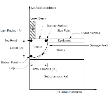
Figure 2.1: Schematic diagram of 2-D three layer skin model.
The “equation 2.1” is solved using finite volume method for 2-D biological tissue slab. There are many other
analytical methods to solve this equation but their computational time hinders the determination of the
temperature during the transient state. To minimise the computational time, numerical solutions which can
approximate the exact solution and determine the transient temperatures in short time, can be used.
The cross-section of 2-dimensional three-layer skin model with the basal cell carcinoma penetrating inside the
dermis, on which the laser beam is irradiated, is shown in “Fig. 2.1”. The depth (D) is taken from the level of
normal skin surface. The tumour depth (D) is considered equal to tumour radius (Rt) for this study. The beam radius (Rl) is kept 1.75 times the tumour radius, i.e. Beam Radius (Rl) = 1.75×Tumour Radius (Rt). The three black dots marked as top point, side point, and bottom point are shown for which the temperature is monitored
throughout the laser exposure and even after the laser irradiation is terminated. The TABLE 1 shows the series
the temperature of the tissue reaches up to 100oC, so the biological effects considered are bio stimulation,
hyperthermia, and reduction in enzyme activity, protein denaturation and vaporisation.
TABLE 1: Thermal effect of laser radiation [25].
Temperature (°C) Biological Effect
37 Normal
<43 Biostimulation
43-45 Hyperthermia
50 Reduction in enzyme activity
60 Protein Denaturation (Coagulation)
70-80 Welding
80 Permeabilisation of Cell Membranes
100 Vaporisation
>150 Carbonisation
>300 Rapid cutting and Ablation
III. RESULTS AND DISCUSSION
The bio-heat equation is solved using finite volume method for 2-D axisymmetric three-layered Skin model with
tumour of specific depth starting from the surface of the skin. The tissue optical properties are: µa.epi = 571.29cm−1, µa.derm= 41.24cm−1, µa.sub f at= 10.0cm−1, µs.derm= 732.31cm−1 and µs.sub f at = 918.58cm−1, and the laser parameters for CW Nd:YAG laser are: beam radii =2.625mm, 3.0625mm and 3.5mm for tumour radii =
1.5mm, 1.75mm and 2.0mm respectively. The average power is varied between 5.0−10.0W; the laser powers are
taken at a step size of 1.0 for this study. The values of key thermal properties of tissue like density (r), specific
heat (cp), and thermal conductivity (k) depend on the water content which can be calculated using “equations 2.3, 2.4, and 2.5” respectively. The initial temperature of the tissue is taken as 37oC. The laser irradiation time
for which the damage front moves 0.2mm beyond the tumour interface into the surrounding healthy tissue is
considered to be the optimised irradiation time. This margin is taken into account so as to minimise the
recurrence rate. In the plots where gap is plotted, the profiles are shown for the five time instances: (i) at the half
of the irradiation time, (ii) at the time instance when laser is terminated, (iii) at the time instance where the
damage front just touches the tumour interface near the q = 0o, (iv) at the time instance after which the damage
front almost stops to propagate further and, (v) at time instance after which it totally stops further penetration.
1.4.Temperature profiles with respect to time at top, side and bottom points near the tumour interface:
The variation of temperature with respect to time at three different locations, viz. top, side, and bottom points as
shown in “Fig. 2.1”, for three different tumour radii is depicted in “Fig. 3.1”. The laser power for this case is
9W. At the point of interaction, temperature went up to 100oC and then it remained constant for some time, as
the water inside the tissue started to vaporise and the temperature dropped drastically when the laser exposure is
terminated. This is happening because the heat conduction into the tissue and heat lost into the air are
counterbalanced by the heat supplied by the laser source. Once the laser exposure is terminated, two phenomena
there is drastic decrease in the surface temperature. As the time progresses, after the laser termination, the
temperature decreases at a slower rate because with the passage of time temperature gradient at the surface of
the tissue decreases. As a consequence, the rate of heat diffusion decreases. The side and bottom points being at
some distance from the point of interaction of laser beam with the tissue, the value of maximum temperature is
around 70oC and 65oC for the side and bottom points respectively. It is also interesting to observe that the
temperature reduction, after the termination of laser exposure, is rapid for side point as compared to bottom
point. The reason for this is that, side point is close to the surface therefore, heat transfer will be governed by
two phenomena:
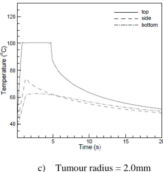
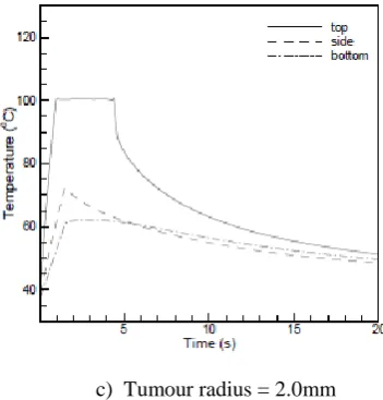
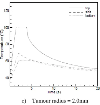
a) Tumour radius = 1.5mm
c) Tumour radius = 2.0mm
Figure 3.1: Temperature profiles for average Laser Power = 9W
heat diffusion into the surrounding tissue and heat dissipation into air. However, for the bottom point only one
phenomenon, i.e. heat diffusion into the surrounding tissues, accounts for the heat loss, which is why the
temperature decreases at a faster rate for side point as compared to the rate of temperature decrease for the
bottom point. In “Fig 3.1(b)”, it can be seen that the temperature profile stays at 100oC for a longer duration as
compared to “Fig 3.1(a)”. This stay is even longer in the profile of temperature plotted for 2.0mm tumour radius as shown in “Fig 3.1(c)”. This is because of the larger irradiation time taken to achieve complete necrosis of the
tumourous tissue with the increase in tumour size.
Furthermore, owing to the presence of unvaporised water in the tissue, there is no drastic rise in temperature. It
can also be observed from these plots that the difference in the maximum temperatures achieved for side and
bottom points is low for tumour radius of 1.5mm, and as the tumour radius increases, the maximum temperature
difference between these two points also increases. This is due to the fact that the side point is experiencing a
higher temperature because of the higher scattering coefficient of the tissue while, with the increase in the
tumour radius, the distance between the bottom point and the interaction point increases which results in less
temperature rise at this location.
c) Tumour radius = 2.0mm
Figure 3.2: Temperature profiles for average Laser Power = 7W
The temperature variations for the laser power of 7W are shown in “Fig. 3.2”. In these plots it can be noticed
that maximum temperature for the side point and bottom point for a tumour radius of 1.5mm is 68oC and 65oC;
it is 73oC and 65oC for a tumour radius of 1.75mm, and 73oC and 60oC for a tumour radius of 2.0mm
respectively. As noted earlier, the difference between the maximum temperatures at the two points is increasing
with the increase in the tumour radius.
a) Tumour radius = 1.5mm
c) Tumour radius = 2.0mm
Figure 3.3: Temperature profiles for average Laser Power = 5W
Depicted in “Fig. 3.3” are the profiles of temperature for 5W laser power for different tumour radii. The
maximum temperature for tumour radius of 1.5mm at side and bottom points is almost same and is equal to
66oC. The maximum temperature achieved for these two points for 1.75mm tumour radius is 70oC and 64oC
respectively; for tumour radius 2.0mm, it is 70oC and 60oC respectively. Also, it is illustrated in these profiles
that the rate of temperature rise for all the three points decreases as the tumour radius is increased for a
particular power because energy per unit area per unit time decreases.
The laser irradiation time for which efficient tumour necrosis is obtained using laser powers between 5.0W
−10.0Wfor three different tumour radii is listed in “TABLE 2”. The laser powers are taken at a step size of 1.0
for this study. It can be noticed that as the laser power increases for a given radius, the irradiation time decreases
almost linearly.
TABLE 2: Optimised exposure duration for lasers of different average powers for various tumour radii
Tumour Radius (mm) Average Power (Watts)
5 6 7 8 9 10
1.5 1.0 0.90 0.70 0.65 0.55 0.50
1.75 1.60 1.20 1.10 0.90 0.85 0.80
2.0 2.10 1.80 1.60 1.40 1.20 1.0
IV. CONCLUSION
A mathematical model that predicts the temperature and monitors the propagation of thermal damage front
during the thermal therapy for a particular tissue has been developed. This model reflects the understanding of
thermal events like coagulation and vaporisation when the laser of a specific power is irradiated on a tumourous
tissue. In this study, the optimised irradiation time has been predicted for a particular tumour radius, laser beam
radius, laser power and specific skin type. It can be concluded from the results that irradiation time decreases
almost linearly with increase in the laser power for a given tumour radius. The laser beam radius also affects the
damage extent and the beam radius has to be optimised for a particular tumour radius so that damage front
power for a particular tumour radius, which is useful to the dermatologist in deciding the proper process
parameters for an efficient treatment of basal cell carcinoma.
REFERENCES
[1.] C. E. Lindholm, E. Kjellen, P. Nilsson, and S. Hertzman, “Microwave-induced hyperthermia and
radiotherapy in human superficial tumours: clinical results with a comparative study of combined
treatment versus radiotherapy alone”, International journal of hyperthermia: the official journal of
European Society for Hyperthermic Oncology, North American Hyperthermia Group, vol. 3, no. 5, pp.
393–411, 1987.
[2.] F. K. Storm, L. R. Kaiser, J. E. Goodnight,W. H. Harrison, R. S. Elliott, A. S. Gomes, and D. L. Morton,
“Thermochemotherapy for melanoma metastases in liver.”, Cancer, vol. 49, no. 6, pp. 1243–1248, 1982.
[3.] Prezi, Modelling and measurements of laser-skin thermal interaction, 2010.
[4.] P. Kiefhaber, G. Nath, and K. Moritz, “Endoscopical control of massive gastrointestinal hemorrhage by
irradiation with a high-power Neodymium-Yag laser”, Progress in surgery, vol. 15, pp. 140–155, 1977.
[5.] R. R. Anderson and J. A. Parrish, “Microvasculature can be selectively damaged using dye lasers: a basic
theory and experimental evidence in human skin”, Lasers in surgery and medicine, vol. 1, no. 3, pp. 263–
276, 1981.
[6.] M. El-Tonsy, Hany, M. El-Domyati, Mostafa, A. El-Sawy, Esmat, W. El-Din, Hosam, T. E.-D. A.-S.
Anbar, and A. Raouf, Hamza, “Continuous-wave Nd:Yag laser hyperthermia: a successful modality in
treatment of basal cell carcinoma”, Dermatology online journal, vol. 10, no. 2, p. 3, 2004.
[7.] A. Y. Citkaya and S. S. Seker, “Modeling and Simulation of Temperature Distribution in Laser-tissue
Interaction”, Session 2P7 Computational Electromagnetics, Hybrid Methods, pp. 844–847, 2011.
[8.] A. J. Welch, J. A. Pearce, K. R. Diller, G. Yoon, and W. F. Cheong, “Heat generation in laser irradiated
tissue”, Journal of biomechanical engineering, vol. 111, no. 1, pp. 62–68, 1989.
[9.] A. Hofstetter, E. Keiditsch, T. Halldorsson, H. Bulow, and F. Frank, The neodymium-YAG laser in
urology, Editiones Roche c/o F. Hoffmann-La Roche, 1980.
[10.]A. J. Welch, “Laser Irradiation of Tissue”, In A. Shitzer and R. C. Eberhart (eds.), Heat Transfer in
Medicine and Biology, Springer US, pp. 135–184, 1985.
[11.]A. N. Takata, L. Zaneveld, andW. Richter, “Laser-induced thermal damage of skin”, Technical report,
DTIC Document, 1977.
[12.]T. Halldorson and J. Langerholc, “Thermodynamic analysis of laser irradiation of biological tissue”,
Applied optics, vol. 17, no. 24, pp. 3948–3958, 1978.
[13.]L. Cummins and M. Nauenberg, “Thermal effects of laser radiation in biological tissue.”, Biophysical
Journal, vol. 42, no. 1, pp. 99–102, 1983.
[14.]T. Halldorson, W. Rother, J. Langerholc, and F. Frank, “Theoretical and experimental investigations prove
Nd: YAG laser treatment to be safe”, Lasers in surgery and medicine, vol. 1, no. 3, pp. 253–262, 1981.
[15.]S. L. Jacques and S. A. Prahl, “Modeling optical and thermal distributions in tissue during laser
[16.]A. Banerjee, A. A. Ogale, C. Das, K. Mitra, and C. Subramanian, “Temperature Distribution in Different
Materials Due to Short Pulse Laser Irradaition.”, Heat Transfer Engineering, vol. 26, no. 8, 2005.
[17.]R. Muthukumaran and C. S. Mishra, “Analysis of theTransport of a Train of Short-Pulse Rdiation of
Gaussian Temporal Profile Through a 2-D ParticipatingMedium”, Heat Transfer Engineering, vol. 30, no.
14, 2009.
[18.]S. Kumar and A. Srivastava, “Thermal analysis of laser-irradiated tissue phantoms using dual phase lag
model coupled with transient radiative transfer equation”, International Journal of Heat and Mass Transfer,
vol. 90, pp. 466–479, November 2015.
[19.]G. W. Yoon, The thermal effect of laser beam scattering in biological medium, University of Texas at
Austin, 1984.
[20.]Z. Gourgouliatos, S. Ghaffari, A. Welch, K. Diller, and R. Straight, “Measurements of argon laser light
attenuation in the skin „in vivo‟ using a unique animal model”, In Engineering in Medicine and Biology
Society, 1989. Images of the Twenty-First Century., Proceedings of the Annual International Conference
of the IEEE Engineering in, vol. 6, pp. 1749–1750, 1989.
[21.]R. Splinter, R. H. Svenson, L. Littmann, J. R. Tuntelder, C. H. Chuang, G. P. Tatsis, and M. Thompson,
“Optical properties of normal, diseased, and laser photocoagulated myocardium at the nd: YAG wavelength.”, Lasers in surgery and medicine., vol. 11, no. 2, pp. 117–124, 1991.
[22.]J. T. J. Walsh and T. F. Deutsch, “Pulsed CO2 laser tissue ablation: measurement of the ablation rate”,
Lasers in surgery and medicine, vol. 8, no. 3, pp. 264–275, 1988.
[23.]R. Dua and S. Chakraborty, “A novelmodeling and simulation technique of photo -thermal interactions
between lasers and living biological tissues undergoing multiple changes in phase”, Computers in biology
and medicine, vol. 35, no. 5, pp. 447–462, 2005.
[24.]M. J. Brugmans, J. Kemper, G. H. Gijsbers, F. W. van der Meulen, and M. J. van Gemert, “Temperature
response of biological materials to pulsed non-ablative CO2 laser irradiation”, Lasers in surgery and
medicine, vol. 11, no. 6, pp. 587–594, 1991.
[25.]L. J. Miserendino, G. C. Levy, and I. M. Rizoiu, “Effects of Nd:YAG laser on the permeability of root
![TABLE 1: Thermal effect of laser radiation [25].](https://thumb-us.123doks.com/thumbv2/123dok_us/9258130.1463740/5.595.98.496.152.352/table-thermal-effect-laser-radiation.webp)