Disclaimer: this is not the definitive version of record of this article. This manuscript has been accepted for publication in Reproduction, but the version presented here has not yet been copy-edited, formatted or proofed. Consequently, Bioscientifica accepts no responsibility for any errors or omissions it may contain. The definitive version is now freely available at
http://dx.doi.org/10.1530/JOE-17-0042, April 2017.
The full details of the publication are as follows:
TITLE: Activation of the P2Y2 receptor regulates bone cell function by enhancing ATP release
AUTHORS: Isabel R Orriss, Dilek Gueneri, Mark Or Hajjawi, Kristy Shaw, Jessal J Patel, and Tim R Arnett
JOURNAL TITLE: Journal of Endocrinology
PUBLICATION DATE: 18 April 2017 (online)
PUBLISHER: BioScientifica
For Review Only
ACTIVATION OF THE P2Y
2RECEPTOR REGULATES BONE
1
CELL FUNCTION BY ENHANCING ATP RELEASE
2
3
4
Isabel R Orriss1, Dilek Guneri1, Mark OR Hajjawi2, Kristy Shaw1,
5
Jessal J Patel1, Timothy R Arnett2
6
7
8
1
Department of Comparative Biomedical Sciences, Royal Veterinary College, London
9
2
Department of Cell & Developmental Biology, University College London, London
10
11
12
13
14
Address correspondence to: Isabel Orriss
15
Department of Comparative Biomedical Science
16
Royal Veterinary College
17
London, NW1 0TU
18
Tel: 020 7468 1238 (ext 5468)
19
Email: [email protected]
20
21
Short title: P2Y2 receptor activation induces ATP release in bone 22
23
Key words: P2Y2 receptor, UTP, ATP release, bone resorption, bone mineralisation 24
25
26
Conflict of Interest: The authors have no conflict of interest
27
28
29
Word count: 4727
30
31
For Review Only
ABSTRACT
33
Bone cells constitutively release ATP into the extracellular environment where it acts locally via
34
P2 receptors to regulate bone cell function. Whilst P2Y2 receptor stimulation regulates bone 35
mineralisation the functional effects of this receptor in osteoclasts remain unknown. This
36
investigation used the P2Y2 receptor knockout (P2Y2R
-/-) mouse model to investigate the role of
37
this receptor in bone. MicroCT analysis of P2Y2R-/- mice demonstrated age-related increases in
38
trabecular bone volume (≤48%), number (≤30%) and thickness (≤17%). In vitro P2Y2R
-/-39
osteoblasts displayed a 3-fold increase in bone formation and alkaline phosphatase activity
40
whilst P2Y2R-/-osteoclasts exhibited a 65% reduction in resorptive activity. Serum cross-linked
41
c-telopeptide levels (CTX, resorption marker) were also decreased (≤35%). The resorption
42
defect in P2Y2R
osteoclasts was rescued by the addition of exogenous ATP, suggesting that
43
an ATP deficit could be a key factor in the reduced function of these cells. In agreement, we
44
found that basal ATP release was reduced up to 53% in P2Y2R-/- osteoclasts. The P2Y2 45
receptor agonists, UTP and 2-thioUTP, increased osteoclast activity and ATP release in
46
wildtype but not P2Y2R-/-cells. This indicates that the P2Y2 receptor may regulate osteoclast 47
function indirectly by promoting ATP release. UTP and 2-thioUTP also stimulate ATP release
48
from osteoblasts suggesting that the P2Y2 receptor exerts a similar function in these cells. 49
Taken together, our findings are consistent with the notion that the primary action of P2Y2 50
receptor signalling in bone is to regulate extracellular ATP levels.
51
52
53
54
55
56
57
58
For Review Only
INTRODUCTION
60
Adenosine triphosphate (ATP) has long been recognized for its role in intracellular energy
61
metabolism; however, it is also exported to the extracellular environment where it acts as an
62
important signalling molecule (Burnstock 2007a). Outside cells, ATP and related compounds
63
act via purinergic receptors to modulate a range of biological processes. These receptors are
64
classified into two groups; P1 and P2 receptors. There are four P1 receptors (A1,A2a,A2b,A3), 65
which are activated by adenosine. The P2 receptors are further subdivided into the P2X
ligand-66
gated ion channels and the P2Y G-protein-coupled receptors. P2X receptors are activated by
67
ATP whilst P2Y receptors respond to nucleotides including ATP, adenosine diphosphate
68
(ADP), uridine triphosphate (UTP) and uridine diphosphate (UDP) (Abbracchio and Burnstock
69
1994; Burnstock 2007b). Currently, seven P2X receptors (P2X1-7) and eight P2Y receptors
70
(P2Y1,2,4,6,11-14) have been identified (Burnstock 2007b). 71
The P2Y receptors display distinct pharmacology with some being activated by
adenine-72
containing nucleotides (P2Y1, P2Y12, P2Y13), whilst others are stimulated by uridine-containing 73
nucleotides (P2Y2, P2Y4, P2Y6, P2Y14) (Burnstock 2007a, b). The primary agonist at the P2Y2 74
receptor is UTP but it is also activated by ATP. Selective synthetic agonists (e.g. 2-thioUTP)
75
are also available. Receptor stimulation activates phospholipase C and results in Ca2+ release
76
from internal stores. Expression of the P2Y2 receptor has been reported in many tissues 77
including heart, blood vessels, lung, kidney and skeletal muscle (Burnstock 2007a).
78
Bone cells express multiple P2 receptor subtypes and knowledge of the functional effects of
79
extracellular nucleotides in bone has increased significantly in recent years (Burnstock, et al.
80
2013; Gartland, et al. 2012; Noronha-Matos and Correia-de-Sa 2016; Orriss 2015). P2Y2 81
receptor expression by osteoclasts has been widely reported (Bowler, et al. 1995; Buckley, et
82
al. 2002; Hoebertz, et al. 2000; Orriss, et al. 2011b). Early work using cells from a human
83
osteoclastoma suggested that ATP could act via the P2Y2 receptor to promote bone resorption 84
(Bowler et al. 1995). However, in a follow up study UTP failed to stimulate resorption,
85
suggesting this was not the case (Bowler, et al. 1998). To date, there are no studies directly
For Review Only
describing the functional effects of P2Y2 receptor activation on osteoclasts. In contrast, 87
activation of several other P2Y receptor subtypes (P2Y1, P2Y6, P2Y12, P2Y14) has been 88
associated with increased osteoclast formation and/or activity (Hoebertz, et al. 2001; Lee, et al.
89
2013; Orriss et al. 2011b; Su, et al. 2012; Syberg, et al. 2012b).
90
The role of the P2Y2 receptor in osteoblasts has been more extensively investigated. P2Y2 91
receptor expression by osteoblasts has been extensively reported (Bowler et al. 1995;
92
Hoebertz et al. 2000; Maier, et al. 1997), with several studies describing that expression is
93
differentiation-dependent with the highest levels seen in mature, bone forming cells
(Noronha-94
Matos, et al. 2012; Orriss, et al. 2006). P2Y2 receptor activation in osteoblast-like cells 95
activates several intracellular signalling pathways including protein kinase C, p38
mitogen-96
activated protein kinase, c-Jun NH2-terminal protein kinase and RhoA GTPase (Costessi, et al. 97
2005; Gardinier, et al. 2014; Katz, et al. 2006, 2008; Pines, et al. 2005). The P2Y2 receptor has 98
also been shown to mediate the Ca2+ mobilisation induced by oscillatory fluid flow (You, et al.
99
2002).
100
One of the first functional effects to be attributed to the P2Y2 receptor was the inhibition of 101
bone mineralisation by ATP and UTP (Hoebertz, et al. 2002; Orriss, et al. 2013; Orriss, et al.
102
2007). Consistent with this,initial skeletal analysis of 8-week old P2Y2 receptor knockout mice
103
(P2Y2R-/-) demonstrated large increases in trabecular and cortical bone parameters in the long
104
bones (Orriss et al. 2007; Orriss, et al. 2011a). Furthermore, P2Y2 overexpression leads to 105
decreased bone formation (Syberg, et al. 2012a) and polymorphisms in the P2Y2 receptor gene 106
are associated with increased bone mineral density and a decreased risk of osteoporosis
107
(Wesselius, et al. 2013). In contrast, a recent study using P2Y2R-/- mice on a different genetic
108
background, described small decreases in the trabecular bone in knockout animals (Xing, et al.
109
2014), this work additionally reported that the P2Y2 receptor promotes bone mineralisation. 110
The P2Y2 receptor may also have a functional role in mediating osteoblast 111
mechanosensitivity. Studies suggest that the P2Y2 receptor promotes mechanotransduction 112
(Xing et al. 2014) and increases cell stiffness and cytoskeletal rearrangement in response to
113
fluid shear stress (Gardinier et al. 2014).
For Review Only
Expression of the P2Y2 receptor has also been reported in MLO-Y4 osteocyte-like cells 115
(Kringelbach, et al. 2014). The same study also demonstrated controlled ATP release from
116
these cells and reported that UTP, probably acting via the P2Y2 or P2Y4 receptors, increased 117
this ATP release.
118
Available evidence thus indicates that the P2Y2 receptor plays significant, although not yet 119
fully defined roles in regulating bone remodelling. This study used the P2Y2R
mouse, which
120
was first generated almost 2 decades ago (Cressman, et al. 1999), to determine how P2Y2 121
receptor-mediated signalling influences bone cell function in vitro and in vivo, with a particular
122
focus on its effects in osteoclasts.
123
124
125
126
127
128
129
130
131
132
133
134
135
136
137
138
139
For Review Only
METHODS
141
Reagents 142
Tissue culture reagents were purchased from Life Technologies (Paisley, UK); unless
143
mentioned, all chemicals were purchased from Sigma Aldrich (Poole, Dorset, UK). UTP and
2-144
thioUTP were purchased from Tocris Bioscience (Bristol, UK).
145
Animals 146
Mice lacking the P2Y2 receptor gene (P2Y2R-/-) were obtained from Jackson Laboratories (Bar
147
Harbor, Maine, USA). The generation and characterisation of P2Y2R
mice (C57BL/6J
148
background) has been previously described (Homolya, et al. 1999). All animals were housed
149
under standard conditions with free access to food and water. Animals were bred from
150
homozygote (P2Y2R
-/-) and parental strain wildtype (P2Y2R +/+
) breeding pairs. All procedures
151
complied with the UK animals (Scientific Procedures) Act 1986 and were reviewed and
152
approved by the Royal Veterinary College Research Ethics Committee.
153
Microcomputed x-ray tomographic (µCT) analysis of P2Y2R
mice 154
The tibiae and femora were isolated from male 4, 8, 16 and 24-week old P2Y2R-/- and P2Y2R+/+
155
mice (n=10), fixed in 10% neutral buffered formalin (NBF) for 24 hours and stored in 70%
156
ethanol until scanning. µCT analysis of trabecular and cortical bone parameters was
157
performed on the tibial and femoral metaphysis (SkyScan 1172, Bruker, Belgium). The
158
appearance of the first cartilage bridge was used as a reference point, with an offset of 0.4mm
159
and 2.5mm for trabecular and cortical bone, respectively. In all cases the length of bone
160
analysed was 1mm. The µCT scanner was set at 50Kv and 200µA using a 0.5mm Al filter and
161
a resolution of 4.3µm. Analysis of isolated bones was performed blind. The images were
162
reconstructed, analysed and visualised using SkyScan NRecon, CTAn and CTVol software.
163
Bone mineral density (BMD) was calibrated and calculated using hydroxyapatite phantoms with
164
a known density.
165
For Review Only
Osteoblast formation assay 167
Osteoblasts were isolated from the calvariae of 3-5 day old P2Y2R +/+
or P2Y2R -/-
mice by
168
trypsin/collagenase digestion as previously described (Orriss, et al. 2012b; Taylor, et al. 2014).
169
Cells were cultured for up to 21 days in alpha Minimum Essential Medium, (αMEM)
170
supplemented with 2mM β-glycerophosphate and 50µg/ml ascorbic acid, with half medium
171
changes every 3 days. The total area of bone nodules formed was quantified by image
172
analysis, as described previously (Orriss et al. 2012b).
173
Primary osteoblasts of bone marrow/stromal cell origin were obtained from the long bones of
174
6-week old male P2Y2R+/+ or P2Y2R-/- animals. The collected cells were suspended in α-MEM
175
and pre-cultured in a 75 cm2 flask in 5% CO2 at 37ºC. After 24 hours the α-MEM was replaced 176
in order to eliminate non-adherent cells; adherent stromal cells were cultured for a further 7
177
days. When confluent, cells were plated into 6-well trays and cultured as above.
178
Alkaline phosphatase (TNAP) activity 179
Osteoblast TNAP activity was measured in cell lysates taken at defined stages of osteoblast
180
differentiation as previously described (Orriss et al. 2012b; Taylor et al. 2014). TNAP activity
181
was normalised to cell protein using Bradford reagent. Time points in osteoblast cultures were
182
defined thus: proliferating (day 4, calvarial only); differentiating (day 7); mature (day 14) and
183
mature, bone-forming (day 21)
184
Osteoclast formation assay 185
Osteoclasts were isolated from the long bones of 6-8 week-old male P2Y2R+/+or P2Y2R-/-mice
186
as described previously (Orriss and Arnett 2012). Cells were plated onto 5mm diameter ivory
187
discs (106 cells) in 96-multiwells in αMEM supplemented with 10% FCS, 5% gentamicin, 100nM
188
PGE2, 200ng/ml M-CSF and 3ng/ml receptor activator of nuclear factor ΚB ligand (RANKL, R&D 189
Systems Europe Ltd, Abingdon, UK). After 24 hours, discs containing adherent osteoclast
190
precursors were transferred to 6-well trays (4 discs/well in 4ml medium) for a further 6 days.
191
Culture medium was acidified to pH~7.0 by the addition 10meq/l H+ (as HCL) on day 7 to
192
For Review Only
thioUTP) were added from day 3 of culture. Apyrase (a broad spectrum ecto-nucleotidase) was
194
used to determine the effects of endogenous ATP.
195
Osteoclasts were fixed in 2.5% glutaraldehyde and stained to demonstrate tartrate-resistant
196
acid phosphatase (TRAP). Osteoclasts were defined as TRAP-positive cells with 2 or more
197
nuclei and/or clear evidence of resorption. The total number of osteoclasts and the plan surface
198
area of resorption pits on each disc was assessed ‘blind’ by transmitted light microscopy and
199
reflective light microscopy and dot-counting morphometry, respectively.
200
Measurement of serum bone markers 201
Blood was collected from 4, 8, 16 and 24-week old male P2Y2R-/- and P2Y2R+/+mice by cardiac
202
puncture immediately after termination. Following clotting, samples were centrifuged at 500g
203
and the serum frozen until analysis. Levels of the bone formation marker, N-terminal propeptide
204
of type I collagen (P1NP) and the bone resorption marker, cross-linked C-telopeptide (CTX)
205
were assayed using the P1NP and RatLaps™ ELISAs, respectively (Immunodiagnostics
206
Systems Ltd, UK).
207
Histology 208
Histological analysis was performed on the femur of 8 and 24-week old male P2Y2R +/+
or
209
P2Y2R-/-mice. Tissues were fixed in 10% NBF, decalcified in 10% EDTA for three weeks and
210
embedded in paraffin wax blocks. Serial sections were cut every 5µm and slides stained with
211
TRAP counterstained with haematoxylin to visualise osteoclasts.
212
Total RNA extraction and DNase treatment 213
P2Y2R+/+and P2Y2R-/- osteoclasts were cultured on dentine discs for 9 days (mature, resorbing
214
cells) before total RNA was extracted using TRIZOL reagent (Invitrogen, Paisley, UK)
215
according to the manufacturer’s instructions. Osteoblasts were cultured for 14 days (mature,
216
bone-forming cells) before RNA collection. Extracted RNA was treated with RNase-free DNase
217
I (35U/ml) for 30 min at 37°C. The reaction was terminated by heat inactivation at 65°C for 10
For Review Only
min. Total RNA was quantified spectrophotometrically by measuring absorbance at 260nM.
219
RNA was stored at –80°C until amplification by qRT-PCR.
220
Quantitative real time polymerase chain reaction (qRT-PCR) 221
Osteoclast and osteoblast RNA (50ng) was transcribed and amplified using the qPCRBIO
222
SyGreen one-step qRT-PCR kit (PCR Biosystems, London, UK), which allows cDNA synthesis
223
and PCR amplification to be carried out sequentially. qRT-PCR was performed according to
224
manufacturer’s instructions with initial cDNA synthesis (45°C for 10 min) and reverse
225
transcriptase inactivation (95°C for 2 min) followed by 40 cycles of denaturation (95°C for 5
226
sec) and detection (60°C for 30 sec). All reactions were carried out in triplicate using RNAs
227
derived from 4 different cultures. Data were analysed using the Pfaffl method of relative
228
quantification (Pfaffl 2001). Primers were obtained from Qiagen Ltd (Manchester, UK).
229
Measurement of ATP release 230
Prior to measurement of ATP release, culture medium was removed, cell layers washed and
231
cells incubated with serum-free DMEM (phenol red free). To measure the effects of P2Y2 232
receptor deletion on basal ATP release, samples were collected after 1 hour and immediately
233
measured luminometrically using the luciferin-luciferase assay, as described previously
234
(Orriss, et al. 2009). All ATP measurements were normalised to cell number. Cell viability and
235
cell number were determined using the CytoTox 96® colorimetric cytotoxicity assay (Promega
236
UK, Southampton UK).
237
To examine the effects of acute exposure to UTP or 2-thioUTP (0.1-50µM) agonists were
238
added to the serum-free DMEM and samples taken for quantification after 10, 30, 60 and 90
239
minutes. The luminescence of the DMEM (± UTP/2-thioUTP) was used as a background
240
reading and subtracted from the relevant measurements. Standard curves used to calculate the
241
ATP concentrations in the presence or absence of UTP/2-thioUTP are shown in Fig. 5. To
242
investigate the effects of long-term treatment with P2Y2 receptor agonists, osteoclasts and 243
osteoblasts were cultured with UTP or 2-thioUTP (0.1-100µM) for 7 or 14 days, respectively.
244
Fresh UTP/2-thioUTP was added at each medium exchange. On the day of assay culture
For Review Only
medium was removed and cells incubated with serum-free DMEM without agonists. Samples
246
were collected after 1 hour and measured immediately.
247
To determine the effects of P2Y2 deletion on ATP breakdown, cells were swapped to DMEM 248
containing 1µM ATP and samples taken after 2, 5, 10, 30 and 60 minutes.
249
Statistical analysis 250
Data were analysed using GraphPad Prism 6 software (San Diego, CA). Results are
251
expressed as means ± SEM for between 6-12 biological replicates. Statistical analyses of bone
252
parameters were performed by two-tailed unpaired student’s t-test. In vitro data were analysed
253
using an unpaired student’s t-test, one-way or two-way ANOVA, followed by a Bonferroni post
254
hoc test. For all in vitro work, results are representative of experiments performed at least
255
three times, using cells isolated from different animals.
256
257
258
259
260
261
262
263
264
265
266
267
268
269
270
271
For Review Only
RESULTS
273
P2Y2R-/-mice show age-related increases in trabecular bone
274
High resolution µCT analysis revealed that P2Y2R-/- mice display increased levels of trabecular
275
bone compared to age-matched P2Y2R+/+ controls. These differences appear to be age-related
276
with the biggest changes observed in the 24-week animals. Trabecular bone volume (BV/TV)
277
was increased ≤46% in the femur and ≤48% in the tibia of P2Y2R-/-mice (Fig. 1A-1B, 1O).
278
Trabecular number (Tb.N) was increased ≤27% in the femora (Fig. 1C, 1O) and ≤30% in the
279
tibiae (Fig. 1D, 1O). Trabecular thickness (Tb.Th) was unchanged up to 8 weeks of age but
280
increased ≤10% and ≤17% at 16 and 24 weeks, respectively (Fig. 1E-1F, 1O). Trabecular
281
bone mineral density (Tb.BMD) was ≤12% higher in P2Y2R-/- mice (Fig. 1G-1H). No differences
282
were observed in the cortical bone volume (Fig. 1K-1L, 1O), cortical thickness (Fig. 1K-1L),
283
endosteal and periosteal diameter (Fig. 1M-1N) and bone length at any age.
284
Increased bone formation by osteoblasts from P2Y2R-/- mice
285
The level of mineralised bone nodule formation was increased ~3-fold in P2Y2R
calvarial
286
osteoblasts (Fig. 2A, 2G) and 5-fold in P2Y2R-/- long bone osteoblasts (Fig. 2B). P2Y2 receptor 287
deletion increased basal TNAP activity (≤3-fold) in calvarial and long bone osteoblasts at all
288
stages of differentiation with the largest effects being observed in the mineralising cells (Fig.
289
2C-2D). Serum TNAP activity was up to 60% higher in P2Y2R-/- animals (Fig. 2E); no
290
differences were observed in the serum P1NP levels (Fig. 2F). No differences in total protein
291
content were observed in any TNAP activity experiments.
292
Osteoclasts from P2Y2R-/- mice exhibit defective resorption
293
Whilst no differences in osteoclast numbers were observed (Fig. 3A, 3D), the level of
294
resorption per osteoclast was decreased 75% in P2Y2R
cultures (Fig. 3B, 3D). Serum CTX
295
levels were reduced up to 35% in P2Y2R-/- mice (Fig. 3C). Qualitative histology suggested that
296
decreased numbers of osteoclasts were evident on the trabecular and endocortical bone
297
surfaces of 24-week old P2Y2R-/-; however, no differences were observed in 8-week old
298
animals (Fig. 3E).
For Review Only
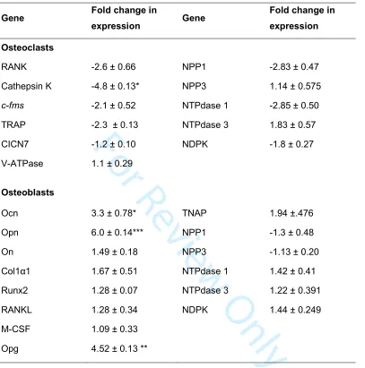
Changes in gene expression in P2Y2R-/- osteoclasts and osteoblasts
300
The effect of P2Y2 receptor deletion on the expression of resorption associated genes and 301
ecto-nucleotidases was investigated in mature, resorbing osteoclasts. mRNA expression of
302
many genes (TRAP, CICN7, RANK, c-fms) showed a downward trend but only cathepsin K
303
expression was significantly reduced (4.8-fold). Osteoclasts express a range of
ecto-304
nucleotidases that hydrolyse ATP (Hajjawi, et al. 2014) and NDPK (nucleoside
305
disphosphokinase), which can regenerate ATP from ADP. P2Y2 receptor deletion did not 306
influence the expression of any of these genes (Table 1).
307
In osteoblasts, deletion of the P2Y2 receptor increased osteocalcin (Ocn), osteopontin (Opn) 308
and osteoprotegerin (OPG) expression 3.3, 6 and 4.5-fold, respectively. The mRNA expression
309
of Col1α1, Runx2, TNAP, osteonectin, RANKL, MCSF and the ecto-nucelotidases was
310
unchanged (Table 1).
311
Activation of the P2Y2 receptor increases bone resorption
312
Treatment with UTP and 2-thioUTP had no effect on osteoclast formation in P2Y2R+/+ or P2Y2R
-313
cells (Fig. 4A-4B). However, the area resorbed per osteoclast was dose-dependently
314
increased by up to 80% and 45% in P2Y2R +/+
cells treated with UTP and 2-thioUTP (≥100nM),
315
respectively. No effects on resorption were seen in P2Y2R-/- osteoclasts (Fig. 4C-4D).
316
Reversal of resorption defect in P2Y2R-/- osteoclasts by extracellular ATP
317
P2Y2R
osteoclasts displayed a 53% reduction in ATP release (Fig. 4E) but showed no
318
difference in the rate of ATP breakdown (Fig. 4F). Apyrase (≥1U/ml), a broad spectrum
ecto-319
nucleotidase that rapidly degrades ATP and ADP, inhibited bone resorption by up to 55% (Fig.
320
4G). To determine if reduced extracellular ATP was the cause of the decreased resorption seen
321
in P2Y2R-/- osteoclasts, cells were cultured with exogenous ATP (1-10µM). Treatment with ATP
322
(≥1µM) fully rescued the resorption defect see in P2Y2R-/- osteoclasts (Fig. 4H).
323
P2Y2 receptor agonists increase ATP release from osteoclasts
324
In P2Y2R+/+ cells, 10 minutes after addition of UTP (≥1µM) extracellular ATP levels were
325
doubled; the increase in ATP levels was sustained for up to 90 minutes post treatment (Fig.
For Review Only
5A). No effect of UTP on ATP release was seen in P2Y2R-/-osteoclasts at any stage (Fig.
5B-327
5D). Treatment with 2-thioUTP (≥0.1µM) also dose dependently increased extracellular ATP
328
levels by ≤50% for up to 90 minutes in P2Y2R+/+ osteoclasts (Fig. 5E); 2-thioUTP was without
329
effect in P2Y2R-/- cells (Fig. 5F-5H).
330
The effect of long-term treatment (7 days) with P2Y2 receptor agonists on basal ATP release 331
was also investigated in mature osteoclasts. In P2Y2R+/+ cells, UTP and 2-thioUTP (≥1µM)
332
increased ATP release by up to 70% and 65% respectively (Fig.5I-5J). No increase in ATP
333
release was seen in P2Y2R-/- osteoclasts. Standard curves used to calculate ATP levels are
334
shown in Fig. 5K-5L. In all experiments, cell viability was unchanged (not shown).
335
ATP release from osteoblasts is stimulated by UTP and 2-thioUTP 336
The rate of ATP breakdown was unchanged in P2Y2R-/- osteoblasts (Fig. 6A). ATP release
337
from P2Y2R-/- cells was decreased (≤60%) at all stages of differentiation (Fig. 6B). Long-term
338
treatment (14 days) with UTP and 2-thioUTP increased the levels of ATP release by up to
4-339
fold and 3-fold, respectively, in P2Y2R+/+osteoblasts (Fig. 6C-6D). No effects were seen in
340
P2Y2R-/- osteoblasts.
341
Acute UTP treatment increased ATP release from P2Y2R+/+ osteoblasts up to 4-fold within
342
10 minutes; stimulatory effects were sustained for up to 60 minutes (Fig. 6E). UTP was without
343
effect in P2Y2R
osteoblasts (Fig. 6F-6H). 2-thioUTP also enhanced ATP release (≤4-fold)
344
from P2Y2R+/+, but not P2Y2R-/- osteoblasts (Fig. 6I-6L).
345
346
347
348
349
350
For Review Only
DISCUSSION
352
This study examined the role of P2Y2 receptor-mediated signalling in osteoclasts and 353
osteoblasts. We found that global deletion of the P2Y2 receptor resulted in greater amounts of 354
trabecular bone and increased BMD. Culture of cells derived from P2Y2R-/- mice revealed that
355
osteoclast resorptive activity was decreased whilst bone mineralisation was increased.
356
Mechanistic analysis revealed that P2Y2 receptor activation (acute and prolonged) promotes 357
ATP release from osteoclasts and osteoblasts.
358
Several P2Y receptors (P2Y1, P2Y6, P2Y12, P2Y14) and extracellular nucleotides (e.g. ATP, 359
ADP, UDP) have been implicated in the regulation of osteoclast formation and activity
360
(Hoebertz et al. 2001; Lee et al. 2013; Orriss et al. 2011b; Su et al. 2012; Syberg et al. 2012b).
361
However, there are no reports directly describing the functional role of the P2Y2 receptor in 362
osteoclasts. This study found that the P2Y2 agonists, UTP and 2-thioUTP, dose-dependently 363
stimulated bone resorption. Consistent with a pro-resorptive role for UTP and the P2Y2 364
receptor, we observed that P2Y2R-/- animals had decreased serum CTX levels and thatcultured
365
P2Y2R
osteoclasts displayed reduced resorptive activity and cathepsin K expression. UDP,
366
the breakdown product of UTP, acts via the P2Y6 receptor to promote osteoclast function 367
(Orriss et al. 2011b). However, since the actions of UTP are lost in P2Y2R-/- osteoclasts, it is
368
unlikely that the effects observed here are due to P2Y6 receptor-mediated signalling. 369
Earlier studies have reported that P2Y2 receptor activation by ATP and UTP can both inhibit 370
(Hoebertz et al. 2002; Orriss et al. 2007; Orriss, et al. 2012a) and promote (Xing et al. 2014)
371
bone mineralisation. Consistent with its role as a negative regulator of bone mineralisation, we
372
observed that P2Y2R-/- osteoblasts exhibited increased levels of bone formation, Ocn
373
expression and TNAP activity. Suprisingly, TNAP mRNA expression was unaffected in P2Y2R
-/-374
osteoblasts. This could indicate that P2Y2 receptor signalling increases enzyme activity by 375
influencing the post-translational modifications of TNAP rather than the overall expression level.
376
We have previously shown that the effects of ATP and UTP are restricted to the mineralisation
For Review Only
process with collagen expression and activity being unaffected (Orriss et al., 2007). The lack of
378
effect of P2Y2 receptor deletion on serum P1NP levels is consistent with these observations. 379
In agreement with the in vitro findings, our longitudinal µCT study revealed that P2Y2 380
deletion led to age-related increases in trabecular bone and BMD. These data are also
381
consistent with our earlier description of the bone phenotype of 8-week old P2Y2R-/- animals
382
(Orriss et al. 2011a), and the observation that P2Y2 receptor overexpression leads to 383
decreased bone formation (Syberg et al. 2012a). However, they are at variance to a recent
384
report of reduced bone levels in P2Y2R-/- mice (Xing et al. 2014). The reasons for these
385
divergent results are unclear but given that parental strain has been shown to affect the
386
phenotype of the P2X7 receptor knockout (Syberg, et al. 2012a), the differing genetic
387
background of the animals studied (C57BL/6 compared to SV129 (Xing et al. 2014)) could be a
388
factor. Variations in µCT methodology could also contribute; for example, this study analysed a
389
1mm region of the trabecular bone within the metaphyseal portion of the long bones at a
390
resolution of 4.3µm. In contrast, Xing et al measured the trabecular bone within a narrow
391
region of the diaphysis at a lower resolution (10.5µm) (Xing et al. 2014).
392
Unlike the observed effects in the trabecular bone, in both this study and that of Xing et al
393
(Xing et al. 2014), cortical bone parameters were unaffected in P2Y2R-/- mice. This suggests
394
that P2Y2 receptor deletion does not have significant effects on bone growth. Thus, P2Y2 395
receptor-mediated signalling appears to be more important in bone undergoing rapid turnover.
396
In vivo, osteoblast and ostoclast function are tightly coupled with osteoclast activation being
397
dependent on osteoblasts. Gene expression analysis revealed a significant increase in
398
osteoblast expression of OPG whilst RANKL expression was unchanged. If reflected in vivo
399
this would reduce osteoclast formation and activity and could contribute to the decreased bone
400
resorption seen in P2Y2R
mice. In agreement, qualitative observations showed that
401
osteoclast numbers on the trabecular and endocortical bone surfaces appeared reduced in
402
these animals. Further bone histomorphometric analysis of in vivo parameters such as bone
403
formation rate and osteoclast number would confirm this and build on the findings reported
404
here.
For Review Only
Controlled ATP release has been demonstrated from numerous cell types including bone
406
cells. Several studies have indicated that the primary method of ATP release from osteoblasts
407
is vesicular exocytosis (Genetos, et al. 2005; Orriss et al. 2009; Romanello, et al. 2001),
408
although the P2X7 receptor may also be involved (Brandao-Burch, et al. 2012). In osteoclasts,
409
ATP release involves the P2X7 receptor (Brandao-Burch et al. 2012; Pellegatti, et al. 2011).
410
Increasing evidence now suggests that ATP can act to enhance its own release; ATP or
UTP-411
induced ATP release has been demonstrated from MLO-Y4 osteocyte-like cells (Kringelbach et
412
al. 2014), leukocytes (De Ita, et al. 2016), urothelial cells (Mansfield and Hughes 2014) and
413
cells from the carotid body (Zhang, et al. 2012). The P2Y2 receptor is thought to mediate this 414
increased ATP release in cells including osteocytes (Kringelbach et al. 2014) and leukocytes
415
(De Ita et al. 2016). Therefore we investigated whether UTP could exert its functional effects
416
on bone cells indirectly i.e. acting via the P2Y2 receptor to induce ATP release. We found that 417
P2Y2R-/- osteoblasts and osteoclasts showed reduced levels of basal ATP release.
418
Furthermore, UTP and 2-thioUTP increased ATP release from these cells following both acute
419
(≤90 minutes) and long-term (≤14 days) treatment. These stimulatory effects were lost in
420
P2Y2R-/- cells suggesting that the increased extracellular ATP levels were mediated via P2Y2 421
receptor signalling. For the long-term experiments, UTP and 2-thioUTP were present in the
422
culture medium for the 7 or 14 days days prior to testing but not in the medium used for the
423
subsequent ATP release assay. This suggests that repeated P2Y2 receptor stimulation could 424
induce changes to the cellular processes which regulate ATP efflux from bone cells. However,
425
at present, the mechanisms by which this could occur are unknown. Interestingly, P2Y2 426
receptor activation in osteoblast-like cells has been shown to induce to actin fibre formation in
427
response to fluid shear stress (Gardinier et al. 2014). This ability to regulate cytoskeletal
428
rearrangement could result in alterations in the vesicular release pathway.
429
Extracellularly, ATP is rapidly broken down by ecto-nucleotidases, restricting its actions to
430
cells close to the release site (Zimmermann, et al. 2012). The rate of ATP breakdown and the
431
mRNA expression of ecto-nucleotidases (NPPs, NTPdases) were unchanged in P2Y2R-/- cells.
432
For Review Only
level of ATP release from bone cells rather than influence the rate of ATP degradation or
434
regeneration.
435
Following release, ATP can act on other P2 receptors to influence the function of
436
surrounding cells. In osteoclasts, ATP and its breakdown product ADP act via the P2Y1 and/or 437
P2Y12 receptors to promote bone resorption (Hoebertz et al. 2001; Su et al. 2012). Thus, our 438
finding that P2Y2 receptor activation promotes ATP release suggest indrect actions of UTP on 439
bone resorption (a potential mechanism of action is shown in Fig. 7). Consistent with this idea,
440
we observed that addition of exogenous ATP rescued the resorption defect in P2Y2R
-/-441
osteoclasts; although not studied here ADP would be expected to have a similar effect.
442
Furthermore, apyrase, which breaksdown all endogenous ATP, inhibited osteoclast activity.
443
The use of apyrase is likely to cause a rapid accumulation of adenosine. We have shown that
444
adenosine has no effect on osteoclast function (Hajjawi, et al. 2016) whilst others report it
445
promotes resorption (Kara, et al. 2010). If the actions of apyrase were a consequence of
446
higher adenosine levels, an increase (or no effect) in resorption would be expected. However,
447
since we observed the opposite it is more likely that the functional effects of apyrase are due to
448
reduced extracellular ATP levels.
449
The role of purinergic signalling in osteoblasts has been widely studied and for some P2
450
receptors multiple functional effects have been described (Burnstock et al. 2013; Gartland et al.
451
2012; Noronha-Matos and Correia-de-Sa 2016; Orriss 2015). The diverse range of
452
experimental models and culture conditions employed in vitro has often resulted in conflicting or
453
confounding results regarding these actions. This is particurly evident for the P2Y2 and P2X7 454
receptors, stimulation of which has been shown to both inhibit and promote bone mineralisation
455
(Noronha-Matos, et al. 2014; Orriss et al. 2012a; Orriss et al. 2007; Panupinthu, et al. 2007;
456
Xing et al. 2014). The data presented here show that P2Y2 deletion leads to increased levels of 457
bone mineralisation. Based on our findings one potential mechanism of action is summarised
458
in Fig. 7. We suggest that UTP acts at the P2Y2 receptor to stimulate ATP release, once 459
released ATP can then act via other P2 receptors to block bone mineralisation (Orriss et al.
For Review Only
2012a), as well as exerting a direct physiochemical blockade via its breakdown product,
461
pyrophosphate (Orriss et al. 2007; Orriss, et al. 2016).
462
Fluid flow and mechanical stress are well known stimulators of osteoblast ATP release
463
(Genetos et al. 2005; Romanello et al. 2001; Rumney, et al. 2012). This enhanced release of
464
ATP has been implicated in mechanically-induced bone formation via increased prostaglandin
465
E2 (PGE2) secretion (Genetos et al. 2005). However, the ATP levels required to induce PGE2 466
production are 10-fold higher than those needed to inhibit mineralisation and may only occur
467
following mechanical stress. These potentially confounding actions serve to illustrate the highly
468
complex, local effects of purinergic signalling on bone cell function. Thus, how a bone cell
469
responds to these signals is likely to be influenced by factors including local nucleotide
470
concentration, receptor expression profile, ecto-nucleotidase expression and activity, and, for
471
osteoblasts and osteocytes, degree of mechanical stress experienced.
472
In conclusion, this study describes, for the first time, a role for the P2Y2 receptor in 473
regulating osteoclast function. The in vitro findings also provide further support for the inhibitory
474
actions of P2Y2 receptor signalling on bone mineralisation under normal conditions. Taken 475
together our findings indicate that the P2Y2 receptor modulates bone homeostasis by regulating 476
extracellular ATP levels and, consequently, local purinergic signalling.
477
ACKNOWLEDGEMENTS
478
The authors are grateful for the support of Arthritis Research UK (grant number 19205).
479
AUTHOR CONTRIBUTIONS
480
Experimental design, IRO, TRA; performed experimental work, IRO, DG, KS, MORH, JJP;
481
wrote and revised manuscript, IRO, TRA.
482
483
484
485
486
For Review Only
REFERENCES
488
Abbracchio MP & Burnstock G 1994 Purinoceptors: are there families of P2X and P2Y
489
purinoceptors? Pharmacol.Ther.64 445-475.
490
Bowler WB, Birch MA, Gallagher JA & Bilbe G 1995 Identification and cloning of human P2U
491
purinoceptor present in osteoclastoma, bone, and osteoblasts. J.Bone Miner.Res. 10
1137-492
1145.
493
Bowler WB, Littlewood-Evans A, Bilbe G, Gallagher JA & Dixon CJ 1998 P2Y2 receptors are
494
expressed by human osteoclasts of giant cell tumor but do not mediate ATP-induced bone
495
resorption. Bone22 195-200.
496
Brandao-Burch A, Key ML, Patel JJ, Arnett TR & Orriss IR 2012 The P2X7 Receptor is an
497
Important Regulator of Extracellular ATP Levels. Front Endocrinol.(Lausanne)3 41.
498
Buckley KA, Hipskind RA, Gartland A, Bowler WB & Gallagher JA 2002 Adenosine
499
triphosphate stimulates human osteoclast activity via upregulation of osteoblast-expressed
500
receptor activator of nuclear factor-kappa B ligand. Bone31 582-590.
501
Burnstock G 2007a Physiology and pathophysiology of purinergic neurotransmission. Physiol
502
Rev.87 659-797.
503
Burnstock G 2007b Purine and pyrimidine receptors. Cell Mol.Life Sci.64 1471-1483.
504
Burnstock G, Arnett TR & Orriss IR 2013 Purinergic signalling in the musculoskeletal system.
505
Purinergic Signal9 541-572.
506
Costessi A, Pines A, D'andrea P, Romanello M, Damante G, Cesaratto L, Quadrifoglio F, Moro
507
L & Tell G 2005 Extracellular nucleotides activate Runx2 in the osteoblast-like HOBIT cell line:
508
a possible molecular link between mechanical stress and osteoblasts' response. Bone. 36
418-509
432.
510
Cressman VL, Lazarowski E, Homolya L, Boucher RC, Koller BH & Grubb BR 1999 Effect of
511
loss of P2Y(2) receptor gene expression on nucleotide regulation of murine epithelial Cl(-)
512
transport. J.Biol.Chem.274 26461-26468.
For Review Only
De Ita M, Vargas MH, Carbajal V, Ortiz-Quintero B, Lopez-Lopez C, Miranda-Morales M,
514
Barajas-Lopez C & Montano LM 2016 ATP releases ATP or other nucleotides from human
515
peripheral blood leukocytes through purinergic P2 receptors. Life Sci145 85-92.
516
Gardinier J, Yang W, Madden GR, Kronbergs A, Gangadharan V, Adams E, Czymmek K &
517
Duncan RL 2014 P2Y2 receptors regulate osteoblast mechanosensitivity during fluid flow. Am J
518
Physiol Cell Physiol306 C1058-1067.
519
Gartland A, Orriss IR, Rumney RM, Bond AP, Arnett TR & Gallagher JA 2012 Purinergic
520
signalling in osteoblasts. Front Biosci.17 16-29.
521
Genetos DC, Geist DJ, Liu D, Donahue HJ & Duncan RL 2005 Fluid Shear-Induced ATP
522
Secretion Mediates Prostaglandin Release in MC3T3-E1 Osteoblasts. J.Bone Miner.Res. 20
523
41-49.
524
Hajjawi MO, MacRae VE, Huesa C, Boyde A, Millan JL, Arnett TR & Orriss IR 2014
525
Mineralisation of collagen rich soft tissues and osteocyte lacunae in Enpp1-/- mice. Bone 69C
526
139-147.
527
Hajjawi MO, Patel JJ, Corcelli M, Arnett TR & Orriss IR 2016 Lack of effect of adenosine on the
528
function of rodent osteoblasts and osteoclasts in vitro. Purinergic Signal12 247-258.
529
Hoebertz A, Mahendran S, Burnstock G & Arnett TR 2002 ATP and UTP at low concentrations
530
strongly inhibit bone formation by osteoblasts: a novel role for the P2Y2 receptor in bone 531
remodeling. J.Cell Biochem.86 413-419.
532
Hoebertz A, Meghji S, Burnstock G & Arnett TR 2001 Extracellular ADP is a powerful osteolytic
533
agent: evidence for signaling through the P2Y1 receptor on bone cells. FASEB J. 15 1139-534
1148.
535
Hoebertz A, Townsend-Nicholson A, Glass R, Burnstock G & Arnett TR 2000 Expression of P2
536
receptors in bone and cultured bone cells. Bone27 503-510.
For Review Only
Homolya L, Watt WC, Lazarowski ER, Koller BH & Boucher RC 1999 Nucleotide-regulated
538
calcium signaling in lung fibroblasts and epithelial cells from normal and P2Y(2) receptor (-/-)
539
mice. J.Biol.Chem.274 26454-26460.
540
Kara FM, Chitu V, Sloane J, Axelrod M, Fredholm BB, Stanley ER & Cronstein BN 2010
541
Adenosine A1 receptors (A1Rs) play a critical role in osteoclast formation and function. FASEB
542
J.24 2325-2333.
543
Katz S, Boland R & Santillan G 2006 Modulation of ERK 1/2 and p38 MAPK signaling pathways
544
by ATP in osteoblasts: involvement of mechanical stress-activated calcium influx, PKC and Src
545
activation. Int.J.Biochem.Cell Biol.38 2082-2091.
546
Katz S, Boland R & Santillan G 2008 Purinergic (ATP) signaling stimulates JNK1 but not JNK2
547
MAPK in osteoblast-like cells: contribution of intracellular Ca2+ release, stress activated and
L-548
voltage-dependent calcium influx, PKC and Src kinases. Arch.Biochem.Biophys.477 244-252.
549
Kringelbach TM, Aslan D, Novak I, Schwarz P & Jorgensen NR 2014 UTP-induced ATP
550
release is a fine-tuned signalling pathway in osteocytes. Purinergic Signal10 337-347.
551
Lee SA, Park JH & Lee SY 2013 Selective induction of P2Y14 receptor by RANKL promotes 552
osteoclast formation. Mol Cells36 273-277.
553
Maier R, Glatz A, Mosbacher J & Bilbe G 1997 Cloning of P2Y6 cDNAs and identification of a 554
pseudogene: comparison of P2Y receptor subtype expression in bone and brain tissues.
555
Biochem.Biophys.Res.Commun.240 298-302.
556
Mansfield KJ & Hughes JR 2014 P2Y receptor modulation of ATP release in the urothelium.
557
Biomed Res Int2014 830374.
558
Noronha-Matos JB, Coimbra J, Sa-e-Sousa A, Rocha R, Marinhas J, Freitas R, Guerra-Gomes
559
S, Ferreirinha F, Costa MA & Correia-de-Sa P 2014 P2X7-induced zeiosis promotes
560
osteogenic differentiation and mineralization of postmenopausal bone marrow-derived
561
mesenchymal stem cells. FASEB J28 5208-5222.
For Review Only
Noronha-Matos JB & Correia-de-Sa P 2016 Mesenchymal Stem Cells Ageing: Targeting the
563
"Purinome" to Promote Osteogenic Differentiation and Bone Repair. J Cell Physiol 231
1852-564
1861.
565
Noronha-Matos JB, Costa MA, Magalhaes-Cardoso MT, Ferreirinha F, Pelletier J, Freitas R,
566
Neves JM, Sevigny J & Correia-de-Sa P 2012 Role of ecto-NTPDases on UDP-sensitive P2Y6 567
receptor activation during osteogenic differentiation of primary bone marrow stromal cells from
568
postmenopausal women. J Cell Physiol227 2694-2709.
569
Orriss I, Syberg S, Wang N, Robaye B, Gartland A, Jorgensen N, Arnett T & Boeynaems JM
570
2011a Bone phenotypes of P2 receptor knockout mice. Front Biosci (Schol Ed)3 1038-1046.
571
Orriss IR 2015 The role of purinergic signalling in the musculoskeletal system. Auton Neurosci
572
124-134.
573
Orriss IR & Arnett TR 2012 Rodent osteoclast cultures. Methods Mol.Biol.816 103-117.
574
Orriss IR, Arnett TR & Russell RG 2016 Pyrophosphate: a key inhibitor of mineralisation. Curr
575
Opin Pharmacol28 57-68.
576
Orriss IR, Key ML, Brandao-Burch A, Patel JJ, Burnstock G & Arnett TR 2012a The regulation
577
of osteoblast function and bone mineralisation by extracellular nucleotides: The role of P2X
578
receptors. Bone51 389-400.
579
Orriss IR, Key ML, Hajjawi MO & Arnett TR 2013 Extracellular ATP released by osteoblasts is a
580
key local inhibitor of bone mineralisation. PLoS One8 e69057.
581
Orriss IR, Knight GE, Ranasinghe S, Burnstock G & Arnett TR 2006 Osteoblast responses to
582
nucleotides increase during differentiation. Bone39 300-309.
583
Orriss IR, Knight GE, Utting JC, Taylor SE, Burnstock G & Arnett TR 2009 Hypoxia stimulates
584
vesicular ATP release from rat osteoblasts. J Cell Physiol220 155-162.
585
Orriss IR, Taylor SE & Arnett TR 2012b Rat osteoblast cultures. Methods Mol.Biol.816 31-41.
For Review Only
Orriss IR, Utting JC, Brandao-Burch A, Colston K, Grubb BR, Burnstock G & Arnett TR 2007
587
Extracellular nucleotides block bone mineralization in vitro: evidence for dual inhibitory
588
mechanisms involving both P2Y2 receptors and pyrophosphate. Endocrinology148 4208-4216. 589
Orriss IR, Wang N, Burnstock G, Arnett TR, Gartland A, Robaye B & Boeynaems JM 2011b
590
The P2Y6 receptor stimulates bone resorption by osteoclasts. Endocrinology152 3706-3716. 591
Panupinthu N, Zhao L, Possmayer F, Ke HZ, Sims SM & Dixon SJ 2007 P2X7 nucleotide
592
receptors mediate blebbing in osteoblasts through a pathway involving lysophosphatidic acid.
593
J.Biol.Chem.282 3403-3412.
594
Pellegatti P, Falzoni S, Donvito G, Lemaire I & Di Virgilio F 2011 P2X7 receptor drives
595
osteoclast fusion by increasing the extracellular adenosine concentration. FASEB J. 25
1264-596
1274.
597
Pfaffl MW 2001 A new mathematical model for relative quantification in real-time RT-PCR.
598
Nucleic Acids Res29 e45.
599
Pines A, Bivi N, Romanello M, Damante G, Kelley MR, Adamson ED, D'andrea P, Quadrifoglio
600
F, Moro L & Tell G 2005 Cross-regulation between Egr-1 and APE/Ref-1 during early response
601
to oxidative stress in the human osteoblastic HOBIT cell line: evidence for an autoregulatory
602
loop. Free Radic.Res.39 269-281.
603
Romanello M, Pani B, Bicego M & D'andrea P 2001 Mechanically induced ATP release from
604
human osteoblastic cells. Biochem.Biophys.Res.Commun.289 1275-1281.
605
Rumney RM, Sunters A, Reilly GC & Gartland A 2012 Application of multiple forms of
606
mechanical loading to human osteoblasts reveals increased ATP release in response to fluid
607
flow in 3D cultures and differential regulation of immediate early genes. J Biomech45 549-554.
608
Su X, Floyd DH, Hughes A, Xiang J, Schneider JG, Uluckan O, Heller E, Deng H, Zou W, Craft
609
CS, et al. 2012 The ADP receptor P2RY12 regulates osteoclast function and pathologic bone 610
remodeling. J Clin Invest122 3579-3592.
For Review Only
Syberg S, Agca C, Wang N, Petersen S, Gartland A, Schwarz P, Jorgensen N & Agca Y 2012a
612
P2Y2 receptor overexpression results in decreased bone formation. Bone50 S85-S86. 613
Syberg S, Brandao-Burch A, Patel JJ, Hajjawi M, Arnett TR, Schwarz P, Jorgensen NR &
614
Orriss IR 2012b Clopidogrel (Plavix(R)), a P2Y12 receptor antagonist, inhibits bone cell function 615
in vitro and decreases trabecular bone in vivo. J.Bone Miner.Res. 27 2373-2386.
616
Syberg S, Petersen S, Beck Jensen JE, Gartland A, Teilmann J, Chessell I, Steinberg TH,
617
Schwarz P & Jorgensen NR 2012a Genetic Background Strongly Influences the Bone
618
Phenotype of P2X7 Receptor Knockout Mice. J Osteoporos2012 391097.
619
Taylor SE, Shah M & Orriss IR 2014 Generation of rodent and human osteoblasts. BoneKey
620
Rep3 585.
621
Wesselius A, Bours MJ, Henriksen Z, Syberg S, Petersen S, Schwarz P, Jorgensen NR, van
622
Helden S & Dagnelie PC 2013 Association of P2Y2 receptor SNPs with bone mineral density 623
and osteoporosis risk in a cohort of Dutch fracture patients. Purinergic Signal9 41-49.
624
Xing Y, Gu Y, Bresnahan JJ, Paul EM, Donahue HJ & You J 2014 The roles of P2Y2 purinergic 625
receptors in osteoblasts and mechanotransduction. PLoS One9 e108417.
626
You J, Jacobs CR, Steinberg TH & Donahue HJ 2002 P2Y purinoceptors are responsible for
627
oscillatory fluid flow-induced intracellular calcium mobilization in osteoblastic cells. J.Biol.Chem.
628
277 48724-48729.
629
Zhang M, Piskuric NA, Vollmer C & Nurse CA 2012 P2Y2 receptor activation opens pannexin-1 630
channels in rat carotid body type II cells: potential role in amplifying the neurotransmitter ATP. J
631
Physiol590 4335-4350.
632
Zimmermann H, Zebisch M & Strater N 2012 Cellular function and molecular structure of
ecto-633
nucleotidases. Purinergic Signal8 437-502.
634
For Review Only
FIGURE LEGENDS
Figure 1. P2Y2R-/- mice display age-related increases in trabecular bone.
Trabecular bone volume (BV/TV) was increased by ≤46% and ≤48% in the (A) femur and
(B) tibiae of P2Y2R
mice, respectively. Trabecular number (Tb.N) was increased (C) ≤27%
in the femur and (D) ≤30% in the tibia. Trabecular thickness (Tb.Th) was ≤17% and ≤10%
higher in the (E) femur and (F) tibia, respectively. (G, H) Trabecular BMD was increased
≤12%. (I, J) Cortical bone volume, (K, L) cortical thickness, (M) periosteal diameter and (N)
endosteal diameter were unchanged. Values are means ± SEM (n=10), significantly different
from controls: * = p<0.05, ** = p<0.01, *** = p<0.001. (O) Representative 3D volumetric
images of the trabecular and cortical bone of 24-week old P2Y2R
-/-and P2Y2R +/+
mice
Figure 2. Increased bone formation by osteoblasts from P2Y2R-/- mice
In cultures of (A) calvarial and (B) long-bone osteoblasts from P2Y2R
mice the level of
mineralised bone nodule formation was increased 3-fold and 5-fold, respectively. Basal
TNAP activity was increased by ≤3-fold in P2Y2R
(C) calvarial and (D) long bone
osteoblasts (n = 6). (F) Serum TNAP activity was increased up to 60% (n = 10). (E) Serum
P1NP levels were unchanged in P2Y2R
mice (n = 10). Values are means ± SEM,
significantly different from controls: * = p<0.05, ** = p<0.01, *** = p<0.001. (G)
Representative whole well scans (unstained) and phase contrast microscopy images
(alizarin red stained) showing the increased bone formation in cultures of P2Y2R
calvarial
osteoblasts. Scale bars: whole well = 0.5cm, microscopy images = 50µm.
Figure 3. Osteoclasts from P2Y2R
mice exhibit defective resorption
P2Y2 receptor deletion (A) had no effect on osteoclast number but (B) decreased resorption
per osteoclast by 75% (n = 8). (C) Serum CTX levels were up to 35% lower in P2Y2R
mice
(n = 10). Values are means ± SEM, significantly different from controls: * = p<0.05, *** =
p<0.001. (D) Representative transmitted and reflective light microscopy images showing
the decreased resorption seen in P2Y2R
For Review Only
Qualitative histology suggested that the number of TRAP-positive osteoclasts was reduced
on the endocortical and trabecular bone surfaces in 24-week but not 8-week old P2Y2R
-/-mice. Scale bar = 100µm
Figure 4. The role of the P2Y2 receptor and extracellular ATP in regulating bone
resorption
Treatment with (A) UTP (B) 2-thioUTP had no effect on osteoclast formation. The area
resorbed per osteoclast was increased up to (C) 80% by UTP and (D) 45% by 2-thioUTP
(≥10nM) in P2Y2R +/+
but not P2Y2R
osteoclasts, (E) P2Y2R
osteoclasts mice displayed a
53% reduction in basal ATP release. (F) ATP breakdown was unchanged in P2Y2R
-/-osteoclasts. (G) Culture with apyrase inhibited bone resorption in normal osteoclasts by up
to 55%. (H) Addition of exogenous ATP (≥1µM) returned the level of resorption in P2Y2R
-/-osteoclast cultures to normal. Values are means ± SEM (n = 8), significantly different from
controls: * = p<0.05, ** = p<0.01, *** = p<0.001.
Figure 5. The effect of UTP and 2-thioUTP on ATP release from osteoclasts
(A) UTP (≥1µM) increased extracellular ATP release by ≤2-fold for up to 90 minutes
post-treatment. (B,C,D) No effects of UTP on ATP released were seen P2Y2R
cells. (E)
2-thioUTP (≥0.1µM) dose-dependently increased extracellular ATP levels by up to 50% (F, G,
H) but had no effect in P2Y2R
osteoclasts. Long-term treatment (7days) with (I) UTP and
(J) 2-thioUTP treatment enhanced ATP release by up to 70% and 65%, respectively in
P2Y2R +/+
but not P2Y2R
osteoclasts. Values are means ± SEM (n = 10), significantly
different from controls: * = p<0.05, ** = p<0.01, *** = p<0.001. Differences between P2Y2R +/+
and P2Y2R
-/-: # = p<0.05, ## = p<0.01, ### = p<0.001. Standard curves used to calculate
ATP concentrations in acute (K) UTP and (L) 2-thioUTP experiments.
Figure 6. The role of the P2Y2 receptor in ATP release from osteoblasts
(A) No differences were observed in the rate of ATP breakdown between P2Y2R +/+
and
P2Y2R
osteoblasts. (B) Basal ATP release was up to 60% lower from P2Y2R
For Review Only
Increased ATP release from P2Y2R +/+
but not P2Y2R
osteoblasts treated for 14 days with
(C) UTP (≤4-fold) and (D) 2-thioUTP (≤3-fold). (E) Acute treatment with UTP (≥10µM)
increased ATP release by ≤4-fold for up to 60 minutes. (F,G,H) No effect of UTP (10µM) on
ATP release from P2Y2R
osteoblasts. (I) ≥1µM 2-thioUTP also enhanced ATP release
(≤4-fold) from P2Y2R +/+
osteoblasts but was without effect in P2Y2R
cells (J,K,L). Values are
means ± SEM (n = 12), significantly different from controls: * = p<0.05, ** = p<0.01, *** =
p<0.001. Differences between P2Y2R +/+
and P2Y2R
-/-: # = p<0.05, ## = p<0.01, ### =
p<0.001.
Figure 7. Proposed role of the P2Y2 receptor in osteoclast and osteoblast function
In osteoclasts, UTP acts via the P2Y2 receptor to promote the release of ATP (via the P2X7
receptor). Once released ATP (and ADP) can act via the P2Y1 and / or P2Y12 receptors to
stimulate bone resorption. UTP can also act via the P2Y2 receptor to stimulate ATP release
from osteoblasts (via vesicular exocytosis). ATP can then act via other P2 receptors (e.g.
P2X1 or P2X7) to inhibit bone mineralisation. ATP can also be broken down by NPP1 to
For Review Only
P2Y
2R
+/+P2Y
2R
For Review Only
P2Y
2R
+/+P2Y
2
R
For Review Only
D
Transmitted light
P2Y
2R
-/-P2Y
2R
+/+P2Y
2
R
+/+P2Y
2R
-/-Reflected light
Cultured
osteoclasts
Trabecular bone surface
Endocortical bone surface
8-week
femur
For Review Only
UTP / ATP
P2Y2 receptor
OSTEOCLAST
ATP
P2X7 receptor
P2Y1 / P2Y12 receptor
P
i+
ADP
OSTEOBLASTS
bone resorption
ATP
AMP +
PP
ibone mineralisation
NPP1
NTPD1
P2X1 / P2X7 receptor
ATP breakdown Acting on receptors ATP release Functional effects
BONE MATRIX
For Review Only
Table 1: The effect of P2Y2 receptor deletion on gene expression in osteoblasts and
osteoclasts
Gene Fold change in
expression Gene
Fold change in
expression
Osteoclasts
RANK -2.6 ± 0.66 NPP1 -2.83 ± 0.47
Cathepsin K -4.8 ± 0.13* NPP3 1.14 ± 0.575
c-fms -2.1 ± 0.52 NTPdase 1 -2.85 ± 0.50
TRAP -2.3 ± 0.13 NTPdase 3 1.83 ± 0.57
CICN7 -1.2 ± 0.10 NDPK -1.8 ± 0.27
V-ATPase 1.1 ± 0.29
Osteoblasts
Ocn 3.3 ± 0.78* TNAP 1.94 ±.476
Opn 6.0 ± 0.14*** NPP1 -1.3 ± 0.48
On 1.49 ± 0.18 NPP3 -1.13 ± 0.20
Col1α1 1.67 ± 0.51 NTPdase 1 1.42 ± 0.41
Runx2 1.28 ± 0.07 NTPdase 3 1.22 ± 0.391
RANKL 1.28 ± 0.34 NDPK 1.44 ± 0.249
M-CSF 1.09 ± 0.33
Opg 4.52 ± 0.13 **
Data obtained from qPCR. Values are means ± SEM (n = 4). Significantly different from
controls * = p<0.05, ** = p<0.01, ** = p<0.001.
RANK = receptor activator of nuclear factor ΚB, c-fms = M-CSF receptor, TRAP = tartrate
resistant acid phosphatase, CICN7 = chloride channel CICN7, NPP1/3 = ecto-nucleotide
pyrophosphatase/phosphodiesterase 1/3, NTPdase = ecto-nucleoside triphosphate
diphosphohydrolase, NDPK = nucleoside diphosphokinase, Ocn = osteocalcin, Opn =
For Review Only
Runx2= runt related transcription factor 2, RANKL = receptor activator of nuclear factor ΚB