for carpal tunnel syndrome:
a case series
David J Brunarski,
DC, MSc, FCCS(C)*
Brian A Kleinberg,
DC**
Kathryn R Wilkins,
MD, FRCP(C)†
Four patients with clinical and electrodiagnostic evidence of carpal tunnel syndrome underwent
intermittent axial wrist traction with a pneumatic device which applied a controlled traction force of forty to sixty pounds per square inch along the axis of the forearm. Traction cycled intermittently five seconds on and five seconds off. Treatment duration was five minutes. Patients in this study received between five and twelve treatment sessions over a three month period. All neurophysiological tests were performed at an independent site without knowledge of treatment plan before treatment commenced and then repeated after the last treatment three months later. Clinical tests were performed initially, after three months and after one year. Significant subjective improvement in all cases were accompanied by objective improvement and
normalization of the nerve conduction studies. (JCCA 2004; 48(3):211–216)
k e y wo r d s : carpal tunnel syndrome, case series, conservative therapy, decompression, traction.
Quatre patients atteints du syndrome du canal carpien, observés en milieu clinique et dont le diagnostic a été confirmé par électrodiagnostic, ont reçu un traitement intermittent de tractions axiales du poignet. On a utilisé un dispositif pneumatique exerçant une traction
contrôlée d’une force de 40 à 60 livres par pouce carré dans l’axe de l’avant-bras. Une traction de cinq secondes était suivie d'un arrêt de cinq secondes. Le traitement durait cinq minutes. Les patients dans cette étude ont reçu entre cinq et douze traitements échelonnés sur une période de trois mois. Tous les tests neurophysiologiques ont été menés dans un laboratoire neutre. Les personnes effectuant ces tests ne connaissaient pas le but recherché au début du traitement. Les traitements ont été repris trois mois après la fin du dernier traitement. Des tests cliniques ont été effectués au début de l’étude, après trois mois et après un an. Dans tous les cas, on a constaté des améliorations subjectives notables et observé des améliorations réelles. On a également noté une normalisation de la conduction nerveuse. (JCCA 2004; 48(3):211–216)
m o t s c l é s : syndrome du canal carpien, série de cas, traitement conservateur, décompression, traction.
** Research Associate, Department of Neurology, McMaster University Medical Centre, Hamilton, Ontario, Canada. ** Director, Dufferin Steeles Chiropractic Health Centre, Concord, Ontario, Canada.
*† Chief of Rehabilitation Medicine and Medical Director of Rehabilitation, Royal Victoria Hospital, Barrie, Ontario, Canada. Communication and reprint requests to: Dr. David J Brunarski, P.O. Box 663, Simcoe, Ontario, Canada N3Y 4T2. Tel: 519-426-8656; Fax: 519-426-8140; E-mail: [email protected]
Introduction
Carpal tunnel syndrome (CTS) affects approximately 2% of the adult population.1,2 Females present fifteen times more frequently than males.3–5 It is considered an entrap-ment neuropathy because the transverse carpal ligaentrap-ment compresses fibers of the median nerve against the palmar surface of the carpal bones at the wrist.6 The mechanism is : carpal bone hypomobility, tendon and ligament thick-ening, and localized edema.7 Symptoms usually begin with nocturnal dysesthesias and then advance to include pain, sensory loss and hand weakness.8 It has been shown that factors such as wrist position, angulation, excess flexion or extension, contribute to increases in the intrac-arpal pressure which can cause ischemic change and lo-calized edema of the median nerve.9 Repetitious manual work and time under compression accentuate the local changes.10,11 Surgery has achieved a measure of success in treating carpal tunnel syndrome.12,13 Conservative management, including alteration of the ergonomics, anti-inflammatory medication, diuretics, steroid injec-tions, and electrotherapy may be helpful.14–16 Mobiliza-tion, yoga type stretching and the use of splints has seen renewed interest.17–23
The purpose of this case series is to demonstrate the utility of intermittent pneumatic decompressive wrist traction in cases with clinical and electrodiagnostic evi-dence of carpal tunnel syndrome.
Material and methods
Four consecutive patients were studied in this case series. Clinical evidence of carpal tunnel syndrome was estab-lished according to the criteria of Levine, et al.24 and Davis, et al.25 The study followed the ethical guidelines mandated by the authors’ respective licencing bodies re-garding experimental techniques, technologies, devices and procedures. Written informed consent was obtained from each patient. All four female patients experienced nocturnal dysesthesias. Tinel’s sign was present over the median nerve at the wrist, in two cases. Three patients displayed a positive Phalen’s test. There was no evidence of muscle wasting or diminished grip strength in any of the patients. All four patients underwent nerve conduc-tion studies and electromyography according to a stand-ardized protocol.26,27
Motor distal latencies were performed using abductor pollicis brevis as a monitor. Stimulations originated eight
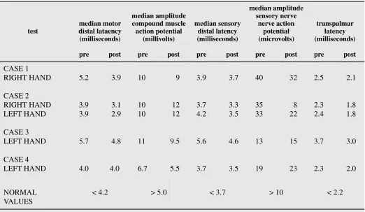
centimeters proximal at the wrist. Sensory studies used digits two and five and were recorded antidromically. Transpalmar recordings were made by stimulating at mid-palm and recording eight centimeters proximally from the wrist. Abductor pollicis brevis and the first dor-sal interosseous muscles were recorded with monopolar needle electromyography. All electrophysiological stud-ies were done on the Neuromax EMG System from Excel Tech Limited (a). All neurophysiological tests were per-formed initially and repeated three months later. (Table 1) All clinical tests were done prior to treatment and repeat-ed at three months and one year.
Once it had been established that there was no evi-dence of acute or chronic denervation, the patient was started on the machine-assisted wrist traction. The device (b) applies a controlled traction force of forty to sixty pounds per square inch (30–50 foot pounds) along the axis of the forearm. The patient is seated next to the trac-tion device and the elbow of the affected wrist is posi-tioned in an upholstered mobile bracket on top of the unit. Once positioned, the patient’s elbow is wrapped above and below the joint with velcro straps. The wrist is then secured with its own strap. The wrist strap is itself attached by two six inch velcro straps to a mobile T-bar which is part of the machine’s pneumatic activator. The pneumatic activator moves in a linear track propelled by the machine’s air compression system. When all three straps have been secured, the elbow bracket is fixed to the machine. The immobilized elbow is now the fixed point during wrist traction. We followed a standardized appli-cation derived from the only published single case study to utilize this machine.28 Upon activation, the T-bar gently tractions the wrist for five seconds followed by a five second rest interval. This intermittent cycle of trac-tion and rest is repeated thirty times over a five minute period. The T-bar is set in neutral alignment for the first ten cycles. It is then angled to traction in a pronated posi-tion for ten cycles and finally in a supinated posiposi-tion for ten cycles. (Figure 1)
Results
Case 1
employed making lipstick moulds at a cosmetics factory for thirteen years. The work was repetitive.
Examination revealed normal strength with no evi-dence of muscle wasting. Both hands felt cold to the touch. There was reduction to light touch and pin prick in a median nerve distribution. Before treatment, this pa-tient’s motor and sensory conduction velocities were sig-nificantly prolonged for the right median nerve at 5.2 and 3.9 milliseconds, respectively. The transpalmar latency was also delayed to 2.5 milliseconds. Evoked compound muscle action potential amplitudes were normal as were tests for the ulnar nerve.
After five traction treatments within a three week
peri-od, the motor distal latency improved to 3.9 milliseconds and the transpalmar latency measured 2.1 milliseconds. There was still some borderline delay in the sensory dis-tal latency (Table 1). The patient is working at modifed duties as a receptionist because she is still at risk for re-currence but she no longer has any numbness, tingling or pain.
Case 2
This forty-six-year-old financial administrator had hand numbness and pain for eighteen months. Most of her work involved writing, and typing on an adding machine or computer. The main complaints were pain in the wrists and hands, radiating into the thumbs. She would often wake up at night with numb hands. Indocid (indometh-acin) did not provide any relief.
Examination revealed evidence of decreased sensation to light touch and pin prick in a median nerve distribu-tion, as well as positive Tinel’s sign at the wrist and Pha-len’s test, bilaterally. There appeared to be no muscle wasting nor any decrease in muscle strength. This patient had been assessed by her personal medical physician and provided ultrasound and radiographic information. Diag-nostic ultrasound revealed mild fluid accumulation with-in the tendon sheaths bilaterally but no solid or cystic abnormality. Bony spurs on the antero-inferior margins of the third through sixth cervical vertebrae were evident on the cervical spine radiographs suggestive of mild de-generative joint disease but no evidence to suggest tho-racic outlet syndrome or double crush.29
The initial nerve conduction studies identified bilater-ally prolonged median nerve sensory distal latencies of 3.71 milliseconds on the right and 4.15 milliseconds on the left. Transpalmar latencies were 2.31 milleseconds for the right and 2.41 milliseconds for the left hand. The patient underwent twelve wrist traction sessions over a two month period. All nerve conduction values returned to normal limits. This patient’s wrists were less tender than previously and she enjoyed good ranges of wrist mo-tion and hand sensamo-tion (Table 1).
Case 3
This fifty-one-year-old teaching nurse had previous right carpal tunnel surgery which proved to be successful. Over the past year she has had increasing numbness and discomfort on the left, non-dominant hand. It disturbed
her sleep and interfered with simple tasks like holding a book. She did not do any repetitive activity.
This patient has a history of hypo-thyroidism which has been well controlled with Eltroxin (levothyroxineso-dium). There was no other pertinent medical history. Ex-amination revealed numbness to light touch and pin prick in a median nerve distribution on the left. Tinel’s sign at the wrist was negative but Phalen’s test was positive. There was no muscle wasting and her hand strength was Grade 5/5. This patient received twelve sessions of mech-anized intermittent wrist traction over a three month peri-od. She no longer suffered with nocturnal pain or awakening and she felt that her hands felt normal and of equal strength. The motor nerve conduction improved from 5.7 milliseconds to 4.8 milleseconds and the
senso-ry conduction went from 5.6 milliseconds to 4.6 millisec-onds (Table 1). Surgery was avoided.
Case 4
A thirty-six year old left-handed school teacher presented with one year of numbness, tingling and pain in the right hand. She had tried a night splint but found it to be too uncomfortable. Examination revealed a positive Phalen’s test and Tinel’s sign at the right wrist. There was reduc-tion of sensareduc-tion to light touch and pin prick in a median nerve distribution over the right wrist and hand. Her strength was intact. The electro-diagnostic studies identi-fied minor prolongations for the right median nerve distal latencies and transpalmar distal latency but the patient was quite symptomatic (Table 1).
Table 1
Pre and post electromyographic results
test
median motor distal lataency (milliseconds)
pre post
median amplitude compound muscle action potential
(millivolts)
pre post
median sensory distal latency (milliseconds)
pre post
median amplitude sensory nerve
nerve action potential (microvolts)
pre post
transpalmar latency (milliseconds)
pre post
CASE 1 RIGHT HAND
CASE 2 RIGHT HAND LEFT HAND
CASE 3 LEFT HAND
CASE 4 LEFT HAND
5.2 3.9
3.9 3.1 3.9 2.9
5.7 4.8
4.0 4.0
10 9
10 12
10 12
11 9.5 6.7 5.5 3.9 3.7 3.7 3.3 4.2 3.5 5.6 4.6 3.7 3.5 40 32
35 8
33 22
13 15
19 23
2.5 2.1
2.3 1.8 2.4 1.8
3.7 3.0
2.3 2.0
NORMAL VALUES
< 4.2 > 5.0 < 3.7 > 10 < 2.2
After three months and 12 sessions of wrist traction, she stated that she no longer awoke at night with tingling and she had no paraesthesiae, numbness or pain during the day.
Discussion
In many patients with carpal tunnel syndrome, the elec-trodiagnostic changes are mild, even though the symp-toms may be quite significant and severe.30 Some studies of conservative treatment report subjective relief as well as improvement in sensory and motor nerve conduction velocities.31 However, many of these patients have their symptoms recur and relatively few individuals enjoy long term relief. This new method of treatment employs a me-chanical means of long-axis traction which we speculate influences the local mobility enough to affect the intrac-arpal canal pressure and thereby allow for nerve recovery and reversal of nerve ischemia.
In this case series we observed subjective improvement in all cases as well as objective changes and significant normalization of some nerve conduction studies. It is not known how long these positive effects will be maintained but after two years of follow-up, none of these patients have required repeat traction or surgery. We are unable to explain with this sample size the significance of sympto-matic improvement despite continued abnormalities in the electrophysiological data. Many unanswered ques-tions remain regarding recurrence rates after surgery and conservative therapy.32 There is currently no gold stand-ard for diagnosing carpal tunnel syndrome.33 The natural history has not been adequately established in a large prospective study; so a combination of electrophysiologi-cal and clinielectrophysiologi-cal parameters were used to establish guide-lines for comparison in this study. These results apply only to the clinical diagnosis of carpal tunnel syndrome without evidence of weakness, atrophy or urgency for surgical decompressive surgery.
Future studies should consider selecting cohorts of asymptomatic and symptomatic subjects, stratifying ac-cording to age, sex, hand temperature and anthropomor-phic measurements recorded bilaterally.34
A large sample, randomized controlled clinical trial with comparison to conservative treatment and sham traction would be useful in evaluating this new and prom-ising method of non-invasive conservative therapy for carpal tunnel syndrome.
References
1 Stevens JC, Sun S, Beard CM, O’Fallon WM, Kurland LT. Carpal tunnel syndrome in Rochester, Minnesota, 1961 to 1980. Neurology 1988; 38:134–138.
2 Franklin GM, Haug J, Heyer N, Checkowey H, Peck N. Occupational carpal tunnel syndrome in Washington State, 1984–1988. Am J Publ Health 1991; 81:741–746.
3 Atroshi I, Gummersson C, Johnsson R, Ornstein C, Ranstam J, Rosen I. Prevalence of carpal tunnel syndrome in a general population. JAMA 1999; 282:153–158. 4 Hanrahan LP, Higgins D, Anderson H, Haskins L, Tai S.
Project sensor: Wisconsin Surveillance of occupational carpal tunnel syndrome. Wis Med J 1991; 90(2):80, 82–83. 5 DeKrom MCTFM, Knipschild PG, Kester ADM, Thijs CT,
Boekkooi PF, Spans F. Carpal Tunnel Syndrome: prevalence in the general population. J Clin Epidemiol 1992; 45(4):373–376.
6 Pecina MM, Krmpotic’-Nemanic’ J, Markiewitz. Tunnel syndromes. Boca Raton: CRC Press, 1991: 55–67. 7 Heywood PL. Through the carpal tunnel. BMJ 1987;
294:660–661.
8 Franzblau A, Werner RA. What is carpal tunnel syndrome? JAMA 1999; 282(2):186–187.
9 Szabo RM, Madison M. Carpal tunnel syndrome. Orthop Clin NA 1992; 23(1):103–109.
10 Werner RA, Armstrong TJ. Carpal tunnel syndrome – ergonomic risk factors and intracarpal canal pressure in carpal tunnel syndrome. Phys Med Rehabil Clin North Am 1997; 8(3):555–567.
11 Simpson RL, Fern SA. Multiple compression neuropathies and the double-crush syndrome. Orthop Clin North Am 1996; 27:381–388.
12 Scholten RJPM, Gerritsen AAM, Uitdehaag BMJ, Van Geldere D, DeVet HCU, Bouter LM. Surgical treatment options for carpal tunnel syndrome (Cochrane Review). In: The Cochrane Library 2, 2003. Oxford: Update Software. 13 Harter BT Jr, McKiernan JE Jr., Kirzinger SS, Archer
FW, Peters CK, Harter CH. Carpal tunnel syndrome : surgical and nonsurgical treatment. J Hand Surg 1993; 18A:734–739.
14 O’Connor D, Marshall S, Massy-Westropp N. Non-surgical treatement (other than steroid injection) for carpal tunnel syndrome (Cochrane Review). In: The Cochrane Library, Issue 3, 2003. Oxford: Update Software.
15 Ebenbichler GR, Resch KL, Nicolakis P, Wiesinger GF, Uhl F, Ghanem A, Fialka V. Ultrasound treatment for carpal tunnel syndrome: randomized ‘sham’ controlled trial. BMJ 1998; 316:731–735.
17 Davis PT, Hulbert JR, Kassak KM, Meyer JJ. Comparative efficacy of conservative medical and chiropractic
treatments for carpal tunnel syndrome: a randomized clinical trial. JMPT 1998; 21:317–326.
18 Davis PT, Hulbert JR. Carpal tunnel syndrome :
conservative and nonconservative treatment. A chiropractic physician’s perspective. J Manip Physiol Ther 1998; 21(5):356–362.
19 Tal-Akabi A, Rushton A. An investigation to compare the effectiveness of carpal bone mobilization and neurodynamic mobilization as methods of treatment for carpal tunnel syndrome. Manual Therapy 2000; 5:214–222. 20 Manente G, Torrieri F, Pineto F, Uncini A. A relief
maneuver in carpal tunnel syndrome. Muscle Nerve 1999; 22:1587–1589.
21 Garfinkel MS, Singhal A, Katz WA, Allan DA, Reshetar R, Schumacher HR Jr. Yoga-based intervention for carpal tunnel syndrome: a randomized trial. JAMA 1998; 280:1601–1603.
22 Walker WC, Metzler M, Cifu DX, Swartz Z. Neutral wrist splinting in carpal tunnel syndrome: a comparison of night-only versus full-time wear instructions. Arch Phy Med Rehabil 2000; 81:424–429.
23 Krueger VL, Kraft GH, Deitz JC, Ameis J, Polissar L. Carpal tunnel syndrome : objective measures and splint use. Arch Phys Med Rehabil 1991; 72:517–520. 24 Levine DW, Simmons BP, Koris MJ, Daltroy LH, Hohi
GG, Fossel AH, Katz JN. A self-administered
questionnaire for the assessment of severity of symptoms and functional status in carpal tunnel syndrome. J Bone Joint Surg 1993; 75–A(11):1585–1591.
25 Homan MM, Franzblau A, Werner RA, Albers JW, Armstrong TJ, Bromberg MB. Agreement between symptom surveys, physical examination procedures and electrodiagnostic findings for the carpal tunnel syndrome. Scand J Work Environ Health 1999; 25(2):115–124. 26 Stevens JC.. AAEM minimonograph #26: The
electrodiagnosis of carpal tunnel syndrome. Muscle Nerve 1997; 20:1477–1486.
27 American Association of Electrodiagnostic Medicine. Practice parameters for electrodiagnostic studies in carpal tunnel syndrome: summary statement. Muscle Nerve 1993; 16:1390–1391.
28 Petruska, G. Carpal tunnel syndrome: a new perspective that blends active and passive care. Sports Chiropractic and Rehabilitation 1997; 11(2):57–60.
29 Upton ARM, McComas AJ. The double crush in nerve-entrapment syndromes. Lancet 1973; 359–362.
30 Concannon MJ, Gainer B, Petroski GF, Puckett CL. The predictive value of electrodiagnostic studies in carpal tunnel syndrome. Plast Reconstr Surg 1997; 100:1452–1458.
31 Dawson DM. Entrapment neuropathies of the upper extremities. N Engl J Med 1993; 329:2013–2018. 32 Werner RA, Franzblau A, Albers JW, Buchele H,
Armstrong TJ. Use of screening nerve conduction studies for predicting future carpal tunnel syndrome. Occup Environ Med 1997; 54:96–100.
33 Rempel D, Evanoff B, Amadio PC, de Krom M, Franklin G, Franzblau A, Gray R, Gerr F, Hagberg M, Hales T, Katz JN, Pransky G. Consensus criteria for the classification of carpal tunnel syndrome in epidemiological studies. Am J Public Health 1998; 88:1447–1451.
34 Salerno DF, Salerno MS, Franzblau A, Werner RA, Bromberg MB, Armstrong TJ, Albers JW. Median and ulnar nerve conduction studies among workers: normative values. Muscle and Nerve 1998; 21:999–1005.
Suppliers
a Excel Tech Limited. XLTEK 2568 Bristol Circle, Oakville, Ontario, Canada L6H 5S1