The Impact of Physical Activity on
Brain Structure and Function in Youth:
A Systematic Review
Sarah Ruth Valkenborghs, PhD,aMichael Noetel, PhD,bCharles H. Hillman, PhD,c,dMichael Nilsson, PhD,eJordan J. Smith, PhD,a Francisco B. Ortega, PhD,fDavid Revalds Lubans, PhDa
abstract
CONTEXT:Advances in neuroimaging techniques have resulted in an exponential increase in thenumber of studies investigating the effects of physical activity on brain structure and function. Authors of studies have linked physical activity andfitness with brain regions and networks integral to cognitive function and scholastic performance in children and adolescents but findings have not been synthesized.
OBJECTIVE:To conduct a systematic review of studies in which the impact of physical activity on brain structure and function in children and adolescents is examined.
DATA SOURCES:Six electronic databases (PubMed, PsychINFO, Scopus, Ovid Medline, SportDiscus, and Embase) were systematically searched for experimental studies published between 2002 and March 1, 2019.
STUDY SELECTION:Two reviewers independently screened studies for inclusion according to predetermined criteria.
DATA EXTRACTION:Two reviewers independently extracted data for key variables and synthesized
findings qualitatively.
RESULTS:Nine studies were included (task-based functional MRI [n= 4], diffusion tensor imaging [n= 3], arterial spin labeling [n= 1], and resting-state functional MRI [n= 1]) in which results for 5 distinct and 4 similar study samples aged 8.76 0.6 to 10.261.0 years and typically of relatively low socioeconomic status were reported. Effects were reported for 12 regions, including frontal lobe (n= 3), parietal lobe (n= 3), anterior cingulate cortex (n= 2), hippocampus (n= 1), and several white matter tracts and functional networks.
LIMITATIONS:Findings need to be interpreted with caution as quantitative syntheses were not possible because of study heterogeneity.
CONCLUSIONS:There is evidence from randomized controlled trials that participation in physical activity may modify white matter integrity and activation of regions key to cognitive processes. Additional larger hypothesis-driven studies are needed to replicatefindings.
aPriority Research Centre for Physical Activity and Nutrition, University of Newcastle, University Drive, Callaghan, New South Wales, Australia;bFaculty of Health Sciences, School of
Behavioural and Health Sciences, Australian Catholic University, Banyo, Queensland, Australia;cDepartments of Psychology anddPhysical Therapy, Movement, and Rehabilitation Sciences,
Northeastern University, Boston, Massachusetts;eCentre for Rehab Innovations, University of Newcastle and Hunter Medical Research Institute, New Lambton Heights, New South Wales, Australia; andfDepartment of Physical Education and Sports, Faculty of Sports Sciences, University of Granada, Granada, Spain
Dr Valkenborghs conducted the search, screening, extraction, and synthesis processes in addition to drafting the manuscript; Dr Noetel screened articles, extracted data, and critically reviewed the manuscript; Drs Hillman, Nilsson, and Smith contributed to the conceptualization of the review and critically reviewed (Continued)
Many children and adolescents are not sufficiently active to accrue the extensive cardiovascular, metabolic, musculoskeletal, and mental health benefits of physical activity.1,2
Habitual physical activity is associated with a variety of health-relatedfitness traits (ie,
cardiorespiratory, morphologic, muscular, motor, and metabolic),3and emerging evidence suggests that participation in physical activity and improving physicalfitness may enhance cognitive health across the life span.4–9
Specifically, acute physical activity can enhance children’s attention (g = 0.43; 95% confidence interval [CI] = 0.09–0.77) and on-task behavior in the classroom (d = 0.77; 95% CI = 0.22–1.32).10–12Similarly, authors of experimental studies have demonstrated longer-term benefits of physical activity for executive functions (g = 0.24; 95% CI = 0.09–0.39),11attention (g= 0.90; 95% CI = 0.56–1.24),11and academic performance (g= 0.26; 95% CI = 0.02–0.49).5,11,13Higher levels of cardiorespiratoryfitness are also positively associated with young people’s academic achievement.13 Although awareness of the positive effects of physical activity on cognitive and/or academic outcomes has increased rapidly in the last 5 years, the mechanisms responsible remain relatively untested in young people.14
Animal studies have provided initial insight into the neurobiological changes induced by physical activity. Molecular effects include epigenetic regulation of gene expression and related changes in concentrations of factors such as brain-derived neurotrophic factor (BDNF) and vascular endothelial growth factor, known to underpin brain plasticity and cellular changes such as neurogenesis, synaptogenesis, and angiogenesis.15–19There is now empirical evidence that the same molecular effects exist in humans (eg,
increases in BDNF and vascular endothelial growth factor) and may be responsible for the positive effects of physical activity on cognitive health.20–23
In addition, a seminal randomized controlled trial (RCT) in older adults demonstrated that 12 months of aerobic exercise increased
hippocampal volume and improved memory, with these improvements being mediated by increases in BDNF.24Since the publication of these findings, there has been an
exponential increase in the number of studies employing MRI techniques to examine associations and explore the impact of physical activity on brain structure and function in humans. Authors of many cross-sectional studies have linked physical activity with brain regions and networks integral to cognitive function and scholastic performance in children and adolescents.25–28
To date, there has been no systematic review of experimental MRI studies in which the impact of physical activity on brain structure and function in children and adolescents is investigated. A recent review of 84 studies in which the effects of physical activity on cognitive functioning and neuroimaging findings were investigated only included 5 MRI studies because the search was conducted in July 2017 and it only included RCTs.29To provide a more in-depth and up-to-date summary of evidence of MRI studies specifically, our review included all designs of experimental studies. Given the importance of cognitive development, clarifying the effects of physical activity on brain structure and function may motivate key stakeholders to address the current physical inactivity pandemic. Therefore, our aim with this study was to conduct a systematic review of MRI studies in which the impact of physical activity on brain structure and function in school-aged children have been examined.
METHODS
The conduct and reporting of this review adhere to the guidelines outlined in the Preferred Reporting Items for Systematic Reviews and Meta-Analysis statement.30The review protocol was registered with the International Prospective Register of Systematic Reviews
(CRD42017081804).
Study Eligibility Criteria
1. Types of participants: participants were typically developing school-aged children (usually 5–18 years of age; however, children outside this age range were included if they were recruited within schools). Studies including populations with learning difficulties, cognitive deficits, and developmental disorders were excluded.
2. Types of studies: experimental studies were eligible if the authors reported statistical analyses of changes in brain structure or function before and after a physical activity intervention.
3. Measure of physical activity, cardiorespiratoryfitness, or muscularfitness: studies with objective (eg, accelerometers and pedometers) or subjective measures of physical activity (eg, exercise session attendance and self-report questionnaires); cardiorespiratoryfitness (eg, maximum oxygen consumption [VO2max] test, Progressive Aerobic
Cardiovascular Endurance Run, and predictive equations); and/or muscularfitness (eg,
dynamometry, standing long jump, and push up test) were eligible.
mechanisms that may explain the relationship between physical activity, cardiorespiratoryfitness or muscularfitness, and cognition or academic achievement were eligible.
Information Sources and Search Strategy
Six electronic databases (PubMed, PsychINFO, Scopus, Ovid Medline, SportDiscus, and Embase) were searched for studies published within the last 16 years (2002–March 1, 2019) (Supplemental Table 4). Additional searches of recently published systematic reviews in which the associations between physical activity, cardiorespiratory fitness or muscularfitness, and cognitive outcomes were examined were conducted, and the reference lists of all retrieved studies were reviewed. The search was restricted to articles published in the English language.
Study Selection
The study screening and selection process was performed on
Covidence.31One reviewer screened the titles and abstracts of records retrieved by the search strategy and classified these as possibly relevant or definitely irrelevant. The full-text articles of records classified as possibly relevant were retrieved and independently reviewed by 2 reviewers. Studies were classified as include or exclude. If there was disagreement between reviewers, consensus was sought through discussion. Reasons were provided for excluding any possibly relevant studies.
Data Extraction
Both reviewers independently extracted data from included studies into a purpose-built data extraction template in excel. Data extraction included (1) sample data (including sample size, age, sex, and education); (2) study details (location, design, setting, duration, and assessment
points); (3) assessment of physical activity, cardiorespiratory, and/or muscularfitness (objective,
subjective, laboratory-based, orfi eld-based); (4) neuroimaging data (MRI modality, analysis methods, regions of interest, etc); (5) data analysis (statistical methods used,
confounders adjusted for, etc); and (6) studyfindings (quantitative and qualitative).
Risk of Bias Assessment
All studies were independently assessed by 2 reviewers and were scored as low, high, or unclear for 8 criteria according to the Cochrane collaboration risk of bias tool and scoring.32Any disagreement concerning risk of bias assessment between the 2 reviewers was resolved through discussion.
Consensus was reached on all articles included in the review.
RESULTS
Overview of Studies
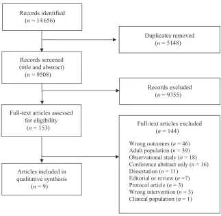
Figure 1 displays theflow of studies through the review process. After the exclusion of duplicates, the
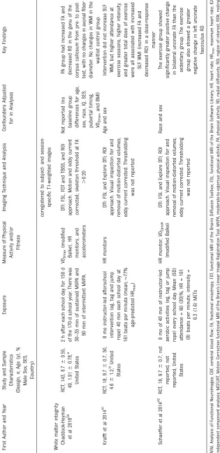
systematic search yielded 9508 potentially relevant citations, of which 153 were retained for full-text review. There was almost-perfect interrater agreement for the full-text review (k= 0.97).33A total of 9 articles satisfied the inclusion criteria and were included in the review, reporting results for 5 distinct and 4 similar samples of participants ranging from 8.760.634to 10.26 1.035years of age and typically of relatively low socioeconomic status. The sample size ranged from 936to 143.34The studies were conducted in North America (8 in the United States) and Asia (1 in China). Of the included studies, 7 were RCTs and 2 were acute before and after studies. Detailed information about each included study is presented in Table 1.
Risk of Bias
Detailed information about the risk of bias for the included studies is presented in Table 2. In summary, all 9 (100%) were deemed to be at unclear risk of selection bias, with unclear description of (1) sequence generation process, (2) concealed allocation processes, and [in 5 (56%) studies] (3) subgroup selection processes. Seven (78%) studies were deemed at unclear risk of reporting bias because of lack of availability of a protocol published by means of either an article or trial registration. Six (67%) studies were deemed at high risk of attrition bias because of significant dropout with inadequate analyses. Overall, only 2 (22%) studies scored as low risk of bias for $3 (of the 8 criteria.34,35There was substantial interrater agreement for the risk of bias assessment (k= 0.61).33
Measures of Brain Structure and Function
Four different MRI modalities were used across the 9 included studies. Four (44%) studies used task-based fMRI, 3 (33%) studies used DTI, 1 (11%) study used ASL, and 1 (11%) study used resting-state fMRI. Data for 12 regions were reported across the 9 included studies: anterior cingulate cortex, cerebellum, corpus callosum, frontal lobe, hippocampus, parietal lobe, superior longitudinal fasciculus, uncinate fasciculus, cognitive control network, default mode network, executive control network, and motor network.
Measures of Physical Activity and Fitness
Authors of seven (78%) studies provided physical activity
[HRmax]) either twice a week or each
school day for 20 to 120 minutes. Of these, 4 studies measured
cardiorespiratoryfitness by means of oxygen uptake during a maximal graded treadmill test (modified Balke protocol).34,37,39,42Authors of 2 studies investigated changes in brain function in response to acute bouts of aerobic exercise at 60% to 70% HRmax.35,36Details of all interventions
are outlined in Table 1.
The Impact of Physical Activity on Brain Structure or Function
Findings from each included study are presented by brain region below and Table 1, with effects further summarized in Table 3.
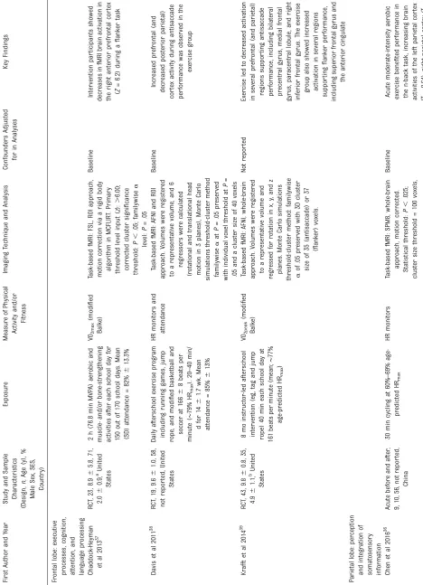
Frontal Lobe
Authors of 3 RCTs with distinct but similarly aged samples reported results for changes in activation of the frontal lobe in response to physical
activity interventions which ranged from 20 to 77 minutes each school day over 3 to 9 months. Authors of 2 of the RCTs assessed prefrontal activation during cognitive tasks (antisaccade [n= 2] andflanker [n= 2]) and found changes pre- and post- intervention but the effects were in opposite directions in both cases. Davis et al38reported that increased bilateral prefrontal (and decreased posterior parietal) cortex activity was observed during antisaccade performance in the physical activity group, whereas Krafft et al39reported decreased activation during antisaccade performance in several prefrontal (and parietal) regions including medial frontal gyrus, right inferior frontal gyrus, and bilateral precentral gyrus. Krafft et al39 also observed increased activation of the superior frontal gyrus of the prefrontal cortex during incongruent trials of theflanker task in the
physical activity group, whereas authors of the third RCT (Chaddock-Heyman et al37) observed decreased activation in the right anterior prefrontal cortex during incongruent trials of theflanker task in the physical activity intervention group but no changes in the control group. Note that although both Chaddock-Heyman et al37and Davis et al38 adjusted for baseline during their region of interest analyses, Krafft et al39did not report if/what covariates were adjusted for and employed a whole-brain analysis approach, which could contribute to the disparate results.
Parietal Lobe
Authors of 3 studies reported results for the parietal lobe from task-based fMRI paradigms. Authors of 2 RCTs with similarly aged samples and relatively similar type, frequency, intensity, and duration of physical activity interventions found decreased parietal cortex activity during antisaccade performance after a physical activity intervention.38,39 Both studies used comparable cluster size thresholds but it should be noted that while Davis et al38adjusted for baseline in their analyses, Krafft et al39did not report if and what covariates were adjusted for.
Chen et al36investigated the acute effects of a 30-minute bout of cycling (60%–69% HRmax) during task-based
fMRI and reported improved n-back performance and increased activation of bilateral parietal cortices (as well as the left hippocampus and bilateral cerebellum).
Anterior Cingulate Cortex
Authors of 2 RCTs reported task-based fMRI results for the anterior cingulate cortex. Authors of 1 RCT found that participation in a physical activity intervention did not change activation of anterior cingulate cortex during neutral or incongruent conditions of aflanker task.37The other RCT found that although there FIGURE 1
were no significant correlations between changes in cardiorespiratory fitness and brain activation during task-based fMRI,39the physical activity intervention led to differential activation across 2 inhibition tasks, with decreased activation of the anterior cingulate cortex during an antisaccade task and increased activation of the cingulate gyrus during the incongruent condition of aflanker task.39 Comparatively, the control group
showed decreased activation during theflanker task.39Such differences across inhibition tasks highlights the complexity of brain activation during performance of tasks that tap different aspects of a similar cognitive construct.
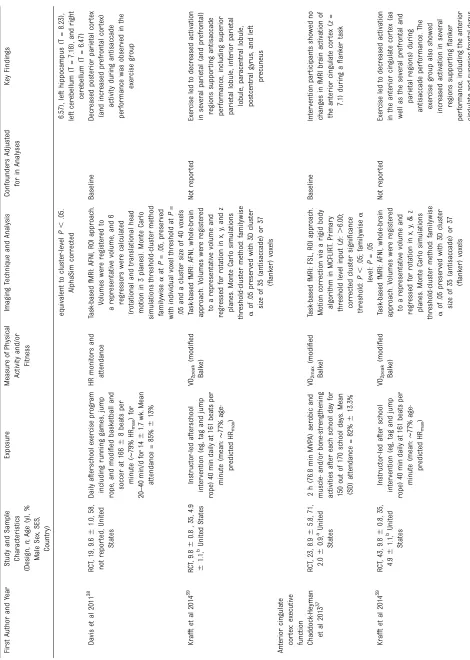
Hippocampus
Authors of 1 acute before and after study reported enhanced
performance in an n-back task and increased brain activity (task-based
fMRI) of the left hippocampus in response to an acute 30-minute bout of cycling (60%–69% HRmax).36
Cerebellum
Authors of 1 acute experimental study investigated the effects of a 30-minute bout of cycling (60%–69% HRmax) during
task-based fMRI and reported
improved n-back performance and increased activation of bilateral cerebellum.36
Functional Networks
Authors of 2 experimental studies reported results for specific functional brain networks. Authors of 1 RCT used an independent component analysis approach and reported that a physical activity intervention caused decreased synchrony between the default mode network and the cognitive control network with brain regions outside of those networks during resting-state fMRI.40 There was no change in synchrony of the salience network, whereas the motor network had decreased synchrony with the left cuneus but increased synchrony with certain frontal regions.40
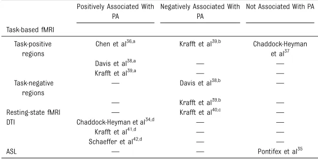
TABLE 3Summary of Studies Which Have Examined the Impact of Physical Activity on Brain Structure or Brain Function
Positively Associated With PA
Negatively Associated With PA
Not Associated With PA
Task-based fMRI
Task-positive regions
Chen et al36,a Krafft et al39,b Chaddock-Heyman et al37
Davis et al38,a — —
Krafft et al39,a — —
Task-negative regions
— Davis et al38,b —
— Krafft et al39,b —
Resting-state fMRI — Krafft et al40,c —
DTI Chaddock-Heyman et al34,d — —
Krafft et al41,d — —
Schaeffer et al42,d — —
ASL — — Pontifex et al35
PA, physical activity;—, not applicable.
aIncreased activation. bDecreased activation.
cDecreased synchrony of resting-state networks with regions outside those networks. dIncreased white matter integrity.
TABLE 2Risk of Bias Assessment
Study Sequence
Generation
Allocation Concealment
Participant Blinding
Assessor Blinding
Personnel Blinding
Selective Outcome Reporting
Incomplete Outcome Data
Other Sources of Bias
Chaddock-Heyman et al34
?a ?a ?a 1 1 1 2b 1
Chaddock-Heyman et al37
?a ?a ?a ?a ?a 1 2b 1
Chen et al36 2 2 2 2 2c ?d ? ?a
Davis et al38 ?a ?a 2 1 2c ?d 1 2e
Krafft et al39 ?a ?a ?a ?a ?a ?d 2b 2e,f
Krafft et al41 ?a ?a ?a ?a ?a ?d 2b 2e,f
Krafft et al40 ?a ?a ?a ?a ?a ?d 2b 2e,f
Pontifex et al35 ?a ?a 1 ?a 1 ?d 2b 1
Schaeffer et al42 ?a ?a ?a ?a ?a ?c 1 2e,f
1represents low risk of bias, ? represents unclear risk of bias, and2represents high risk of bias.
aUnclear description in article.
bSignificant dropout with inadequate analyses. cAuthors appeared to provide intervention and control. dNo protocol.
Pontifex et al35investigated the acute effects of a 20-minute bout of fast walking and/or slow jogging (70% HRmax) on cerebral bloodflow in
10.261.0 year old (n= 41) and found no differences across any of the networks examined (frontoparietal, executive control, and motor networks).
White Matter Integrity
Authors of 3 studies reported results of 2 RCTs that had examined the effects of physical activity on white matter tracts in similarly aged children using regions of
interest analyses.34,41,42One large RCT (n= 143) revealed that 2 hours of physical activity each school day for 8 months improved white matter integrity (ie, increased fractional anisotropy, which indicates the orientation of diffusion and is higher along well-defined pathways) and decreased radial diffusivity (a marker of myelin disintegration) in the genu of the corpus callosum from pretest to post-test, with no changes in estimates of axonalfiber diameter (axial diffusivity).34There were no changes in the white matter integrity of the wait list control group, reflective of typical development. The other RCT (n= 18) also delivered an 8-month intervention consisting of a 40-minute session each school day. Authors of 1 study reported that the physical activity group showed greater increases in bilateral uncinate fasciculus fractional anisotropy and greater decreases in left uncinate fasciculus radial diffusivity compared with the control group.42In the second report from this RCT, the physical activity intervention did not significantly increase white matter integrity in the superior longitudinal fasciculus. However, higher
attendance in the exercise intervention, higher intensity, and greater total dose of exercise were all associated with increased fractional anisotropy and decreased radial diffusivity of the superior longitudinal
fasciculus in a dose-response manner.41
DISCUSSION
In this systematic review, we examined evidence of the impact of physical activity on brain structure and function in youth from MRI studies. Nine experimental studies were included in the review, of which 7 were RCTs and 2 were acute before and after studies, reporting data for 12 regions acquired with 4 MRI modalities. All 7 RCT studies (4 samples) reported significant changes in either brain structure or function after a physical activity intervention in young people.37–42
To date, the parietal cortex is the only specific region that has had.1 RCT report in which authors found an impact of physical activity on brain structure or function and for the effects to be in the same direction (ie, authors of both RCTs found
decreased posterior parietal cortex activity during antisaccade
performance after a physical activity intervention38,39). Otherwise, RCT findings for the impact of physical activity on activation during task-based fMRI were inconsistent (ie, authors of 1 study found an association and another did not) for the anterior cingulate cortex,37,39or conflicting (ie, physical activity had an impact on activation, but authors of 1 study reported increased activation and authors of another study reported a decreased activation in the case of each task paradigm
[antisaccade and incongruent condition of aflanker task]) for frontal regions.37–39It should be noted that although the sample ages and cluster size thresholds were similar in these studies, the interventions varied from 20 to 77 minutes per session over 3 to 9 months which presents considerable heterogeneity.
The desired direction of the effect of physical activity on activation will
differ depending on the region and context (eg, task and rest) of interest. However, positive associations between physical activity and activation of task-positive regions during performance of task paradigms is interpreted as a greater ability to use resources in some studies,36,43,44whereas negative associations (ie, less activation) are considered to represent a more efficient use of resources in others.37,45There is evidence to support decreased activation of a task-positive region during task performance being reflective of a more mature and adult-like brain46–48but this should be interpreted with caution until thefindings have been replicated by studies adequately powered to perform mediation analyses.49
Authors of 2 RCTs found that physical activity caused decreased activation of the posterior parietal cortex during antisaccade task performance. Although this did not reflect a difference in antisaccade performance between the physical activity and control group in 1 study,39authors of the other study did not report data for antisaccade task performance.38The inferior parietal lobule, located within the posterior parietal cortex, forms part of the default mode (task-negative) network,50–52which is known to decouple from the cognitive control network during successful
demonstrated in a cross-sectional pediatric physical activity study. Despite similar memory performance to their inactive peers, during encoding of later remembered versus forgotten word pairs, participants with high levels of physical activity displayed (1) robust deactivation of the default mode network, (2) strong negative coupling with the
hippocampus, and (3) a more focal increase in activation of the left hippocampus only.45
Decreased synchrony between a given network and regions outside of that network is usually an indication of a more focal, coherent, and specialized pattern of
activation.58,59Authors of 1 RCT in this review examined deactivation and activation of functional networks during resting-state fMRI and found that physical activity may be conducive of a more mature efficient brain by causing decreased synchrony of the default mode network and cognitive control network with brain regions outside of those networks during resting-state fMRI.40
In terms of structural changes, 1 large RCT (FITKids2;n= 143) revealed that participation in physical activity can improve white matter integrity of the corpus callosum; a region important for cognitive processing.34A second RCT investigated effects of physical activity on white matter integrity and detected significant improvements in the bilateral uncinate fasciculus (which usually matures later than many other tracts60).42This was particularly evident in the left uncinate fasciculus, which is linked with auditory-verbal memory proficiency, verbal IQ, and full-scale IQ.42,61,62
In a second study from the same RCT,41changes in white matter integrity of the superior longitudinal fasciculus were not significantly different between the groups. However, higher attendance at exercise sessions, higher intensity,
and greater total dose of exercise were positively associated with changes white matter integrity.41 Similarly, white matter integrity did not change among adults
participating in a 1-year exercise intervention, but changes in fitness were positively associated with white matter integrity of prefrontal and temporal regions (which are linked by the uncinate fasciculus).63 Improvements in fitness were also associated with changes in short-term memory, but increases in white matter integrity were not associated with short‐term memory improvement. In another larger-scale study involving adults, white matter integrity in multiple tracts (including those that connect medial temporal and prefrontal cortices) mediated the relationship betweenfitness and spatial working memory.64 Additional support for the importance offitness in terms of white matter integrity also exists in pediatric cross-sectional studies, which have found positive associations betweenfitness and fractional anisotropy in several of the same white matter tracts in children.65
Future Directions
To date, no RCT has examined the impact of a physical activity intervention on volumes of brain regions in children or adolescents. This is surprising given that a recent meta-analysis on the effect of aerobic exercise on hippocampal volume in adults included 14 studies.66This review revealed a significant effect of aerobic exercise on both left and right hippocampal volume in comparison with control conditions in healthy older adults. The effects were driven by exercise attenuating normal age-related neurodegeneration, which has been shown to precede and lead to cognitive decline and Alzheimer disease.67,68Whether exercise can increase the volumetric growth of the hippocampus and whether these increases in volume subsequently
confer benefits to cognition, memory, and/or academic performance during childhood and adolescence has not been established.
More studies in adolescents are needed because all experimental studies included in this review were conducted with children. Future researchers should also measure cardiorespiratory and muscular fitness so that (1) baselinefitness can be adjusted for in analyses and (2) changes infitness due to physical activity interventions can be analyzed for correlations with changes in brain structure or function. There is considerable scope for different intensities, frequencies, and types of physical activity such as high-intensity interval training, resistance exercise, exergaming, and cognitively demanding physical activity to be explored.69
Limitations
Although this is thefirst systematic review of MRI studies in the area of pediatric physical activity, there are some limitations that should be noted. Most notably, because of the small number of RCTs and
considerable heterogeneity of included studies, we were unable to conduct meta-analyses. In addition, we did not check for afile drawer effect so the risk of publication bias cannot be ruled out.
to adhere to the Consolidated Standards of Reporting Trials guidelines72to reduce the risk of bias, particularly in terms of selection bias and reporting bias.73 Findings need to be interpreted with caution until additional RCTs can (1) replicatefindings and (2) establish whether exercise-induced changes in brain structure or function mediate the cognitive and/or academic benefits of physical activity.
Conclusions
There is some evidence from RCTs that participation in physical activity may enhance brain structure and function in terms of white matter integrity and activation of regions key to cognitive processes, respectively. No RCT researchers have reported on the impact of physical activity on volumes of brain regions in children or adolescents.
ABBREVIATIONS
ASL: arterial spin labeling BDNF: brain-derived neurotrophic
factor
CI: confidence interval DTI: diffusion tensor imaging fMRI: functional MRI
HRmax: maximum heart rate
RCT: randomized controlled trial VO2max: maximum oxygen
consumption
the manuscript; Dr Ortega critically reviewed the manuscript; Dr Lubans conceptualized the review and contributed to the design, synthesis, and drafting of the manuscript; and all authors approved thefinal manuscript as submitted and agree to be accountable for all aspects of the work.
This trial has been registered with the International Prospective Register of Systematic Reviews (https://www.crd.york.ac.uk/prospero/) (identifier CRD42017081804).
DOI:https://doi.org/10.1542/peds.2018-4032
Accepted for publication Jul 16, 2019
Address correspondence to David Lubans, PhD, Priority Research Centre for Physical Activity and Nutrition, University of Newcastle, University Dr, Callaghan, NSW 2308, Australia. E-mail: [email protected]
PEDIATRICS (ISSN Numbers: Print, 0031-4005; Online, 1098-4275).
Copyright © 2019 by the American Academy of Pediatrics
FINANCIAL DISCLOSURE:The authors have indicated they have nofinancial relationships relevant to this article to disclose.
FUNDING:Supported by an Australian Research Council Future Fellowship grant (FT 140100399).
POTENTIAL CONFLICT OF INTEREST:The authors have indicated they have no potential conflicts of interest to disclose.
REFERENCES
1. Janssen I, Leblanc AG. Systematic
review of the health benefits of physical
activity andfitness in school-aged
children and youth.Int J Behav Nutr
Phys Act. 2010;7(1):40
2. Hallal PC, Andersen LB, Bull FC, et al; Lancet Physical Activity Series Working Group. Global physical activity levels: surveillance progress, pitfalls, and
prospects.Lancet. 2012;380(9838):
247–257
3. Lang JJ, Tomkinson GR, Janssen I, et al. Making a case for cardiorespiratory
fitness surveillance among children
and youth.Exerc Sport Sci Rev. 2018;
46(2):66–75
4. Biddle SJ, Asare M. Physical activity and mental health in children and
adolescents: a review of reviews.Br
J Sports Med. 2011;45(11):886–895
5. Esteban-Cornejo I, Tejero-Gonzalez CM, Sallis JF, Veiga OL. Physical activity and
cognition in adolescents: a systematic
review.J Sci Med Sport. 2015;18(5):
534–539
6. Donnelly JE, Hillman CH, Castelli D, et al.
Physical activity,fitness, cognitive
function, and academic achievement in
children: a systematic review.Med Sci
Sports Exerc. 2016;48(6):1197–1222
7. Ruiz-Ariza A, Grao-Cruces A, de Loureiro
NEM, Martínez-López EJ. Influence of
physicalfitness on cognitive and
academic performance in adolescents:
a systematic review from 2005–2015.
Int Rev Sport Exerc Psychol. 2017;10(1):
108–133
8. Costigan SA, Eather N, Plotnikoff RC, Hillman CH, Lubans DR. High-intensity interval training for cognitive and
mental health in adolescents.Med Sci
Sports Exerc. 2016;48(10):1985–1993
9. Lubans DR, Smith JJ, Morgan PJ, et al. Mediators of psychological well-being
in adolescent boys.J Adolesc Health.
2016;58(2):230–236
10. Álvarez-Bueno C, Pesce C, Cavero-Redondo I, et al. Academic achievement and physical activity: a meta-analysis. Pediatrics. 2017;140(6):e20171498
11. de Greeff JW, Bosker RJ, Oosterlaan J, Visscher C, Hartman E. Effects of physical activity on executive functions, attention and academic performance in preadolescent children: a
meta-analysis.J Sci Med Sport. 2018;21(5):
501–507
12. Daly-Smith AJ, Zwolinsky S, McKenna J, et al. Systematic review of acute physically active learning and classroom movement breaks on
children’s physical activity, cognition,
academic performance and classroom behaviour: understanding critical
design features.BMJ Open Sport Exerc
13. Marques A, Santos DA, Hillman CH, Sardinha LB. How does academic achievement relate to
cardiorespiratoryfitness, self-reported
physical activity and objectively reported physical activity: a systematic review in children and adolescents
aged 6-18 years.Br J Sports Med. 2018;
52(16):1039
14. Lubans D, Richards J, Hillman C, et al. Physical activity for cognitive and mental health in youth: a systematic
review of mechanisms.Pediatrics. 2016;
138(3):e20161642
15. Fernandes J, Arida RM, Gomez-Pinilla F. Physical exercise as an epigenetic modulator of brain plasticity and
cognition.Neurosci Biobehav Rev. 2017;
80:443–456
16. Cooper C, Moon HY, van Praag H. On the
run for hippocampal plasticity.Cold
Spring Harb Perspect Med. 2018;8(4): a029736
17. Lista I, Sorrentino G. Biological mechanisms of physical activity in
preventing cognitive decline.Cell Mol
Neurobiol. 2010;30(4):493–503
18. Vaynman S, Ying Z, Gomez-Pinilla F. Hippocampal BDNF mediates the
efficacy of exercise on synaptic
plasticity and cognition.Eur J Neurosci.
2004;20(10):2580–2590
19. Rich B, Scadeng M, Yamaguchi M,
Wagner PD, Breen EC. Skeletal myofiber
vascular endothelial growth factor is required for the exercise training-induced increase in dentate gyrus
neuronal precursor cells.J Physiol.
2017;595(17):5931–5943
20. Hashimoto T, Tsukamoto H, Takenaka S, et al. Maintained exercise-enhanced brain executive function related to cerebral lactate metabolism in men. FASEB J. 2018;32(3):1417–1427
21. Nascimento CM, Pereira JR, de Andrade LP, et al. Physical exercise in MCI elderly promotes reduction of
pro-inflammatory cytokines and
improvements on cognition and BDNF
peripheral levels.Curr Alzheimer Res.
2014;11(8):799–805
22. Leckie RL, Oberlin LE, Voss MW, et al. BDNF mediates improvements in executive function following a 1-year
exercise intervention.Front Hum
Neurosci. 2014;8:985
23. Voss MW, Erickson KI, Prakash RS, et al. Neurobiological markers of exercise-related brain plasticity in older adults. Brain Behav Immun. 2013;28:90–99
24. Erickson KI, Voss MW, Prakash RS, et al. Exercise training increases size of hippocampus and improves memory. Proc Natl Acad Sci USA. 2011;108(7):
3017–3022
25. Chaddock-Heyman L, Weng TB, Kienzler C, et al. Scholastic performance and functional connectivity of brain
networks in children.PLoS One. 2018;
13(1):e0190073
26. Talukdar T, Nikolaidis A, Zwilling CE,
et al. Aerobicfitness explains individual
differences in the functional brain connectome of healthy young adults. Cereb Cortex. 2018;28(10):3600–3609
27. Chaddock L, Erickson KI, Prakash RS, et al. A neuroimaging investigation of the association between aerobic
fitness, hippocampal volume, and
memory performance in preadolescent
children.Brain Res. 2010;1358:172–183
28. Bunketorp Käll L, Malmgren H, Olsson E, Lindén T, Nilsson M. Effects of a curricular physical activity
intervention on children’s school
performance, wellness, and brain
development.J Sch Health. 2015;85(10):
704–713
29. Gunnell KE, Poitras VJ, LeBlanc A, et al. Physical activity and brain structure, brain function, and cognition in children and youth: a systematic review
of randomized controlled trials.Ment
Health Phys Act. 2019;16:105–127
30. Liberati A, Altman DG, Tetzlaff J, et al. The PRISMA statement for reporting systematic reviews and meta-analyses of studies that evaluate health care interventions: explanation and
elaboration.Ann Intern Med. 2009;
151(4):W65–W94
31. Covidence. Veritas Health Innovation, Melbourne, Australia. Available at: www. covidence.org. Accessed August 16, 2019
32. Higgins JP, Altman DG, Gøtzsche PC, et al; Cochrane Bias Methods Group; Cochrane Statistical Methods Group.
The Cochrane Collaboration’s tool for
assessing risk of bias in randomised
trials.BMJ. 2011;343:d5928
33. Landis JR, Koch GG. The measurement of observer agreement for categorical
data.Biometrics. 1977;33(1):159–174
34. Chaddock-Heyman L, Erickson KI, Kienzler C, et al. Physical activity increases white matter microstructure
in children.Front Neurosci. 2018;
12(950):950
35. Pontifex MB, Gwizdala KL, Weng TB, Zhu
DC, Voss MW. Cerebral bloodflow is not
modulated following acute aerobic
exercise in preadolescent children.Int
J Psychophysiol. 2018;134:44–51
36. Chen AG, Zhu LN, Yan J, Yin HC. Neural basis of working memory enhancement after acute aerobic exercise: fMRI study
of preadolescent children.Front
Psychol. 2016;7:1804
37. Chaddock-Heyman L, Erickson KI, Voss MW, et al. The effects of physical activity on functional MRI activation associated with cognitive control in children: a randomized controlled intervention. Front Hum Neurosci. 2013;7:72
38. Davis CL, Tomporowski PD, McDowell JE, et al. Exercise improves executive function and achievement and alters brain activation in overweight children:
a randomized, controlled trial.Health
Psychol. 2011;30(1):91–98
39. Krafft CE, Schwarz NF, Chi L, et al. An 8-month randomized controlled exercise trial alters brain activation during cognitive tasks in overweight children. Obesity (Silver Spring). 2014;22(1):
232–242
40. Krafft CE, Pierce JE, Schwarz NF, et al. An eight month randomized controlled exercise intervention alters resting state synchrony in overweight children. Neuroscience. 2014;256:445–455
41. Krafft CE, Schaeffer DJ, Schwarz NF, et al. Improved frontoparietal white matter integrity in overweight children is associated with attendance at an
after-school exercise program.Dev
Neurosci. 2014;36(1):1–9
42. Schaeffer DJ, Krafft CE, Schwarz NF, et al. An 8-month exercise intervention alters frontotemporal white matter integrity in overweight children. Psychophysiology. 2014;51(8):728–733
43. Voss MW, Chaddock L, Kim JS, et al.
Aerobicfitness is associated with
greater efficiency of the network
preadolescent children.Neuroscience.
2011;199:166–176
44. Mehta RK, Shortz AE, Benden ME. Standing up for learning: a pilot investigation on the neurocognitive
benefits of stand-biased school desks.
Int J Environ Res Public Health. 2015; 13(1):ijerph13010059
45. Herting MM, Nagel BJ. Differences in brain activity during a verbal associative memory encoding task in
high- and low-fit adolescents.J Cogn
Neurosci. 2013;25(4):595–612
46. Casey BJ, Trainor RJ, Orendi JL, et al. A developmental functional MRI study of prefrontal activation during
performance of a go-no-go task.J Cogn
Neurosci. 1997;9(6):835–847
47. Scherf KS, Sweeney JA, Luna B. Brain basis of developmental change in
visuospatial working memory.J Cogn
Neurosci. 2006;18(7):1045–1058
48. Squire LR, Ojemann JG, Miezin FM, et al. Activation of the hippocampus in normal humans: a functional
anatomical study of memory.Proc Natl
Acad Sci USA. 1992;89(5):1837–1841
49. Stillman CM, Cohen J, Lehman ME, Erickson KI. Mediators of physical activity on neurocognitive function: a review at multiple levels of analysis. Front Hum Neurosci. 2016;10:626
50. Cabeza R, Nyberg L. Imaging cognition II: an empirical review of 275 PET and fMRI
studies.J Cogn Neurosci. 2000;12(1):
1–47
51. McKiernan KA, Kaufman JN, Kucera-Thompson J, Binder JR. A parametric manipulation of factors affecting task-induced deactivation in functional
neuroimaging.J Cogn Neurosci. 2003;
15(3):394–408
52. Shulman GL, Fiez JA, Corbetta M, et al.
Common bloodflow changes across
visual tasks: II. Decreases in cerebral
cortex.J Cogn Neurosci. 1997;9(5):
648–663
53. Putcha D, Ross RS, Cronin-Golomb A, Janes AC, Stern CE. Salience and default mode network coupling predicts
cognition in aging and Parkinson’s
disease.J Int Neuropsychol Soc. 2016;
22(2):205–215
54. Domagalik A, Beldzik E, Fafrowicz M, Oginska H, Marek T. Neural networks related to pro-saccades and anti-saccades revealed by independent
component analysis.Neuroimage. 2012;
62(3):1325–1333
55. Beaty RE, Benedek M, Kaufman SB, Silvia PJ. Default and executive network coupling supports creative idea
production.Sci Rep. 2015;5:10964
56. Raichle ME, MacLeod AM, Snyder AZ, et al. A default mode of brain function. Proc Natl Acad Sci USA. 2001;98(2):
676–682
57. Kim H. Neural activity that predicts subsequent memory and forgetting: a meta-analysis of 74 fMRI studies. Neuroimage. 2011;54(3):2446–2461
58. Luna B, Padmanabhan A, O’Hearn K.
What has fMRI told us about the development of cognitive control
through adolescence?Brain Cogn. 2010;
72(1):101–113
59. Fox MD, Snyder AZ, Vincent JL, et al. The human brain is intrinsically organized into dynamic, anticorrelated functional
networks.Proc Natl Acad Sci USA. 2005;
102(27):9673–9678
60. Lebel C, Walker L, Leemans A, Phillips L, Beaulieu C. Microstructural maturation of the human brain from childhood to
adulthood.Neuroimage. 2008;40(3):
1044–1055
61. Mabbott DJ, Rovet J, Noseworthy MD, Smith ML, Rockel C. The relations between white matter and declarative memory in older children and
adolescents.Brain Res. 2009;1294:
80–90
62. Constable RT, Ment LR, Vohr BR, et al. Prematurely born children
demonstrate white matter
microstructural differences at 12 years of age, relative to term control subjects: an investigation of group and gender
effects.Pediatrics. 2008;121(2):306–316
63. Voss MW, Heo S, Prakash RS, et al. The
influence of aerobicfitness on cerebral
white matter integrity and cognitive function in older adults: results of
a one-year exercise intervention.Hum
Brain Mapp. 2013;34(11):2972–2985
64. Oberlin LE, Verstynen TD, Burzynska AZ, et al. White matter microstructure
mediates the relationship between
cardiorespiratoryfitness and spatial
working memory in older adults. Neuroimage. 2016;131:91–101
65. Chaddock-Heyman L, Erickson KI,
Holtrop JL, et al. Aerobicfitness is
associated with greater white matter
integrity in children.Front Hum
Neurosci. 2014;8:584
66. Firth J, Stubbs B, Vancampfort D, et al. Effect of aerobic exercise on
hippocampal volume in humans: a systematic review and meta-analysis. Neuroimage. 2018;166:230–238
67. Raz N, Lindenberger U, Rodrigue KM, et al. Regional brain changes in aging healthy adults: general trends,
individual differences and modifiers.
Cereb Cortex. 2005;15(11):1676–1689
68. Jack CR Jr, Wiste HJ, Vemuri P, et al;
Alzheimer’s Disease Neuroimaging
Initiative. Brain beta-amyloid measures and magnetic resonance imaging atrophy both predict time-to-progression from mild cognitive
impairment to Alzheimer’s disease.
Brain. 2010;133(11):3336–3348
69. Schmidt M, Jäger K, Egger F, Roebers CM, Conzelmann A. Cognitively engaging chronic physical activity, but not aerobic exercise, affects executive functions in primary school children: a group-randomized controlled trial. J Sport Exerc Psychol. 2015;37(6):
575–591
70. Wasserstein RL, Lazar NA. The ASA’s
statement on p-values: context, process,
and purpose.Am Stat. 2016;70(2):
129–133
71. Chen G, Taylor PA, Cox RW. Is the statistic value all we should care about
in neuroimaging?Neuroimage. 2017;
147:952–959
72. Schulz KF, Altman DG, Moher D; CONSORT Group. CONSORT 2010 statement: updated guidelines for reporting parallel group randomised
trials.BMJ. 2010;340:c332
73. Poldrack RA, Baker CI, Durnez J, et al. Scanning the horizon: towards transparent and reproducible
neuroimaging research.Nat Rev
Supplemental Information
SUPPLEMENTAL TABLE 4Search Strategy Search Terms
(child* OR adolescent OR youth OR young person OR young people OR school* OR teen* OR preadolescent OR kid* OR development OR maturation) AND
(“physical activity”OR“physical exercise”OR sport ORfitness OR recreation OR walk* OR aerobic activity OR aerobicfitness OR“cardiovascular exercise OR
“cardiovascularfitness”OR“cardiorespiratory exercise”OR“cardiorespiratoryfitness”OR“VO2”OR“oxygen consumption”OR“aerobicfitness”OR“aerobic capacity”OR“aerobic exercise”OR“muscularfitness”OR“muscular exercise”OR“resistance training”)
AND
(brain OR“brain structure”OR“brain function”OR“brain plasticity”OR neurogenesis OR“stem cell”OR MRI OR“magnetic resonance imaging”OR fMRI OR
“functional magnetic resonance imaging”OR DTI OR“diffusion tensor imaging”OR BOLD OR“blood oxygen level dependent”OR VBM OR“voxel based morphometry”OR“grey matter”OR“gray matter”OR“white matter integrity”OR volumetry OR“fractional anisotropy”OR“radial diffusivity”OR“resting state”OR“default mode network”OR“spectroscopy”
DOI: 10.1542/peds.2018-4032 originally published online September 25, 2019;
2019;144;
Pediatrics
Jordan J. Smith, Francisco B. Ortega and David Revalds Lubans
Sarah Ruth Valkenborghs, Michael Noetel, Charles H. Hillman, Michael Nilsson,
Systematic Review
The Impact of Physical Activity on Brain Structure and Function in Youth: A
Services
Updated Information &
http://pediatrics.aappublications.org/content/144/4/e20184032
including high resolution figures, can be found at:
References
http://pediatrics.aappublications.org/content/144/4/e20184032#BIBL
This article cites 72 articles, 13 of which you can access for free at:
Subspecialty Collections
cal_fitness_sub
http://www.aappublications.org/cgi/collection/sports_medicine:physi
Sports Medicine/Physical Fitness
dicine_sub
http://www.aappublications.org/cgi/collection/adolescent_health:me
Adolescent Health/Medicine following collection(s):
This article, along with others on similar topics, appears in the
Permissions & Licensing
http://www.aappublications.org/site/misc/Permissions.xhtml
in its entirety can be found online at:
Information about reproducing this article in parts (figures, tables) or
Reprints
http://www.aappublications.org/site/misc/reprints.xhtml
DOI: 10.1542/peds.2018-4032 originally published online September 25, 2019;
2019;144;
Pediatrics
Jordan J. Smith, Francisco B. Ortega and David Revalds Lubans
Sarah Ruth Valkenborghs, Michael Noetel, Charles H. Hillman, Michael Nilsson,
Systematic Review
The Impact of Physical Activity on Brain Structure and Function in Youth: A
http://pediatrics.aappublications.org/content/144/4/e20184032
located on the World Wide Web at:
The online version of this article, along with updated information and services, is
http://pediatrics.aappublications.org/content/suppl/2019/09/18/peds.2018-4032.DCSupplemental
Data Supplement at:
by the American Academy of Pediatrics. All rights reserved. Print ISSN: 1073-0397.