R E V I E W
Dental Pulp Stem Cells: Advances to Applications
This article was published in the following Dove Press journal: Stem Cells and Cloning: Advances and Applications
Takeo W Tsutsui
Department of Pharmacology, School of Life Dentistry at Tokyo, The Nippon Dental University, Tokyo, Japan
Abstract:Dental pulp stem cells (DPSCs) have a high capacity for differentiation and the
ability to regenerate a dentin/pulp-like complex. Numerous studies have provided evidence of DPSCs’ differentiation capacity, such as in neurogenesis, adipogenesis, osteogenesis, chondrogenesis, angiogenesis, and dentinogenesis. The molecular mechanisms and functions of DPSCs’differentiation process are affected by growth factors and scaffolds. For example, growth factors such as basic fibroblast growth factor (bFGF), transforming growth factor-β (TGF-β), nerve growth factor (NGF), platelet-derived growth factor (PDGF), and bone morphogenic proteins (BMPs) influence DPSC fate, including in differentiation, cell prolif-eration, and wound healing. In addition, several types of scaffolds, such as collagen, hydrogel, decellularized bioscaffold, and nanofibrous spongy microspheres, have been used to characterize DPSC cellular attachment, migration, proliferation, differentiation, and func-tions. An appropriate combination of growth factors and scaffolds can enhance the differ-entiation capacity of DPSCs, in terms of optimizing not only dental-related expression but also dental pulp morphology. For a cell-based clinical approach, focus has been placed on the tissue engineering triad [cells/bioactive molecules (growth factors)/scaffolds] to characterize DPSCs. It is clear that a deep understanding of the mechanisms of stem cells, including their aging, self-renewal, microenvironmental homeostasis, and differentiation correlated with cell activity, the energy for which is provided from mitochondria, should provide new approaches for DPSC research and therapeutics. Mitochondrial functions and dynamics are related to the direction of stem cell differentiation, including glycolysis, oxidative phosphorylation, mito-chondrial metabolism, mitomito-chondrial transcription factor A (TFAM), mitomito-chondrial elonga-tion, and mitochondrial fusion and fission proteins. This review summarizes the effects of major growth factors and scaffolds for regenerating dentin/pulp-like complexes, as well as elucidating mitochondrial properties of DPSCs for the development of advanced applications research.
Keywords:dental pulp stem cell, bioactive molecule, growth factor, scaffold, mitochondria,
regenerative therapy
Introduction
Dental pulp stem cells (DPSCs) have great potential for a range of applications in stem cell research and regenerative medicine. In the life science literature, there are numerous reports on DPSC properties from in vitro and in vivo studies, such as cell growth, capacity for differentiation, competence in assays, and potential for
pio-neering stem cell functions. Thefirst report on DPSCs revealed that their stem cell
properties are comparable to those of bone marrow stromal cells (BMSCs) in vitro and in vivo.1
The study of DPSCs by Gronthos’s group1 reported an immunophenotype
similar to that of BMSCs, along with the formation of a calcified nodule upon
treatment with differentiation medium in vitro. This group transplanted DPSCs into
Correspondence: Takeo W Tsutsui Department of Pharmacology, The Nippon Dental University School of Life Dentistry at Tokyo, 1-9-20 Fujimi, Chiyoda-Ku, Tokyo 102-8159, Japan Tel +81 3-3261-8311
Fax +81 3-3264-8399 Email [email protected]
Stem Cells and Cloning: Advances and Applications
Dovepress
open access to scientific and medical research
Open Access Full Text Article
Stem Cells and Cloning: Advances and Applications downloaded from https://www.dovepress.com/ by 118.70.13.36 on 27-Aug-2020
the dorsal surface of immunocompromised mice with hydroxyapatite/tricalcium phosphate (HA/TCP), with the results showing that DPSCs were able to regenerate
a dentin/pulp-like complex. They also showed
a difference in the structures formed after transplantation compared with the case for BMSCs. In the literature, they speculated that adult dental pulp tissue might also contain a population of stem cells.1
The correlation of the presence of these cells in pulp with reparative dentinogenesis has also been explored.2,3 Reparative dentin is also referred to as tertiary, reactive, or irregular secondary dentin. Tertiary dentin is produced in
response to various irritants (attrition, caries, or
a restorative dental procedure) by the stimulus-affected cells. There are two categories of tertiary dentin: reaction-ary dentin, which is deposited by preexisting odontoblasts; and reparative dentin, which is from newly differentiated
odontoblast-like cells.4 The precursors of odontoblasts
have been shown to be regulated by growth factors such as transforming growth factor-β(TGF-β), basicfibroblast growth factor (bFGF), platelet-derived growth factor (PDGF), epidermal growth factor (EGF), tumor necrosis
factor-α (TNF-α), and insulin-like growth factors (IGF)I
and II. PDGF and bFGF were revealed to stimulate [3H]
thymidine incorporation into DNA, while TGF-β, EGF,
and TNF-αhave less of an effect of this kind.5
DPSCs exhibit greater proliferation than other stem cells, BMSCs and adipocyte stem cells (ASCs). Analysis of the cellular growth curve demonstrated that DPSCs remained in the log phase from 3 to 5 days and that BMSCs had a longer population doubling time (PDT) than DPSCs during a 10-day period. A BrdU cell eration assay also showed that DPSCs had higher
prolif-erative ability than BMSCs.6
The immunophenotype of DPSCs has been reported to feature mesenchymal stem cell markers, for example,
CD73,7 CD90,7 and CD105.8 Interestingly, another
mesenchymal stem cell (MSC) marker, STRO-1, was found to be coexpressed with CD146 and pericyte antigen 3G5 in dental pulp, forming a specific niche.9In addition, DPSCs express neural lineage markers including those found on neural stem cells, such as nestin,10,11 musashi-1,10 βIII tubulin11glialfibrillary acidic protein (GFAP),11and neuro-nal nuclei (NeuN).11
DPSCs have the capacity to differentiate into odonto-blast-like cells. This differentiation capacity was revealed
by thefinding that DPSCs mixed with HA/TCP were able
to regenerate a dentin/pulp-like structure by transplantation
into immunocompromised mice.1Other in vivo studies on
regenerating dentin formation also reported a dentin/pulp-like structure.12–15Moreover, several in vivo studies using DPSC transplantation have shown the capacity for differ-entiation in animal models (eg, osteogenic,16angiogenic,17
and neurogenic18functions). For in vivo models, scaffolds
are a key factor for tissue engineering. Several kinds of
material for scaffolds that influence DPSC properties have
been reported.19–26Mitochondria are cytoplasmic organelles that have critical functions in energy metabolism for the regulation of stem cells. Considering analysis of the energy metabolism of stem cells for regenerative research, knowl-edge of mitochondrial properties is important as it should deepen our understanding of the differentiation of these cells. In one study, the differences in energy metabolism in human MSCs were analyzed over the course of their
differentiation.27 Moreover, dysfunction in mitochondrial
membrane potential was observed in cells from the decid-uous teeth of a Rett syndrome patient, showing the func-tional importance of stem cells and the value of mitochondrial analysis to explore the effectiveness of cell-based clinical approaches. This review summarizes major
growth factors (bFGF, TGF-β, NDF, BMPs, and PDGF),
scaffolds, and mitochondrial properties within the research
field of DPSCs.
Growth Factors
Basic Fibroblast Growth Factor (bFGF)
bFGF is a potent modulator of cell proliferation, motility, and differentiation.5,28The bFGF locus is on chromosome429and has been reported to present mRNAs of 4.6 kilobases (kb)
and additionally 2.2 kb in hypothalamus.30 The important
properties of bFGF include a high affinity for heparin/
heparan sulfate (HS) and physiological transfer of glycosa-minoglycans to the extracellular matrix (ECM). bFGF derived from endothelial cells and bone cells may function in the ECM.31,32Moreover, this high affinity for heparin/HS influences the maintenance of many different target tissues.
According to the literature, bFGF has been detected in DPSCs6and after the endodontic procedure of irrigation.33 Basic FGF receptors (FGFR1 and FGFR2) were also
found to be expressed in human dental pulp cells.34
Moreover, treatment of DPSCs with bFGF led to their proliferation and differentiation during neurogenesis35,36
and osteogenesis.37 Furthermore, Lue’s group36 reported
functional recovery in a spinal cord injury rat model upon
the application of heparin–poloxamer hydrogel containing
Stem Cells and Cloning: Advances and Applications downloaded from https://www.dovepress.com/ by 118.70.13.36 on 27-Aug-2020
DPSCs and bFGF. The duration of treatment with bFGF was also shown to affect osteogenic differentiation to DPSCs. In the literature, it is demonstrated that 1 week
of treatment increased osteogenic differentiation.
Interestingly, 2 weeks of treatment actually decreased osteogenic differentiation, with similar results for these
treatment periods being obtained in vitro and in vivo.37
Overall, these findings show that bFGF promotes
prolif-eration and is related to osteogenic and neurogenic differentiation.
Transforming Growth Factor-
β
(TGF-
β
)
The TGF-β subfamily is divided into three isoforms,
TGF-β1, 2, and 3, which are produced as large precursor
molecules constituting mature TGF-β and the
latency-associated peptide (LAP).38LAP is cleaved off by an
endo-protease and remains noncovalently bound to TGF-β,
constituting the small latency complex (SLC).39 The SLC
is associated with latent TGF-β binding proteins (LTBPs)
1–4.39,40 Active TGF-β is a potent regulator in biological processes, including development, carcinogenesis, wound healing, hematopoiesis, and immune responses, as well as
having specific effects on proliferation, differentiation,
migration, and apoptosis in microenvironments related to
particular cell types,41 including stem cells such as bone
marrow-derived MSCs (BM-MSCs), adipose tisuue-derived MSCs (A-MSCs), and MSCs from dental pulp (DP-MSCs) producing TGF-β1.42,43 The TGF-βfamily induces signal-ing through transmembrane type I and type II membrane binding serine/threonine kinase receptors; there are seven type I receptors [activin receptor like kinase (ALK)] and five type II receptors.44Upon the binding of a ligand to type I and type II receptors, type II receptors phosphorylate type I receptors. This leads to the phosphorylation of R-Smads,
which induces a downstream signaling pathway.45
Several studies have demonstrated that treatment with
recombinant TGF-β1 can enhance BMSC and pulp cell
proliferation.5,46 For example, a three-dimensional (3D)
aggregate of DPSCs cultured with TGF-β3 and BMP-2 in
serum-free medium induced calcification.47 In addition,
Song’s group showed that TGF-β1 induced DPSCs to
dif-ferentiate into bladder smooth muscle cells (SMCs).48
Moreover, when DPSCs were exposed to
SMC-conditioned medium with TGF-β1 for 14 days, this led to
the increased expression of SMC-specific gene and protein
markers (alpha-SMA, desmin, and calponin). Furthermore, the mature SMC marker myosin was detected after 11 days
of this exposure.48 TGF-β1 in culture medium was also
shown to upregulate alpha-SMA in the differentiation of
DPSCs into smooth muscle cells.10The supplementation of
TGF-β1/β3 in culture medium induced DPSCs to undergo
chondro-differentiation.10 TGF-βtreatment induced several types of differentiation of DPSCs, including calcification,
SMC-specific gene expression, and chondro-differentiation.
Nerve Growth Factor (NGF)
NGF is an essential regulator in the development, survival, differentiation, and maintenance of neuronal and non-neuronal cells. NGF is a member of the neurotrophin
family, which includes brain-derived growth factor
(BDNF), glial-cell-derived neurotrophic factor (GDNF), neurotrophic-3 (NT-3), and neurotrophic-4/5 (NT4/5).
Two NGF receptors have been identified: the trk
proto-oncogene product p140trk(trkA) and the p75 neurotrophin
receptor (p75NTR).49–51The NGF binding sites of neurons are referred to as high-affinity and low-affinity receptor
binding sites.52 The trkA and p75NTR receptors exhibit
low-affinity NGF binding.52–54High-affinity binding sites
are created when trkA and p75NTR are coexpressed.52The
complex network of signal pathways of trkA includes the
Ras-MAP kinase cascade.55 TrkA and p75NTR are also
expressed in keratinocytes56 and in bone marrow and
lymphoid tissues57 for cell proliferation, differentiation,
and survival. Mitsiadis’ group reported that NGF, trkA,
and p75NTR are expressed in dental tissue and act in cell
proliferation, differentiation, and odontogenesis, while
also being expressed in nervefibers of developing human
teeth.58
Moreover, a tiny group of DPSCs was shown to
express NGF, trkA, and p75NTR, the expression of which
was affected by the presence of β-glycerophosphate in
culture medium, especially in cells forming mineralized
nodules.59 DPSCs have the capacity to differentiate into
neurons and to repair injured neural systems. In the case of a rat model of spinal cord injury (SCI), recovery of hin-dlimb locomotor functions occurred upon the
transplanta-tion of DPSCs with chitosan scaffolds.18 Moreover, in
a comparative study of DPSCs and BMSCs, DPSC secre-tion of NGF, BDNF, and NT-3 was shown to be higher than that of BMSCs. Furthermore, DPSC coculture with βIII-tubulin+retinal cells was associated with a decrease in the number of neurite-bearing cells and the duration of
treatment with Trk receptor blockers.60NGF was found to
be expressed in dental tissue undergoing cell proliferation and odontogenesis. Furthermore, NGF expression was shown to affect the differentiation of DPSCs and their
Stem Cells and Cloning: Advances and Applications downloaded from https://www.dovepress.com/ by 118.70.13.36 on 27-Aug-2020
potential to promote recovery from spinal cord injury via differentiation into neurons.
Platelet-Derived Growth Factor (PDGF)
PDGF was identified in cell-free plasma derived from
serum, a component of whole blood,61 and purified from
human platelets.62,63In terms of its structure, PDGF con-sists of two polypeptide A and B chains combined in three
disulfide-linked dimers (AA, AB, and BB). PDGF-C64and
PDGF-D consist of domains: CUB and PDGF/VEGF and
N-linked glycosylation site.65 The gene encoding the
PDGF-A chain is located on chromosome 7,66 while that
for the PDGF-B chain is located on chromosome 22.66,67
The PDGFC gene and PDGFD gene are located on
chro-mosomes 4 and 11.68
PDGF binds two receptor tyrosine kinases, namely,α
-receptor and β-receptor, which are located on different
chromosomes, 4 and 5. The PDGF α-receptor binds the
PDGF-A chain and the PDGF-B chain with high affinity,
while the PDGF β-receptor binds the PDGF-B chain with
high affinity. PDGF-C binds to theα-receptor but not theβ
-receptor.64 PDGF-D interacts with the β-receptor, but not
the α-receptor.65 PDGF signaling is a key regulator in
mesenchymal cells. PDGF binding to its receptor induces dimerization and autophosphorylation, as well as activation of a signal transduction molecule containing a cytoplasmic
CH2 domain.69
Human DPSCs secrete PDGF-AA and other growth factors, and the titers of NGF, BDNF, and VEGF were revealed by ELISA to be greater than those of human bone marrow-derived mesenchymal stem cells and human
adipose-derived stem cells.70 The overexpression of
PDGF-BB in human DPSCs increases cell proliferation and odontoblastic differentiation in particular. In addition, the secretion of PDGF-BB by DPSCs can increase the likelihood of stem cell homing via the PI3K/Akt pathway
and improve the DPSC-mediated dentin–pulp complex
regeneration in vivo.71 Moreover, separated PDGFRβ+
and PDGFRβ+/c-kit+ dental pulp cells show faster
prolif-eration than whole pulp cells and PDGFRβ− cells.
Furthermore, an in vivo study demonstrated that
trans-planted PDGFRβ+/c-kit+ dental pulp cells with hydrogel
formed globular dentin and pulp-like tissue in rat
incisor.72 According to these findings, PDGF enhances
DPSC proliferation, odontoblast differentiation, and
regeneration of dentin–pulp complex.
Bone Morphogenic Proteins (BMPs)
BMPs, which have been shown to have the ability to induce bone formation, are important in embryo, heart, neural, cartilage, and tooth development. Many studies have reported the characterization73of this protein group,
which belongs to the TGF-βsuperfamily. In terms of the
ligands of BMPs, they bind to type I and type II receptors that signal through canonical and noncanonical pathways. Upon ligand binding, the type II receptor activates the type I receptor by phosphorylation and activates smads.
This signal plays an important role in early
odontogenesis74 and tooth development, including tooth
homeostasis,75number, size, and shape.76
Shi’s group demonstrated BMP signal activation of pre-odontoblasts/odontoblasts, dental pulp, and a small number of transit-amplifying cells (TAC) in 1-month-old mice.
Using 1-month-old Gli1-CreERT2 ;td Tomato mice, Gil1
+(tdTomato+) cells showed that the progeny of Gil1+ cells differentiated into odontoblasts and dental pulp cells and colocalized with phosphorylated Smad1/5/9 (activated BMP signaling) after tamoxifen induction in the preodonto-blast region and dental pulp cells in close proximity to this
region.75Thesefindings suggest that BMP signaling
main-tains tooth morphology and homeostasis.
BMPs influence DPSCs during the processes of
prolif-eration and differentiation. BMP2-transfected DPSCs iso-lated by STRO-1 revealed high levels of alkaline phosphatase (ALP) activity in vitro and the enhancement
of mineralized tissue upon implantation.77 BMP4 affects
the growth of dental pulp cells and enhances the mRNA
expression levels of ALP, DSPP, and DMP-1.78 BMP7
induction resulted in increases in dentin sialophosphoprotein (DSPP), osteocalcin (OCN), dentin matrix protein 1 (DMP-1), and runt-related transcription factor 2 (RUNX2) mRNA expression levels and the formation of mineralized nodules
in DPSCs.79 Through the p38 mitogen-activated protein
kinase (MAPK) and WNT canonical pathway, BMP2 was shown to promote the differentiation and mineralization of
human DPSCs.80Moreover, the incorporation of BMP2 and
VEGF into a three-dimensional culture model (TDM) using human DPSCs enhanced the potency of stem cells to induce
angiogenesis and odontogenesis. Specifically, the human
DPSCs and VEGF were encapsulated in a fibrin gel, and
inserted into BMP2-coated demineralized dentin discs. The qRT-PCR results of this TMD showed higher expression of platelet and endothelial cell adhesion molecule (PECAM), BSP, DMP-1, OCN, and CBFA1 than in a monolayer control
Stem Cells and Cloning: Advances and Applications downloaded from https://www.dovepress.com/ by 118.70.13.36 on 27-Aug-2020
group.81In another study, the autogenous transplantation of BMP2-treated three-dimensional (3D) porcine pulp cell pel-let culture onto amputated pulp induced reparative dentin
formation.82According to these reports, BMPs affect DPSC
proliferation and differentiation, and increase dentinogen-esis-related gene expression; moreover, three-dimensional culture enhances the properties of DPSCs.
Scaffolds
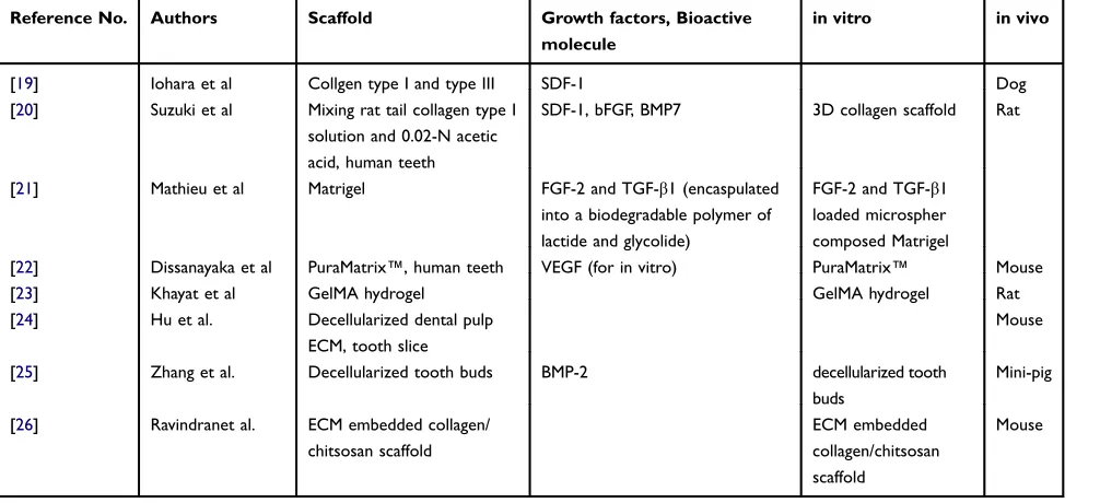
Many different carriers for cells have been reported (Table 1). Scaffolds support appropriate cellular attach-ment, migration, proliferation, differentiation, and function to produce tissue constructs specific to the particular pur-pose. One such purpose would be to provide support for replacement by transplanted cells, but for scaffolds there is concern about the nature of their degradation, cytotoxicity, and immune reactions to them by the recipient.
CD105+ DPSCs were transplanted with stromal
cell-derived factor-1 (SDF-1) and collagen into the mature teeth of dogs that had undergone pulpectomy. This trans-plantation resulted in newly regenerated tissue, which
expressed angiogenic/neurotrophic factors.19 Suzuki’s
group also reported the migration of dental stem cells (DSCs) using collagen gel cylinders. The DSCs were seeded on the surface of these cylinders and cultured
with stromal-derived factor-1α(SDF-1), bFGF, and
BMP7, which induced the recruitment of the cells into the cylinders. SDF-1 or bFGF recruited more cells into collagen gel than the case without cytokines, and BMP7
also recruited few of them.20 Regarding other cytokines,
an investigation of STRO-1-sorted cells (human pulp cells; immature third molars) treated with growth factors (FGF-2
and TGF-β1) in a biodegradable polymer matrix of lactide
and glycolide released using a Matrigel-covered dish was also reported. FGF-2 increased dental pulp proliferation
and TGF-β1 was observed to exert chemotactic potential.
This Matrigel-covered dish culture showed the controlled release of growth factors upon investigating the early stage
of pulp/dentin regeneration.21 In another study,
vascular-ized pulp-like tissue and osteodentin were analyzed upon the transplantation of DPSCs, human umbilical vein endothelial cells (HUVECs), or co-culture of both types
of cell encapsulated in a three-dimensional (3D)
PuraMatrix™in mice. The results showed that
transplan-tation in the co-culture group produced more ECM, vas-cularization, and mineralization than achieved with the
DPSC monocultures in vivo.22 A further study focused
on DPSCs and HUVECs encapsulated in 5% gelatin methacrylate (GelMA) xenogeneic hydrogel and injected into root segments. This transplantation in mice showed
neovasculature formation.23 Another scaffold type in the
form of ECM was supplied by decellularized dental pulp from swine as a bioscaffold for pulp regeneration. The swine pulp was decellularized with a mixed solution of
10% sodium dodecyl sulfate and Triton X-100.
Transplantation of human DPSCs with decellularized den-tal pulp into nude mice demonstrated ECM preservation and a pulp-like tissue structure, as revealed by histological
Table 1Scaffold, Growth Factors, and Bioactive Molecules
Reference No. Authors Scaffold Growth factors, Bioactive molecule
in vitro in vivo
[19] Iohara et al Collgen type I and type III SDF-1 Dog
[20] Suzuki et al Mixing rat tail collagen type I solution and 0.02-N acetic acid, human teeth
SDF-1, bFGF, BMP7 3D collagen scaffold Rat
[21] Mathieu et al Matrigel FGF-2 and TGF-β1 (encaspulated into a biodegradable polymer of lactide and glycolide)
FGF-2 and TGF-β1 loaded microspher composed Matrigel
[22] Dissanayaka et al PuraMatrix™, human teeth VEGF (for in vitro) PuraMatrix™ Mouse
[23] Khayat et al GelMA hydrogel GelMA hydrogel Rat
[24] Hu et al. Decellularized dental pulp ECM, tooth slice
Mouse
[25] Zhang et al. Decellularized tooth buds BMP-2 decellularized tooth
buds
Mini-pig
[26] Ravindranet al. ECM embedded collagen/ chitsosan scaffold
ECM embedded collagen/chitsosan scaffold
Mouse
Stem Cells and Cloning: Advances and Applications downloaded from https://www.dovepress.com/ by 118.70.13.36 on 27-Aug-2020
analysis.24 Decellularized natural porcine tooth bud has also been shown to be useful as a bioengineered scaffold for tooth regeneration, when transplanted with porcine dental epithelial cells, human dental pulp cells, and human umbilical vein endothelial cells. The implantation of samples into the mandibles of mini-pigs revealed
den-tin- and enamel-like tissues.25 In addition, DPSCs were
cultured on a 3D scaffold using a decellularized ECM embedded in a collagen/chitosan scaffold. The subcuta-neous implantation of the scaffold with DPSCs into nude mice resulted in dental pulp-like tissue and the expression
of dentin sialoprotein (DSP) and DSPP.26 To obtain
a deeper understanding of the stemness of DPSCs, there is a need to analyze their activity including in the presence of scaffolds. A key focus for this analysis should be mitochondria, one of the key organelles during the differ-entiation of DPSCs, the energy from which is vital for this process.
Research on Prospective Advanced
Applications for DPSC
Mitochondria
To understand the mechanisms of stem cells including their aging and self-renewal, the establishment of micro-environmental homeostasis and differentiation should be developed as a new approach for DPSC research and therapeutics. According to the literature, mitochondrial functions and dynamics are particularly related to the direction of stem cell differentiation, including for DPSCs. The sequence of human mitochondrial DNA (mtDNA) is 16,569 base pairs long, which includes genes for the 12S and 16S rRNAs, 22 tRNAs, cytochrome c oxidase subunits I, II, and III, ATPase subunit 6, cytochrome b, and eight other
predicted protein-coding genes.83Mitochondria function in
energy metabolism, which regulates the homeostasis of cells including stem cells.
Undifferentiated stem cells show higher levels of
glycolysis compared with stem cells undergoing
differentiation.27,84 Differentiation for osteogenesis has
been shown to be retarded by exogenous H2O2and
mito-chondrial inhibitors. The transition of mitomito-chondrial energy production from glycolysis to oxidative phosphorylation
induces osteogenesis in human MSCs.27Adipogenic
differ-entiation is inhibited by mitochondrially targeted antioxi-dants. During differentiation into adipocytes, there are early increases in mitochondrial metabolism and reactive oxygen species (ROS) generation, which are dependent on mTORC1
signaling in the primary human MSCs.85Furthermore,
dur-ing adipocyte differentiation, PPARγ-dependent
transcrip-tion is dependent on mitochondrial complex III-generated superoxide.85Differentiation for adipogenesis and osteogen-esis has been shown to be correlated with mitochondrial elongation and increases in Mfn1 and 2 (mitochondrial fusion proteins) expression. Forni’s group reported the use of mouse skin mesenchymal stem cells (msMSCs), which are
CD105+ CD90+ CD73+ CD29+ CD34− mesodermal
precursors.86 In addition, chondrogenesis of msMSCs
showed increases of Drp1, Fis1, and Fis2 (fission proteins)
expression and mitophagy enhancement.86The regulation of
processes such as fission/fusion, mitochondrial biogenesis,
and oxidative metabolism of mitochondria is thus key for differentiation and homeostasis in MSCs.
Intriguingly, many studies have reported that mito-chondria are transferred from MSCs to injured cells through tunneling nanotubes. The introduction of MSCs into an infarcted heart mouse model resulted in increased expression of heme oxygenase-1 (HO-1) and peroxisome
proliferator-activator receptor gamma coactivator-1-α
(PGC-1-α) genes in MSCs infused in intact myocardium.
This suggested that HO-1 or mitophagy inhibition was
associated with cardiac apoptosis.87 Heart muscle
expresses a high level of the heart muscle protein (HMP) mitofilin.88 Mitofilin is anchored in the inner
mitochon-drial membrane and is a transmembrane protein.89 The
morphology of cristae is maintained by the mitochondrial inner membrane organizing system (MINOS) including
mitofilin, which is a core component of it along with
Mito10. Mitofilin has been reported to function as
a multifunctional regulator of mitochondrial morphology
and protein biogenesis.90 In MSCs derived from bone
marrow, mitofilin was shown to regulate their homeostasis
and osteogenesis.91 Earlier induction of
osteogenic/denti-nogenic markers in DPSCs was also achieved by the
depletion of mitofilin/3C4 antigens.92 Adipose
(AD)-MSCs and bone marrow (BM)-(AD)-MSCs showed higher mito-chondrial transfer than dental pulp (DP)-MSCs and
Wharton’s jelly (WJ)-MSCs. In addition, DP-MSCs and
WJ-MSCs had reduced mtROS compared with BM-MSCs and AD-MSCs in cardiomyocyte coculture. Moreover, DP-MSCs and WJ-MSCs revealed higher mitochondrial respiratory abilities.93 The initiation of the differentiation of human DPSCs to odontoblasts was also observed to involve mitochondrial elongation with developed cristae, enhancement of the mitochondrial oxygen consumption
rate, increasing mitochondrial ATP production,
Stem Cells and Cloning: Advances and Applications downloaded from https://www.dovepress.com/ by 118.70.13.36 on 27-Aug-2020
upregulation of mitochondrial glycolytic enzyme activ-ities, and increased glycolytic capacity and glycolytic
reserve.94 Disruption of the differentiation of human
DPSCs into odontoblasts was also induced by
lipopolysac-charide (LPS), which decreased HO-1 and PGC-1-α
levels.95 LPS simulation is inhibited by Schisandrin
C and activates mitochondrial biogenesis, which increased
HO-1 and PGC-1-α through the phosphorylated-protein
kinase B (p-AKt) and nuclear factor erythroid 2-related
factor-2 pathway.96 The above findings show that
mito-chondrial dynamics, metabolism, and function are asso-ciated with the fate of stem cells including DPSCs. Stem cell differentiation is also related to mitochondrial activity. Dental pulp stem cells from children, another type of stem cell from human deciduous teeth (SHED), differentiate into neuronal cells, which was shown to increase mito-chondrial membrane potential, mitomito-chondrial DNA, and
elongated mitochondria.97In patients with Rett syndrome,
loss-of-function mutations in MECP2 have been identified,
which is a gene encoding methyl-CpG-binding protein
(MeCP2). Using MeCP2-expressing and MeCP2-deficient
stem cells from exfoliated deciduous teeth, it was shown
that differentiating MeCP2-deficient stem cells exhibited
reductions in mitochondrial membrane potential and ATP production, and restricted mitochondrial distribution in neurites compared with MeCP2-expressing cells. In
addi-tion, central mitochondrial fission factor (dynamin-related
protein1) showed lower expression in MeCP2-deficient
cells than in MeCP2-expressing ones.98 These reports
suggest the importance of mitochondrial function in stem
cells. Understanding the molecular profile and morphology
of mitochondria is thus important to improve the effective-ness of DPSC-based clinical approaches.
Conclusion and Future Challenges
Thefield of research on DPSCs has great potential because
the cells not only have the characteristics of good differ-entiation potential and being easy to culture, but can also be conveniently obtained from extracted teeth, which are usually discarded. Growth factors and scaffolds strongly affect DPSC proliferation and their direction of differentia-tion. DPSCs can aid the regeneration of dentin/pulp-like complex or other tissues in the presence of growth factors
and scaffolds more efficiently than in their absence.
Moreover, upon the combination of DPSCs with other cells and bioactive molecules, enhanced DPSC properties were obtained in vitro and in vivo. Currently, to demon-strate the advantage of combining analyses of the
expression of major genes and proteins, as an example, dentinogenesis-related genes and proteins were mainly ana-lyzed. Analysis of DPSC metabolism in the presence of growth factors and scaffolds should also help us to obtain a deep understanding of their stemness. Such analysis of metabolism is important because mitochondria are the main organelles producing the energy not only for the mainte-nance of homeostasis, but also during differentiation. DPSCs are a promising cell source in the cutting-edge researchfield of stem cells and for developing regenerative medicine applications. Experiments should be performed to evaluate their clinical application, requiring further explora-tion and a deeper understanding of various characteristics of DPSCs.
Acknowledgments
I thank Edanz for editing the English text of a draft of this manuscript.
Disclosure
The author reports no conflicts of interest in this work.
References
1. Gronthos S, Mankani M, Brahim J, Robey PG, Shi S. Postnatal human dental pulp stem cells (DPSCs) in vitro and in vivo.Proc Natl Acad Sci U S A. 2000;97(25):13625–13630. doi:10.1073/pnas.24 0309797
2. Smith AJ, Cassidy N, Perry H, Begue-Kirn C, Ruch JV, Lesot H. Reactionary dentinogenesis.Int J Dev Biol.1995;39(1):273–280. 3. Kitamura C, Kimura K, Nakayama T, Terashita M. Temporal and
spatial expression of c-jun and jun-B proto-oncogenes in pulp cells involved with reparative dentinogenesis after cavity preparation of rat molars.J Dent Res.1999;78(2):673–680. doi:10.1177/002203459907 80020701
4. Bartold PM, Bianco P, Finkelstein MW, et al. Ten Cate’s Oral Histology: Development, Structure, and Function. 6. Mosby;2003. 5. Shiba H, Fujita T, Doi N, et al. Differential effects of various growth
factors and cytokines on the syntheses of DNA, type I collagen, laminin,fibronectin, osteonectin/secreted protein, acidic and rich in cysteine (SPARC), and alkaline phosphatase by human pulp cells in culture. J Cell Physiol. 1998;174(2):194–205. doi:10.1002/(ISSN) 1097-4652
6. Kunimatsu R, Nakajima K, Awada T, et al. Comparative characteriza-tion of stem cells from human exfoliated deciduous teeth, dental pulp, and bone marrow-derived mesenchymal stem cells.Biochem Biophys Res Commun. 2018;501(1):193–198. doi:10.1016/j.bbrc.20 18.04.213
7. Huang GT, Yamaza T, Shea LD, et al. Stem/progenitor cell-mediated de novo regeneration of dental pulp with newly deposited continuous layer of dentin in an in vivo model. Tissue Eng Part A. 2010;16 (2):605–615. doi:10.1089/ten.tea.2009.0518
8. Lindroos B, Maenpaa K, Ylikomi T, Oja H, Suuronen R, Miettinen S. Characterisation of human dental stem cells and buccal mucosa
fibroblasts. Biochem Biophys Res Commun. 2008;368(2):329–335. doi:10.1016/j.bbrc.2008.01.081
Stem Cells and Cloning: Advances and Applications downloaded from https://www.dovepress.com/ by 118.70.13.36 on 27-Aug-2020
9. Shi S, Gronthos S. Perivascular niche of postnatal mesenchymal stem cells in human bone marrow and dental pulp. J Bone Miner Res. 2003;18(4):696–704. doi:10.1359/jbmr.2003.18.4.696
10. Karbanova J, Soukup T, Suchanek J, Pytlik R, Corbeil D, Mokry J. Characterization of dental pulp stem cells from impacted third molars cultured in low serum-containing medium. Cells Tissues Organs. 2011;193(6):344–365. doi:10.1159/000321160
11. Sakai K, Yamamoto A, Matsubara K, et al. Human dental pulp-derived stem cells promote locomotor recovery after complete transection of the rat spinal cord by multiple neuro-regenerative mechanisms. J Clin Invest. 2012;122(1):80–90. doi:10.1172/JCI59 251
12. Batouli S, Miura M, Brahim J, et al. Comparison of stem-cell-mediated osteogenesis and dentinogenesis. J Dent Res. 2003;82 (12):976–981. doi:10.1177/154405910308201208
13. Tsutsui TW, Inaba T, Fisher LW, Robey PG, Tsutsui T. In vitro chromosome aberration tests using human dental pulp cells to detect the carcinogenic potential of chemical agents.Odontology.2006;94 (1):44–50. doi:10.1007/s10266-006-0065-1
14. Matsui M, Kobayashi T, Tsutsui TW. CD146 positive human dental pulp stem cells promote regeneration of dentin/pulp-like structures. Hum Cell. 2018;31(2):127–138. doi:10.1007/s13577-017-0198-2
15. Prescott RS, Alsanea R, Fayad MI, et al. In vivo generation of dental pulp-like tissue by using dental pulp stem cells, a collagen scaffold, and dentin matrix protein 1 after subcutaneous transplantation in mice.J Endod.2008;34(4):421–426. doi:10.1016/j.joen.2008.02.005 16. Fujii Y, Kawase-Koga Y, Hojo H, et al. Bone regeneration by human dental pulp stem cells using a helioxanthin derivative and cell-sheet technology.Stem Cell Res Ther.2018;9(1):24. doi:10.1186/s13287-018-0783-7
17. Angelopoulos I, Brizuela C, Khoury M. Gingival mesenchymal stem cells outperform haploidentical dental pulp-derived mesenchymal stem cells in proliferation rate, migration ability, and angiogenic potential.Cell Transplant.2018;27(6):967–978. doi:10.1177/096368 9718759649
18. Zhang J, Lu X, Feng G, et al. Chitosan scaffolds induce human dental pulp stem cells to neural differentiation: potential roles for spinal cord injury therapy. Cell Tissue Res. 2016;366(1):129–142. doi:10. 1007/s00441-016-2402-1
19. Iohara K, Imabayashi K, Ishizaka R, et al. Complete pulp regenera-tion after pulpectomy by transplantaregenera-tion of CD105+ stem cells with stromal cell-derived factor-1. Tissue Eng Part A. 2011;17(15– -16):1911–1920. doi:10.1089/ten.tea.2010.0615
20. Suzuki T, Lee CH, Chen M, et al. Induced migration of dental pulp stem cells for in vivo pulp regeneration. J Dent Res. 2011;90 (8):1013–1018. doi:10.1177/0022034511408426
21. Mathieu S, Jeanneau C, Sheibat-Othman N, Kalaji N, Fessi H, About I. Usefulness of controlled release of growth factors in inves-tigating the early events of dentin-pulp regeneration. J Endod. 2013;39(2):228–235. doi:10.1016/j.joen.2012.11.007
22. Dissanayaka WL, Hargreaves KM, Jin L, Samaranayake LP, Zhang C. The interplay of dental pulp stem cells and endothelial cells in an injectable peptide hydrogel on angiogenesis and pulp regeneration in vivo. Tissue Eng Part A. 2015;21(3–4):550–563. doi:10.1089/ten.tea.2014.0154
23. Khayat A, Monteiro N, Smith EE, et al. GelMA-encapsulated hDPSCs and HUVECs for dental pulp regeneration. J Dent Res. 2017;96(2):192–199. doi:10.1177/0022034516682005
24. Hu L, Gao Z, Xu J, et al. Decellularized swine dental pulp as a bioscaffold for pulp regeneration. Biomed Res Int. 2017;20 17:9342714. doi:10.1155/2017/9342714
25. Zhang W, Vazquez B, Oreadi D, Yelick PC. Decellularized tooth bud scaffolds for tooth regeneration. J Dent Res.2017;96(5):516–523. doi:10.1177/0022034516689082
26. Ravindran S, Zhang Y, Huang CC, George A. Odontogenic induction of dental stem cells by extracellular matrix-inspired three-dimensional scaffold.Tissue Eng Part A.2014;20(1–2):92–102. doi:10.1089/ten. tea.2013.0192
27. Chen CT, Shih YR, Kuo TK, Lee OK, Wei YH. Coordinated changes of mitochondrial biogenesis and antioxidant enzymes during osteogenic differentiation of human mesenchymal stem cells. Stem Cells. 2008;26(4):960–968. doi:10.1634/stem-cells.2007-0509
28. Klagsbrun M. The fibroblast growth factor family: structural and biological properties.Prog Growth Factor Res.1989;1(4):207–235. doi:10.1016/0955-2235(89)90012-4
29. Mergia A, Eddy R, Abraham JA, Fiddes JC, Shows TB. The genes for basic and acidicfibroblast growth factors are on different human chromosomes. Biochem Biophys Res Commun. 1986;138(2):64 4–651. doi:10.1016/S0006-291X(86)80545-9
30. Abraham JA, Mergia A, Whang JL, et al. Nucleotide sequence of a bovine clone encoding the angiogenic protein, basicfibroblast growth factor. Science.1986;233(4763):545–548. doi:10.1126/science.2425435 31. Vlodavsky I, Folkman J, Sullivan R, et al. Endothelial cell-derived basic
fibroblast growth factor: synthesis and deposition into subendothelial extracellular matrix.Proc Natl Acad Sci U S A.1987;84(8):2292–2296. doi:10.1073/pnas.84.8.2292
32. Globus RK, Plouet J, Gospodarowicz D. Cultured bovine bone cells synthesize basicfibroblast growth factor and store it in their extra-cellular matrix. Endocrinology. 1989;124(3):1539–1547. doi:10.12 10/endo-124-3-1539
33. Zeng Q, Nguyen S, Zhang H, et al. Release of growth factors into root canal by irrigations in regenerative endodontics. J Endod. 2016;42(12):1760–1766. doi:10.1016/j.joen.2016.04.029
34. Chang YC, Chang MC, Chen YJ, et al. Basicfibroblast growth factor regulates gene and protein expression related to proliferation, differentia-tion, and matrix production of human dental pulp cells.J Endod.2017;43 (6):936–942. doi:10.1016/j.joen.2017.01.024
35. Zhang J, Lian M, Cao P, et al. Effects of nerve growth factor and basicfibroblast growth factor promote human dental pulp stem cells to neural differentiation. Neurochem Res. 2017;42(4):1015–1025. doi:10.1007/s11064-016-2134-3
36. Luo L, Albashari AA, Wang X, et al. Effects of transplanted heparin-poloxamer hydrogel combining dental pulp stem cells and bFGF on spinal cord injury repair. Stem Cells Int. 2018;2018: 2398521. doi:10.1155/2018/2398521
37. Qian J, Jiayuan W, Wenkai J, et al. Basicfibroblastic growth factor affects the osteogenic differentiation of dental pulp stem cells in a treatment-dependent manner. Int Endod J. 2015;48(7):690–700. doi:10.1111/iej.2015.48.issue-7
38. Crane JL, Cao X. Bone marrow mesenchymal stem cells and TGF-beta signaling in bone remodeling. J Clin Invest. 2014;124(2):466–472. doi:10.1172/JCI70050
39. de Araujo Farias V, Carrillo-Galvez AB, Martin F, Anderson P. TGF-beta and mesenchymal stromal cells in regenerative medicine, auto-immunity and cancer.Cytokine Growth Factor Rev.2018;43:25–37. doi:10.1016/j.cytogfr.2018.06.002
40. Miyazono K, Olofsson A, Colosetti P, Heldin CH. A role of the TGF-binding protein in the assembly and secretion of TGF-beta 1. EMBO J. 1991;10(5):1091–1101. doi:10.1002/j.1460-2075.1991. tb08049.x
41. Blobe GC, Schiemann WP, Lodish HF. Role of transforming growth factor beta in human disease. N Engl J Med. 2000;342 (18):1350–1358. doi:10.1056/NEJM200005043421807
42. Heo JS, Choi Y, Kim HS, Kim HO. Comparison of molecular profiles of human mesenchymal stem cells derived from bone marrow, umbilical cord blood, placenta and adipose tissue. Int J Mol Med. 2016;37(1):115–125. doi:10.3892/ ijmm.2015.2413
Stem Cells and Cloning: Advances and Applications downloaded from https://www.dovepress.com/ by 118.70.13.36 on 27-Aug-2020
43. Tomic S, Djokic J, Vasilijic S, et al. Immunomodulatory properties of mesenchymal stem cells derived from dental pulp and dental follicle are susceptible to activation by toll-like receptor agonists.Stem Cells Dev. 2011;20(4):695–708. doi:10.1089/scd.2010.0145
44. Schmierer B, Hill CS. TGFbeta-SMAD signal transduction: molecu-lar specificity and functional flexibility. Nat Rev Mol Cell Biol. 2007;8(12):970–982. doi:10.1038/nrm2297
45. Oshimori N, Fuchs E. The harmonies played by TGF-beta in stem cell biology.Cell Stem Cell.2012;11(6):751–764. doi:10.1016/j.stem. 2012.11.001
46. Ng F, Boucher S, Koh S, et al. PDGF, TGF-, and FGF signaling is important for differentiation and growth of mesenchymal stem cells (MSCs): transcriptional profiling can identify markers and signaling pathways important in differentiation of MSCs into adipogenic, chon-drogenic, and oste. Blood. 2008;112:295–307. doi:10.1182/blood-2007-07-103697
47. Karbanova J, Soukup T, Suchanek J, Mokry J. Osteogenic differen-tiation of human dental pulp-derived stem cells under various ex-vivo culture conditions. Acta Medica (Hradec Kralove). 2010;53 (2):79–84. doi:10.14712/18059694.2016.64
48. Song B, Jiang W, Alraies A, et al. Bladder smooth muscle cells differentiation from dental pulp stem cells: future potential for blad-der tissue engineering. Stem Cells Int. 2016;2016:6979368. doi:10.1155/2016/6979368
49. Johnson D, Lanahan A, Buck CR, et al. Expression and structure of the human NGF receptor.Cell. 1986;47(4):545–554. doi:10.1016/ 0092-8674(86)90619-7
50. Kaplan DR, Hempstead BL, Martin-Zanca D, Chao MV, Parada LF. The trk proto-oncogene product: a signal transducing receptor for nerve growth factor.Science.1991;252(5005):554–558. doi:10.1126/ science.1850549
51. Hartman DS, McCormack M, Schubenel R, Hertel C. Multiple trkA proteins in PC12 cells bind NGF with a slow association rate.J Biol Chem.1992;267(34):24516–24522.
52. Neet KE, Campenot RB. Receptor binding, internalization, and retro-grade transport of neurotrophic factors.Cell Mol Life Sci.2001;58 (8):1021–1035. doi:10.1007/PL00000917
53. Woo SB, Timm DE, Neet KE. Alteration of NH2-terminal residues of nerve growth factor affects activity and Trk binding without affecting stability or conformation. J Biol Chem. 1995;270(11):6278–6285. doi:10.1074/jbc.270.11.6278
54. Mahadeo D, Kaplan L, Chao MV, Hempstead BL. High affinity nerve growth factor binding displays a faster rate of association than p140trk binding. Implications for multi-subunit polypeptide receptors.J Biol Chem.1994;269(9):6884–6891.
55. Sofroniew MV, Howe CL, Mobley WC. Nerve growth factor signal-ing, neuroprotection, and neural repair. Annu Rev Neurosci. 2001;24:1217–1281. doi:10.1146/annurev.neuro.24.1.1217
56. Di Marco E, Mathor M, Bondanza S, et al. Nerve growth factor binds to normal human keratinocytes through high and low affinity recep-tors and stimulates their growth by a novel autocrine loop.J Biol Chem.1993;268(30):22838–22846.
57. Vega JA, Garcia-Suarez O, Hannestad J, Perez-Perez M, Germana A. Neurotrophins and the immune system.J Anat. 2003;203(1):1–19. doi:10.1046/j.1469-7580.2003.00203.x
58. Mitsiadis TA, Pagella P. Expression of Nerve Growth Factor (NGF), TrkA, and p75(NTR) in developing human fetal teeth.Front Physiol. 2016;7:338. doi:10.3389/fphys.2016.00338
59. Mitsiadis TA, Magloire H, Pagella P. Nerve growth factor signalling in pathology and regeneration of human teeth. Sci Rep. 2017;7 (1):1327. doi:10.1038/s41598-017-01455-3
60. Mead B, Logan A, Berry M, Leadbeater W, Scheven BA. Intravitreally transplanted dental pulp stem cells promote neuropro-tection and axon regeneration of retinal ganglion cells after optic nerve injury. Invest Ophthalmol Vis Sci. 2013;54(12):7544–7556. doi:10.1167/iovs.13-13045
61. Kohler N, Lipton A. Platelets as a source of fibroblast growth-promoting activity. Exp Cell Res. 1974;87(2):297–301. doi:10.1016/0014-4827(74)90484-4
62. Antoniades HN, Scher CD, Stiles CD. Purification of human platelet-derived growth factor.Proc Natl Acad Sci U S A.1979;76 (4):1809–1813. doi:10.1073/pnas.76.4.1809
63. Heldin CH, Westermark B, Wasteson A. Platelet-derived growth factor: purification and partial characterization.Proc Natl Acad Sci U S A.1979;76(8):3722–3726. doi:10.1073/pnas.76.8.3722 64. Li X, Ponten A, Aase K, et al. PDGF-C is a new protease-activated
ligand for the PDGF alpha-receptor. Nat Cell Biol. 2000;2 (5):302–309. doi:10.1038/35010579
65. LaRochelle WJ, Jeffers M, McDonald WF, et al. PDGF-D, a new protease-activated growth factor.Nat Cell Biol.2001;3(5):517–521. doi:10.1038/35074593
66. Betsholtz C, Johnsson A, Heldin CH, et al. cDNA sequence and chromosomal localization of human platelet-derived growth factor A-chain and its expression in tumour cell lines.Nature.1986;320 (6064):695–699. doi:10.1038/320695a0
67. Swan DC, McBride OW, Robbins KC, Keithley DA, Reddy EP, Aaronson SA. Chromosomal mapping of the simian sarcoma virus onc gene analogue in human cells. Proc Natl Acad Sci U S A. 1982;79(15):4691–4695. doi:10.1073/pnas.79.15.4691
68. Uutela M, Lauren J, Bergsten E, et al. Chromosomal location, exon structure, and vascular expression patterns of the human PDGFC and PDGFD genes.Circulation. 2001;103(18):2242–2247. doi:10.1161/ 01.CIR.103.18.2242
69. Heldin CH, Ostman A, Ronnstrand L. Signal transduction via platelet-derived growth factor receptors. Biochim Biophys Acta. 1998;1378(1):F79–F113. doi:10.1016/s0304-419x(98)00015-8 70. Mead B, Logan A, Berry M, Leadbeater W, Scheven BA.
Paracrine-mediated neuroprotection and neuritogenesis of axotomised retinal gang-lion cells by human dental pulp stem cells: comparison with human bone marrow and adipose-derived mesenchymal stem cells.PLoS One.2014;9 (10):e109305. doi:10.1371/journal.pone.0109305
71. Zhang M, Jiang F, Zhang X, et al. The effects of platelet-derived growth factor-bb on human dental pulp stem cells mediated dentin-pulp complex regeneration.Stem Cells Transl Med.2017;6 (12):2126–2134. doi:10.1002/sctm.17-0033
72. Cai S, Zhang W, Chen W. PDGFRbeta(+)/c-kit(+) pulp cells are odontoblastic progenitors capable of producing dentin-like structure in vitro and in vivo.BMC Oral Health.2016;16(1):113. doi:10.1186/ s12903-016-0307-8
73. Wozney JM, Rosen V, Celeste AJ, et al. Novel regulators of bone formation: molecular clones and activities. Science. 1988;242 (4885):1528–1534. doi:10.1126/science.3201241
74. Yang G, Yuan G, Ye W, Cho KW, Chen Y. An atypical canonical bone morphogenetic protein (BMP) signaling pathway regulates Msh homeobox 1 (Msx1) expression during odontogenesis. J Biol Chem. 2014;289(45):31492–31502. doi:10.1074/jbc.M114. 600064
75. Shi C, Yuan Y, Guo Y, et al. BMP signaling in regulating mesench-ymal stem cells in incisor homeostasis. J Dent Res. 2019;98 (8):904–911. doi:10.1177/0022034519850812
76. Plikus MV, Zeichner-David M, Mayer JA, et al. Morphoregulation of teeth: modulating the number, size, shape and differentiation by tuning Bmp activity. Evol Dev. 2005;7(5):440–457. doi:10.1111/ j.1525-142X.2005.05048.x
77. Yang X, van der Kraan PM, Bian Z, Fan M, Walboomers XF, Jansen JA. Mineralized tissue formation by BMP2-transfected pulp stem cells. J Dent Res. 2009;88(11):1020–1025. doi:10.1177/ 0022034509346258
78. Sun N, Jiang T, Wu C, Sun H, Zhou Q, Lu L. Expression and influence of BMP-4 in human dental pulp cells cultured in vitro. Exp Ther Med. 2018;16(6):5112–5116. doi:10.3892/etm.2018. 6824
Stem Cells and Cloning: Advances and Applications downloaded from https://www.dovepress.com/ by 118.70.13.36 on 27-Aug-2020
79. Zhu L, Na J, Mu R et al. Bone morphogenic protein 7 promotes odontogenic differentiation of dental pulp stem cells in vitro.Life Sci. 2018;202:175–181.
80. Yang J, Ye L, Hui TQ, et al. Bone morphogenetic protein 2-induced human dental pulp cell differentiation involves p38 mitogen-activated protein kinase-activated canonical WNT pathway. Int J Oral Sci. 2015;7(2):95–102. doi:10.1038/ijos.2015.7
81. Aksel H, Ozturk S, Serper A, Ulubayram K. VEGF/BMP-2 loaded three-dimensional model for enhanced angiogenic and odontogenic potential of dental pulp stem cells.Int Endod J.2018;51(4):420–430. doi:10.1111/iej.12869
82. Iohara K, Nakashima M, Ito M, Ishikawa M, Nakasima A, Akamine A. Dentin regeneration by dental pulp stem cell therapy with recombinant human bone morphogenetic protein 2. J Dent Res.2004;83(8):590–595. doi:10.1177/154405910408300802 83. Anderson S, Bankier AT, Barrell BG, et al. Sequence and organization of
the human mitochondrial genome. Nature. 1981;290(5806):457–465. doi:10.1038/290457a0
84. Zhang J, Khvorostov I, Hong JS, et al. UCP2 regulates energy metabolism and differentiation potential of human pluripotent stem cells.EMBO J. 2011;30(24):4860–4873. doi:10.1038/emboj.2011.401
85. Tormos KV, Anso E, Hamanaka RB, et al. Mitochondrial complex III ROS regulate adipocyte differentiation. Cell Metab. 2011;14 (4):537–544. doi:10.1016/j.cmet.2011.08.007
86. Forni MF, Peloggia J, Trudeau K, Shirihai O, Kowaltowski AJ. Murine mesenchymal stem cell commitment to differentiation is regulated by mitochondrial dynamics. Stem Cells. 2016;34(3):74 3–755. doi:10.1002/stem.2248
87. Mahrouf-Yorgov M, Augeul L, Da Silva CC, et al. Mesenchymal stem cells sense mitochondria released from damaged cells as danger signals to activate their rescue properties. Cell Death Differ.2017;24(7):1224–1238. doi:10.1038/cdd.2017.51
88. Icho T, Ikeda T, Matsumoto Y, Hanaoka F, Kaji K, Tsuchida N. A novel human gene that is preferentially transcribed in heart muscle.Gene. 1994;144(2):301–306. doi:10.1016/0378-1119(94)90394-8
89. Gieffers C, Korioth F, Heimann P, Ungermann C, Frey J. Mitofilin is a transmembrane protein of the inner mitochondrial membrane expressed as two isoforms. Exp Cell Res. 1997;232(2):395–399. doi:10.1006/excr.1997.3539
90. von der Malsburg K, Muller JM, Bohnert M, et al. Dual role of mitofilin in mitochondrial membrane organization and protein biogenesis. Dev Cell. 2011;21(4):694–707. doi:10.1016/j.devcel.20 11.08.026
91. Lv YJ, Yang Y, Sui BD, et al. Resveratrol counteracts bone loss via mitofilin-mediated osteogenic improvement of mesenchymal stem cells in senescence-accelerated mice. Theranostics. 2018;8(9):23 87–2406. doi:10.7150/thno.23620
92. Hwang HI, Lee TH, Jang YJ. Cell proliferation-inducing protein 52/ mitofilin is a surface antigen on undifferentiated human dental pulp stem cells. Stem Cells Dev. 2015;24(11):1309–1319. doi:10.1089/ scd.2014.0387
93. Paliwal S, Chaudhuri R, Agrawal A, Mohanty S. Human tissue-specific MSCs demonstrate differential mitochondria transfer abilities that may determine their regenerative abilities.Stem Cell Res Ther.2018;9(1):298. doi:10.1186/s13287-018-1012-0
94. Wang L, Cheng L, Wang H, et al. Glycometabolic reprogramming associated with the initiation of human dental pulp stem cell differentiation.Cell Biol Int.2016;40(3):308–317. doi:10.1002/cbin. v40.3
95. Takanche JS, Kim JS, Kim JE, Han SH, Yi HK. Schisandrin C enhances odontoblastic differentiation through autophagy and mitochondrial biogenesis in human dental pulp cells. Arch Oral Biol.2018;88:60–66. doi:10.1016/j.archoralbio.2018.01.018 96. Takanche JS, Lee YH, Kim JS, et al. Anti-inflammatory and
antiox-idant properties of Schisandrin C promote mitochondrial biogenesis in human dental pulp cells. Int Endod J. 2018;51(4):438–447. doi:10.1111/iej.12861
97. Kato H, Thi Mai Pham T, Yamaza H, et al. Mitochondria regulate the differentiation of stem cells from human exfoliated deciduous teeth. Cell Struct Funct.2017;42(2):105–116. doi:10.1247/csf.17012 98. Hirofuji S, Hirofuji Y, Kato H, et al. Mitochondrial dysfunction in
dopaminergic neurons differentiated from exfoliated deciduous tooth-derived pulp stem cells of a child with Rett syndrome. Biochem Biophys Res Commun.2018;498(4):898–904. doi:10.1016/ j.bbrc.2018.03.077
Stem Cells and Cloning: Advances and Applications
Dovepress
Publish your work in this journal
Stem Cells and Cloning: Advances and Applications is an interna-tional, peer-reviewed, open access journal. Areas of interest in established and emerging concepts in stem cell research include: Embryonic cell stems; Adult stem cells; Blastocysts; Cordblood stem cells; Stem cell transformation and culture; Therapeutic cloning; Umbilical cord blood and bone marrow cells; Laboratory,
animal and human therapeutic studies; Philosophical and ethical issues related to stem cell research. This journal is indexed on CAS. The manuscript management system is completely online and includes a very quick and fair peer-review system, which is all easy to use. Visit http://www.dovepress.com/testimonials.php to read real quotes from published authors.
Submit your manuscript here:https://www.dovepress.com/stem-cells-and-cloning-advances-and-applications-journal
Stem Cells and Cloning: Advances and Applications downloaded from https://www.dovepress.com/ by 118.70.13.36 on 27-Aug-2020