STUDIES OF THE EXPRESSION AND FUNCTION OF THE
RETINOID-X-RECEPTOR y GENE IN RODENT
EMBRYONIC DEVELOPMENT
P a n telis
G eorgiad es
A thesis s ubmi tted for the degree o f Doct or o f Ph il osophy
D e p a r t m e n t o f M o l e c u l a r P a t h o l o g y
ProQ uest Number: 10105680
All rights reserved
INFORMATION TO ALL U SE R S
The quality of this reproduction is d ep en d en t upon the quality of the copy subm itted. In the unlikely even t that the author did not sen d a com plete manuscript
and there are m issing p a g e s, th e se will be noted. Also, if material had to be rem oved, a note will indicate the deletion.
uest.
ProQ uest 10105680
Published by ProQ uest LLC(2016). Copyright of the Dissertation is held by the Author. All rights reserved.
This work is protected against unauthorized copying under Title 17, United S ta tes C ode. Microform Edition © ProQ uest LLC.
ProQ uest LLC
789 East E isenhow er Parkway P.O. Box 1346
D ur i ng this work I was aw ar de d t wo s ch ol ars hi ps : W e l l c o m e P r i z e S t u d e n t s h i p
ABSTRACT
Retinoids, such as 9 cis retinoic acid (9cRA) and all tr a n s retinoic acid (tRA), are signalling molecules that are im portant for the establishm ent of the
c o rre c t b eh av io u r of many d ifferent types of cells during em bryonic
development and adult life. Retinoid X receptors (RXRs) and retinoic acid
receptors (RARs) are transcription factors that m ediate the effects of
retinoids by binding to them. RXRs can also be involved in the mediation of
non-retinoid signalling molecules because they can form heterodimers with other transcription factors such as thyroid hormone receptors and vitamin D r e c e p t o r s .
The objective of the present work was to provide information about the role
of RXRy during rodent development. I isolated a truncated rat RXRy cDNA
clone by screening a rat cDNA library with a chick RXRy cDNA probe. Using
the rat clone as a probe, I examined the expression pattern of RXRy in rat
em bryos and found it to be mainly restricted to specific regions of the
p e rip h e ra l and central nervous systems and in sk eletal m uscle. Its
expression in adult rats was also examined and was found to be expressed in
specific regions. These include skeletal muscle, heart, brain, sciatic nerve
and liver. I also examined the expression of RXRy in mouse embryos and
found it to be essentially the same to that observed in rats.
The role of RXRy in rodent myogenesis was examined by comparing its
spatio-tem poral expression with that of m uscle-specific genes of known
function so as to establish the timing of the initiation of RXRy expression. I
showed that the initiation of RXRy expression was concomitant with muscle
differentiation of limb but not early myotomal myoblasts, suggesting that it may be involved in limb myoblast differentiation and that retinoic acid may
be a myogenic differentiation signal. This hypothesis was tested in vitro,
using a skeletal muscle cell line. As predicted, both 9cRA and tRA were able
to relieve the serum-induced inhibition of myoblast differentiation and RXRy was expressed during this process. RXRy may mediate the differentiation effect of retinoic acid because its expression was not induced by retinoic acid, since it was expressed in myoblasts in the absence of retinoic acid.
5
supports this because I showed that overexpression of RXRy in BHK cells
activates reporter gene expression from a prom oter which is under the
control of upstream regulatory sequences derived from the neuronal nAChR a 2 subunit gene, and 9cRA enhances this activation. Moreover, this effect was cell type-specific because when ND7 cells were used, no activation was observed in the presence of RXRy and 9cRA. This shows that the function of R X R y can be influenced by trans-acting factors.
Finally, I showed that the expression of RXRy in postnatal heart is
age-dependent, suggesting that it may be involved in the regulation of genes whose expression is age-dependent. Moreover, I demonstrated that treatment
of prim ary cultures of neonatal rat cardiom yocytes with either thyroid
hormone or tRA leads to changes in RXRy expression. This finding is
c o n sistent with the proposed role of RXRy because there is evidence
ACKNOWLEDGEMENTS
I thank my supervisor Professor Paul M. B rickell for giving me the
opportunity to work in his group and for his guidance and support
throughout this work. I also thank the Wellcome Trust for awarding me with a Wellcome Prize Studentship, and the A.G. Leventis Foundation for awarding me with a postgraduate scholarship.
I send my gratitude to the following people for their friendship, technical support and helpful discussions; Dr Eduardo Seleiro, Dr Annie Rowe, Dr David Darling, Dr Nicholas Eager, Dr Esther Bell, Dr Philippa Francis-West, Dr Nicholas Lakin, Dr Torben Lund, Mr Julius Kieskiewicz, Dr Hugo Caro, Dr Leslie Robson, Professor Cheryll Tickle, Professor Lewis Wolpert, Dr John
W o o d , M r M arty C ohn, Mr R o n ald N itte n b e rg , M iss K o n s ta n tin a
Kostakopoulou, Dr Debbie Cummings, Mr Andreas Costi, Mr John Estridge, Dr Nathaniel Milton, Dr Paula Timmons, Dr Peter Rigby. I also thank Professor David Latchman and Jan Wenley for their support.
TABLE OF CONTENTS
Title page...1
Abstract... 4
Acknowledgements...6
Table of contents... 7
List of figures...13
CHAPTER I I n t r o d a c t i o n 1.1 - Foreword...19
1.2 - Development, cells and genes... 19
1.3 - Retinoids and vertebrate development... 21
1.3.1 - Retinoid metabolism... 21
1.3.2 - Retinoids and embryos... 22
1.4 - Retinoic acid influences the expression of different genes...25
1.5 - Retinoids and gene expression...27
1.5.1 - Signalling by retinoids... 27
1.5.2 - The steroid hormone receptor superfamily...28
1.5.2.1 - The structure and function of the DNA binding domain... 30
1.5.2.2 - The structure and function of the ligand binding domain... 34
1.5.2.3 - The A and B domains... 35
1.5.2.4 - The D domain... 35
1.5.3 - Retinoid receptors and transcriptional regulation... 37
1.5.5 - Retinoid binding proteins...49
1.6 - Aims of this thesis... 50
CHAPTER II M a t e r i a l s an d m e t h o d s 2.1 - Screening of a cDNA library under low stringency conditions... 52
2.2 - Gene probes... 56
2.3 - Construction of a^ ^-P labelled DNA probes...59
2.4 - Large and small scale preparation of plasmid DNA... 60
2.5 - Phenol/chloroform extraction of nucleic acids...62
2.6 - Ethanol precipitation of nucleic acids... 63
2.7 - DNA purification using the "Gene Clean" method...63
2.8 - Restriction enzyme digestion...64
2.9 - Agarose gel electrophoresis of DNA... 64
2.10 - Subcloning of DNA fragments... 64
2.11 - DNA sequencing... 66
2.12 - Isolation of total RNA... 67
2.13 - Agarose gel electrophoresis of total RNA...68
2.14 - Northern blotting and hybridisation... 69
2.15 - Labelling of RNA probes...70
2.16 - Culture of cell lines... 72
2.17 - Treatment of cultured cells with retinoic acid and thyroid hormone....73
2.18 - Preparation of primary cultures of ventricular rat cardiomyocytes...73
2.19 - Staining of cultured cells...74
2.20 - In situ hybridisation of wax embryonic sections... 74
2.21 - In situ hybridisation of whole embryos... 77
2.22 - Transient transfection of cultured cells with plasmid DNA... 79
2.23 - Luciferase assays...80
9
2.25 - List of general use buffers and culture media...82
CHAPTER III I sol at io n o f a rat RXRy c DNA c l on e and i n v e s t i g a t i o n o f RXRy e x p r e s s i o n in e m b r y o n i c and a d ul t r od en t s 3.1 - Introduction... 84
3.1.1 - Aim... 84
3.2 - Results... 85
3.2.1 - Isolation and characterisation of a rat RXRy cDNA clone...85
3.2.2 - Expression of RXRy mRNA in adult rat tissues and rat cell lines...93
3.2.3 - E x p ressio n o f RXRy tr a n s c r i p ts d u r in g ro d e n t e m b ry o n ic development... 96
3.3 - Discussion... 112
CHAPTER IV R X R y and s k e l e t a l m u s c l e d i f f e r e n t i a t i o n in rat e m b r y o s and c u l tu r ed rat m yo bl as t s 4.1 - Introduction... 119
4.1.1 - Aim... 119
4.1.2 - Cell behaviour during myogenesis...119
4.1.2.1 - Description of myogenesis at the cellular level... 120
4 .1 .2 .2 - C o m m itm e n t of em b ry o n ic cells to w a rd s the m y o g e n ic phenotype...125
4.1.2.2.1. - Prospective skeletal muscle cells are myogenically competent from a very early stage but are not determined...127
4.1.2.2.3 - Signal(s) from the neural tube and/or notochord are necessary for
myogenesis in the myotomes...129
4.1.2.2.4 - M yogenically unspecified m esoderm al cells are m yogenically competent... 130
4.1.2.2.5 - The role of the axial structures in the initial appearance of myotomal muscle... 131
4.1.2.2.6 - The myogenic signal derived from the axial structures is selective rather than instructive and is mediated by diffusible factors...133
4.1.2.2.7 - The development of limb skeletal muscle is independent of axial organs...134
4.1.2.2.8 - The timing of myogenic determination... 135
4.1.3 - Gene action during myogenesis... 137
4.1.3.1 - The contribution of skeletal muscle cell lines to the study of myogenic differentiation...137
4.1.3.2 - Information about the role of members of the MyoD family derived from ectopic expression studies... 140
4.1.3.3 - Developmental expression of the MyoD genes...141
4.1.3.4 - Information about the function of the MyoD genes from gene knockout experiments...144
4.1.3.5 - Regulation of the expression of the genes responsible for the skeletal muscle phenotype... 146
4.1.3.6 - Cell proliferation and skeletal muscle differentiation... 148
4.1.4 - Experimental objectives... 151
4.2 - Results...153
4.2.1 - D evelopm ental expression of RXRy during the primary wave of myogenesis in axial and limb skeletal muscle in rat embryos... 153
4.2.1.1 - Myotomal expression of RXRy during rat development... 153
11
4.2.1.3 - Expression of RXRy in late stages of the primary myogenic wave in
the mouse embryo... 163
4.2.1.4 - Conclusion...163
4.2.2 - Treatment of proliferating H9 myoblasts with either 9cRA or tRA reduces their capacity to incorporate tritiated thym idine... 165
4.2.3 - Myoblasts are induced to differentiate into multinucleated myofibers as a result of retinoid treatment...168
4.2.4 - RXRy and myogenin expression during treatment of myoblasts with retinoic acid... 170
4.3 - Discussion... 173
CHAPTER V The role o f RXRy and 9cis r e t i n o i c a ci d in t he t r a n s c r i p t i o n a l r e g u l a t i o n o f a n e u r o n a l n i c o t i n i c a c e t y l c h o l i n e r e c e p t o r s u b u n i t g e n e 5.1 - Introduction... 180
5.1.1 - Aim... 180
5.1.2 - Neuronal nicotinic acetylcholine receptors... 181
5.1.2.1 - Neuronal nAChRs and the nervous system...181
5.1.2.2 - The molecular function of neuronal nAChRs... 182
5.1.2.3 - Neuronal nicotinic acetylcholine receptor subunits are members of functional neuronal nAChRs...182
5.1.2.4 - D ifferent subunit combinations are im portant for the functional diversity of neuronal nAChRs... 184
5.1.2.5 - Expression of the neuronal nAChR a 2-subunit gene in rats and chicks... 185
5.1.2.6 - Transcriptional regulation of the chick neuronal nAChR a 2 - s u b u n i t gene... 187
1
2
5 .1 .2 .6 .2 - Six 11-base p air m otifs are in v o lv ed in the s ile n c in g
activity... 188 5.1.2.6.3 - The 11-base pair motifs are necessary for both activation and silencing of transcription...189 5.1.2.6.4 - Regulatory sequences of the chick « 2 gene are able to confer neuron-specific expression in transgenic mice...190
5.1.3 - Experimental objectives... 191 5.2 - Results...193 5.2.1 - Identification of a putative retinoid response element within the 5' r e g u l a t o r y re g io n o f the c h ic k n e u r o n a l n A C h R cc 2 s u b u n i t gene... 193 5.2.2 - Construction of a sense RXRy expression vector... 193 5.2.3 - Effect of RXRy and 9cRA on transcription from a heterologous promoter controlled by upstream regulatory regions of the « 2 gene in BHK-21 cells... 198
5.2.4 - The transcriptional effect of RXRy and 9cRA on the a l reg ulatory
region in ND7-23 cells... 200 5.3 - Discussion...200
CHAPTER VI
R X R y e x p r e s s i o n in p o s t n a t a l r a t h e a r t a n d c u l t u r e d n e o n a t e
c a r d i o m y o c y t e s
6.1 - Introduction... 206 6.1.1 - Aim... 206
6.1.2.1 - A g e -re la te d changes in the cell b e h a v io u r o f p o s tn a ta l
cardiomyocytes... 207 6.1.2.2 - Age-related changes in cardiac contractile gene expression during
postnatal life and the role of thyroid hormone and retinoic acid in these
1
3
6.2 - Results... 211
6.2.1 - RXRy and a-cardiac actin RNA levels in intact neonate and adult rat
hearts 211
6.2.1.2 - RXRy and a - c a r d i a c actin expression in cultured rat neonate
cardiomyocytes in response to thyroid hormone and tRA treatment...213 6.3 - Discussion...216 :CHAPTER VII
General Discussion... 220.1
REFERENCES... 221
LIST OF FIGURES
Figure 1.1
Schematic representation of the basic structure of members of the steroid hormone receptor superfamily... 29 Figure 1.2
Schematic representation of the DNA binding domain of R X R a ... 32 Figure 1.3
The chemical structures of all trans and 9 cis retinoic acid... 36 Figure 1.4
N u c l e o t i d e s e q u e n c e s ' o f n a t u r a l l y - o c c u r r i n g h o r m o n e r e s p o n s e
elements... 38 Figure 1.5
Intra- and in ter-sp e cies am ino acid sequence com pariso n of re tin o id
receptors ... 45 Figure 1.6
RAR and RXR subtypes and isoforms... 47 Figure 2.1
Diagrammatic representation of the DNA templates used for the generation of DNA and RNA probes... 58
Restriction map and nucleotide sequence and predicted amino acid sequence of the chick RXRy cDNA clone X.R2... 86 Figure 3.2
Restriction enzyme digests of RXR8 and RXR5 clones...87
Figure 3.3
Diagram matic representation and nucleotide sequence of the RXR8 cDNA clone... 88 Figure 3.4
Nucleotide sequence alignment of the R X R 8 cDNA clone with the chicken RXRy cDNA clone... 90 Figure 3.5
Restriction map, nucleotide and predicted amino acid sequence of the RXR8 cDNA clone... 91 Figure 3.6
Nucleotide and predicted amino acid sequence alignment and comparison of the putative coding region of RXR8 with chicken RXRy and mouse R X R a , p, and y...92 Figure 3.7
A typical example of a formaldehyde/agarose gel containing electrophoresed total RNA... 94 Figure 3.8
Northern blot analysis of rat RXRy expression in adult rat tissues and cell lines... 95
Figure 3.9a
1
5
D is tr ib u t io n of RXRy transcripts in the forelimbs of a 11.5 d.p.c. rat
embryo... 99 Figure 3.10
D istribution of RXRy transcripts in the trunk and posterior end regions of a
10 d.p.c. mouse embryo... 100
Figure 3.11
D istribution of RXRy transcripts in the hindbrain and neural tube of a day 12
p.c. rat embryo...101 Figure 3.12
D istrib u tio n of RXRy transcripts in the neural tube, branchial arches and
forelimb, of a day 10.5 p.c. mouse embryo... 102
Figure 3.13
D is tr ib u t io n o f RXRy transcripts in the forelimb of a day 13 p.c. rat
embryo...103 Figure 3.14
D istribution of RXRy transcripts in the hindbrain region of a day 13 p.c rat embryo...104 Figure 3.15
D istribution of RXRy transcripts in the trunk and neck regions of a day 15 p.c rat embryo...105 Figure 3.16
Distribution of RXRy transcripts in the head and neck regions of a day 15 p.c rat embryo... 106
Figure 3.17
D i s t r i b u t i o n o f RX Ry transcripts in the brain of a day 15 p.c. rat
embryo...107
Figure 3.18
D istribution of RXRy transcripts in the dorsal root ganglia of a day 15 p.c.
D is trib u tio n of RXRy transcripts in the anterior spinal chord and head regions of a day 15 p.c. mouse embryo... 110 Figure 3.20
D is tr ib u t io n o f RXRy transcripts in the brain of a day 15 p.c. mouse
embryo... I l l
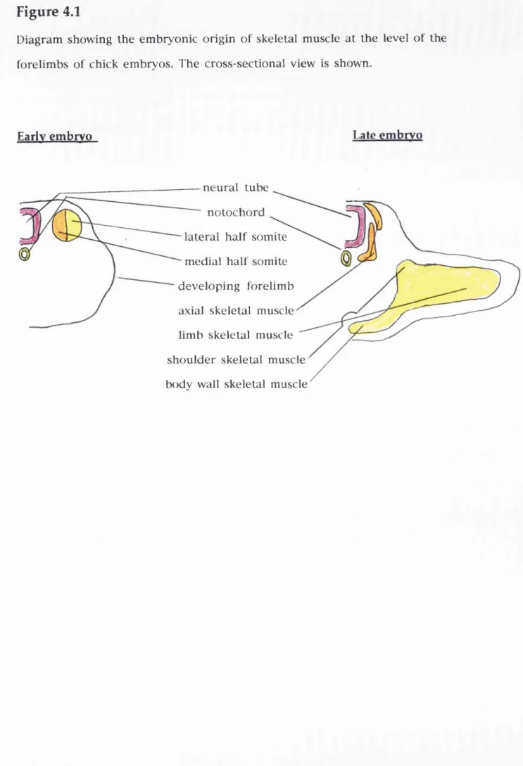
Figure 4.1
Diagram showing the embryonic origin of skeletal muscle at the level of the
forelimbs of chick embryos...122 Figure 4.2
Diagram matic representation of the morphogenetic events which take place
during the development of chick somites at the level of the forelimb 124
Figure 4.3
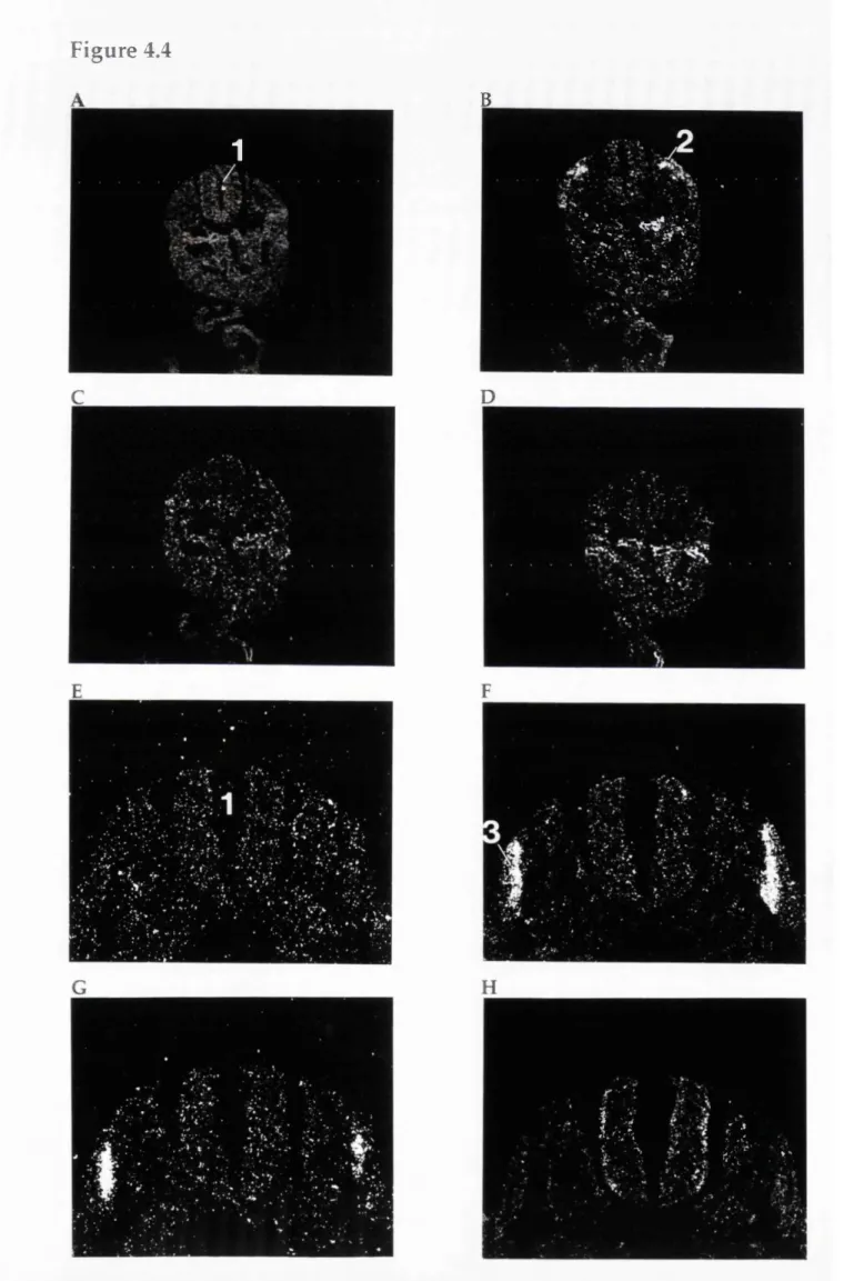
In situ hybridisation analyses of MyoD gene expression in the developing mouse embryo... 142 Figure 4.4
The distribution of RXRy transcripts in the somites and myotome of day 11 and day 11.5 p.c. rat embryos... 154 Figure 4.5
The distribution of RXRy transcripts in the myotome of day 12 p.c. rat embryo and a day 10.5 p.c. mouse embryo... 155
Figure 4.6
The distribution of RXRy transcripts in the myotome of day 13 p.c. rat
embryo... 156 Figure 4.7
The distribution of RXRy transcripts in the forelimb of a day 11.5 p.c. rat
embryo... 158 Figure 4.8
1
7
Figure 4.9
The distribution of RXRy transcripts in the hindlimb of a day 13 p.c. rat embryo... 160
Figure 4.10
The distribution of RXRy transcripts in the myotome and the skeletal muscle of the forelimb of a day 13 p.c. rat embryo...161 Figure 4.11
The distribution of RXRy transcripts in the skeletal muscle of the forelimb and hindlimb of a day 15 p.c. rat embryo...162 Figure 4.12
The distribution of RXRy transcripts in the skeletal muscle of the trunk and
forelimb of a day 15 p.c. mouse embryo... 164
Figure 4.13
The effects of cRA, tRA, and thyroid hormone on the incorporation of tritiated thymidine by proliferating H9 myoblasts... 166 Figure 4.14
H9 cells stained with Leishman's stain after treatment with tRA, cRA, and
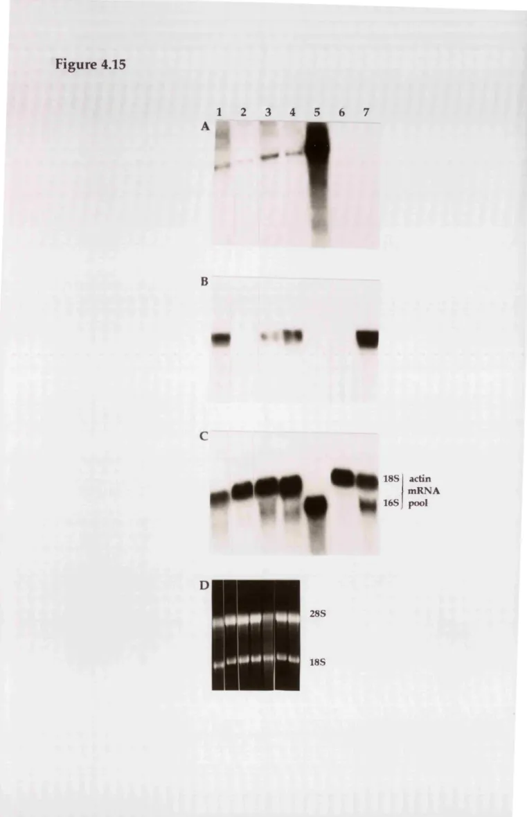
thyroid hormone... 169 Figure 4.15
Autoradiograms of northern blots of total RNA from H9 myoblasts hybridised with probes specific for: RXRy transcripts, myogenin transcripts, and the 'non muscle' and 'muscle' actin mRNA pools...172
Figure 5.1
Nucleotide sequence comparison of the putative retinoid response element present in the regulatory region of the chicken neuronal nAChR a 2 subunit gene with known naturally occurring retinoid response elem ents...194
Figure 5.2
Figure 5.3
C onfirm ation of the identity of the sense RXRy expression vector by
restriction enzyme digests...196
Figure 5.4
D iagram m atic represen tatio n of the luciferase re p o rte r gene constructs
containing upstream regulatory regions of the a 2 subunit gene... 197 Figure 5.5
The transcriptional effects of RXRy and 9cRA on the activity of the a2.6 and a2.4 luciferase constructs in BHK-21 and ND7-23 cells... 199 Figure 6.1
Northern blot analysis of RXRy, a-cardiac actin and the non-muscle' mRNAs in neonate and adult rat hearts...212 Figure 6.2
Primary cultures of neonatal rat ventricular cardiomyocytes... 214
Figure 6.3
Northern blot analysis of RXRy, a-cardiac actin and the non-muscle' mRNAs in neonate rat cardiomyocytes cultured in the presence or absence of thyroid
1 9
CHAPTER I
INTRODUCTION
1.1 - Foreword
The aim o f the present work was to understand the role of the retinoid
re c e p to r RX Ry and its relationship with the retinoid retinoic acid, during
ro dent em bryonic developm ent. Thus, this chapter provides b ackground
information about embryonic development, the role of retinoids and retinoid
receptors in development, and gives information about the action of retinoid
receptors at the molecular level.
1.2 - Developm ent, cells and genes
This section deals in very general terms with w hat happens during
em bryonic development so as to serve as background knowledge for the understanding of the potential role of the transcription factor RXRy and the signalling molecule retinoic acid. Vertebrate embryonic development can be defined as the gradual transformation of the small and structurally simple
fertilised egg cell into a bigger and incredibly com plex m u ltic ellu lar
organism (Slack, 1991; W olpert, 1991). Cell behaviour, such as cell
m ultiplication, cell movement, changes in cell shape and character, brings
about development. Since gene action controls cell behaviour it follows that
gene action governs development.
The early embryo is not transformed into its mature form in one step.
Instead, this transform ation takes place in several stages and involves
position dependent changes in cell commitment and cell potency. These two terms describe two different aspects of the same cell property: (a) cell potency is the total of all the things into which a given embryonic region or
d e v e lo p m e n ta l pathw ays av ailab le to a reg io n or cell at a given developmental stage. Early embryonic cells are totipotent because they can become any cell type. For example, in early mammalian embryos there is a
region called the inner cell mass which gives rise to all the tissues of the
embryo proper. These cells are totipotent. As development proceeds several position-dependent changes in cell potency take place giving rise to a gradual subdivision of the embryo into regions that have different potencies. Thus, an early embryonic region becomes its mature form only after it has
gone through several transform ations, each being a d iffere n t tra n s ie n t
em bryonic structure. These changes in potency are restrictions of potency which ultimately result in a region being unipotent (capable of giving rise to only one type of structure) just prior to its final transformation into its
mature form.
If one wants to understand embryonic development one needs to understand
how pattern formation takes place. That is, understanding why the potency
of a given region becomes restricted in one way and not another and why a specific restriction of potency is experienced by a given region and not another during a given embryonic stage. Insight into this comes when one realises that having the potency to change behaviour is necessary but not sufficient for the change to occur. In order for such changes to occur cells need to receive information in the form of signals. There are two known
types of signalling: cytoplasm ic localisation and induction. C ytoplasm ic
localisation presupposes differential distribution of regulatory molecules in
the cytoplasm of a cell. Then in the course of an asymmetrical cell division
th e two d a u g h te r cells in h e rit d if f e r e n t re g u la to ry m o le c u le s and
consequently behave differently. Induction requires that signalling comes
from outside the region that responds to the signal. The ability of a cell to
2
1
Signals are selective rather than instructive. That is, a signal selects a developm ental pathway out of several pathways available, and does not instruct the cell to follow a pathway which it does not have the potency to
follow. Evidence for this comes from experim ents involving inductions
between tissues from different species where the rule is that the nature of
structures formed by the responding tissue is limited by the range of the
available developm ental pathways present in the responding tissue. For
example, mouse dermis induces hairs from mouse epidermis, but feathers from chick epidermis and scales from lizard epidermis (Slack, 1991).
W hen cells, upon reception of a signal change b ehaviour to becom e
something, they are said to be determined to become that something when
they can do so independently of their embryonic environment. The act of changing cell behaviour is called differentiation and the cell state achieved is called state of commitment. When this state is the one found in the mature organism it is referred to as the terminally differentiated state.
Genes important for pattern formation are those that are responsible for the
generation , reception and interpretation of developmental signals so that a
cellular response is made. Examples of such genes are those which code for m em brane-bound receptors, transcription factors, or secreted proteins that act as signalling molecules. The next section deals with the role of retinoids as signalling molecules during vertebrate embryonic development.
1.3
- R etinoids and vertebrate developm ent
1.3.1
- R etinoid m etabolism
body originate in the diet either as provitamin A carotenoids or as preformed retinoids. The dietary carotenoids undergo a series of metabolic conversions extracellularly in the intestinal lumen and intracellularly in the intestinal
mucosa, which result in the generation of retinol. The absorbed retinol is transported to the liver, where the majority of the body's retinoids are stored as retinyl esters. Retinol enters the circulation, bound to retinol-binding
protein and is subsequently delivered to target tissues. A num ber of
interstitial retinoid binding proteins have been identified in ex tracellular spaces, suggesting that they may be involved in the delivery of retinoids to cells. Once inside the cell, retinol can be metabolised to all trans retinoic acid (tRA), 9 cis retinoic acid (9cRA) and other retinoids (Blomhoff et al., 1990;
Blaner and Olson, 1994). Endogenous retinoids include, tRA, 9cRA and 13 cis
retinoic acid (13cRA).
1.3.2 - Retinoids and embryos
Retinoids are very im portant for the correct em bryonic developm ent of
vertebrate em bryos, in c luding humans and rodents, beca u se they are
present in embryos and their absence or excess results in the generation of developmental defects (Morris-Kay, 1993; Means and Gudas, 1995).
The im portance of retinoids in human em bryos becam e evid en t when
pregnant women took the oral m edication isotretinoin (13cRA) for the
treatment of severe dermatological conditions. This resulted in spontaneous abortions, premature delivery and the generation of malformed foetuses and infants (Lammer and Armstrong, 1992). This condition is known as retinoic acid embryopathy. These defects were detected in autopsies of foetuses and
included brain, craniofacial and cardiovascular m alform ations. Surviving
2 3
structures derived from the hindbrain. These included dilation of the roof of the fourth ventricle and malformations in the cerebellar hemispheres.
S im ila r a b n o rm a litie s w ere ob serv ed in ro d e n t e m b ry o s a fte r oral administration of pregnant females with tRA (Morriss, 1972; Kraft et al., 1989; Horton and Maden, 1995). These defects must be the result of transient
elevations in the concentration of endogenous retinoids. The em bryonic
levels of orally administered tRA, 9cRA and 13cRA, peak at 2 hours after administration and become very low at 4 hours after administration and are
undetectable by 16 hours after adm inistration in both rats (W ard and
Morriss-Kay, 1995) and mice (Satre and Kochhar, 1989).
Evidence supporting the notion that the teratogenic effect of retinoids is direct and not due to a retinoid-induced change of maternal metabolism, came from experiments that involved rodent whole-embryo cultures. These experiments involved the direct exposure of embryos to retinoids. When tRA and 13cRA were added to the medium of mouse or rat whole-embryo cultures, embryonic defects were produced, similar to those found in humans (Kraft,
1992).
The finding that tRA and 13cRA are interconvertible isomers occurring
normally in the body suggested that the tRA and not 13cRA may be the active teratogen. E vidence for this came from experim ents involving w hole-rat embryo cultures showing that tRA is 10 times more teratogenically active than 13cRA. Further evidence came from the finding that tRA can prevent many of the defects in rat embryos induced as a result of maternal vitamin A deficiency during pregnancy (Morriss-Kay, 1993).
some differences in the type of malformations produced (Morriss-Kay, 1993;
Means and Gudas, 1995). Following vitamin A deficiency, developm ental
abnormalities included anophthalmia (absence of the eye), abnormalities in the genito-urinary tract, diaphragm, heart, lung and limbs.
The differences in the type of abnormalities associated with vitamin A deficiency or excess RA are difficult to explain. They can be partly explained
when one realises that the teratogenic phenotype depends on the dose and
timing of RA treatment. For example, treatment of mouse embryos during late gastrulation or early neurulation [7.75 days post coitum (d.p.c.) or 8 d.p.c.] had no effect on the morphology of limbs and trunk, but resulted in defects of the central nervous system (CNS) which included loss of rhombomeres
(repetitive morphological structures of the hindbrain), delayed neurulation
and reduction in the size of the forebrain and midbrain (Murphy et al., 1992).
On the other hand, mouse embryos that were treated with tRA at 10.5 d.p.c.,
had no observable CNS defects but had deformed inner ear structures,
truncated long bones of limbs, digit defects and fused vertebrae (Horton and
M aden, 1995). These tim e-dependent differences in the type of retinoid-
induced developmental defects must be due to differences in the commitment of the responding cells which change as development proceeds. In addition, the fact that during a given embryonic stage, some embryonic regions and
not others are sensitive to retinoids must reflect differences in their
c o m p e t e n c e .
The finding that retinoids are present in embryos strongly suggests that
they must be im portant for correct em bryonic developm ent. Endogenous
retin o id s were detected using high perform ance liquid chro m a to g rap h y
(HPLC). For example, tRA was found in embryonic chick limb buds (Thaller
and Eichele, 1987), Xenopus embryos (Durston et al., 1989). In addition, tRA
2 5
examined (from day 9 to day 14 of development). These included the spinal c h o rd , som ites b ra n c ia l arches, lim b buds, fo re b ra in , m id b ra in and
hindbrain (Horton and Maden, 1995). M oreover, 9cRA was detected in
Xenopus embryos (Kraft et al., 1994).
In conclusion, retinoic acid must be an endogenous signalling molecule that is responsible for the correct development of many different cell types. Thus, it must be able to induce cells to behave in many different ways. This
suggests that it must regulate the expression of many different genes.
Evidence for this is given in the next section.
1.4
-
R etin oic acid in flu en ces
the
exp ression
of
d iffe r e n t
genes
One way that retinoic acid might induce changes in cell behaviour could be
by regula tin g the expression of d evelopm entally im po rtan t genes that
themselves control cell behaviour. Such genes are the vertebrate Hox genes (Carrasco and Lopez, 1994; Krumlauf, 1994). The Hox genes are a family of
regulatory genes whose products are transcription factors. They contain a
region called the homeobox which encodes a 60 amino acid polypeptide, called the homeodomain, that is responsible for DNA binding. They play a crucial role in the generation of cellular differences along the vertebrate
an te rio r/p o s te rio r (A/P) axis because they show genom ic structural and
org an isatio n al sim ilarities with the hom eotic genes of D rosophila, and
because mutations in these genes lead to abnorm alities or absence of
structures, and to hom eotic transform ations along the A/P axis. Hox
expression is established in m esoderm , ectoderm and endoderm during
gastrulation and organogenesis. In the mouse and humans, Hox genes are organised in 4 unlinked clusters. A distinguishing feature of the Hox genes is
chromosome and their spatio-temporal expression along the A/P axis. This property is referred to as colinearity. That is, genes at the extreme 3' end of the clusters are activated the earliest and have the most anterior boundaries of expression. Moving along the clusters in a 5' direction, each successive gene adopts a progressively later, more posterior pattern of expression. Their tra n s c rip tio n a l re g u la tio n is im p o rtan t for the u n d e rs ta n d in g o f the generation of differences along the embryonic A/P axis. There is evidence that tRA may be important for the establishment of their expression pattern. For example, the expression of Hox genes in a human embryonal carcinoma (EC) cell line (see below) are activated sequentially by tRA in a 3' to 5' order
(Boncinelli et al., 1991). This reflects the colinearity observed in vivo,
strongly suggesting that tRA may be involved in the regulation of Hox gene
expression in vivo. Moreover, treatment of mouse embryos with tRA resulted
in alterations in the expression domains of endogenous Hox genes (e.g. Wood et al., 1994). The molecular mechanisms involved in the regulation of Hox genes by retinoic acid are not fully understood.
A good experimental system for the study of genes whose expression is
regulated by retinoic acid during embryonic development is the use of EC
cells (Slack, 1991). EC cells are the stem cells present in teratocarcinomas.
These are tumours of germ cells or early mammalian embryos, consisting of dividing stem cells (EC cells) and a wide range of disorganised differentiated cell types. EC cells resemble cells of the early mammalian embryo in the sense that they are pluripotent and undifferentiated. For example, the P I 9
line of EC cells differentiates into neurons, glia and fibroblast-like cells
when exposed to tRA (McBurney et al., 1988). The F9 cell line is a murine EC
cell line that was used extensively for the identification of genes whose
expression is regulated by tRA (G udas, 1991). These genes include
2 7
structural genes such as laminin B l , Laminin B2, and collagen I V ( a l ) , and growth factors such as transforming growth factors (TGFs) and fibroblast growth factors (FGFs). The expression of the activin receptor type IIB in F9 cells is elevated by both 9cRA and tRA (Wan et al., 1995). In addition, 9cRA
and tRA induce a sim ilar phenotype in human te ra to c a rc in o m a cells,
suggesting that these retinoids have similar effects (Kurie et al., 1993).
It should be noted that retinoic acid influences the expression of genes in
many other cell lines and primary cell cultures. It is clear from the above
that retinoids can influence the expression of many genes. The next section discusses the molecular mechanism through which retinoids regulate gene e x p r e s s i o n .
1.5 - Retinoids and gene expression
1.5.1 - Signalling by retinoids
There are two main mechanisms through which signalling molecules can regulate gene expression: (a) the signalling molecule binds to a cell surface receptor resulting in a cascade of events that transduce the signal to the
nucleus via intracellular second messengers, (b) the signalling m olecule
bin d s to n u c le a r re c e p to rs w hich th e m s e lv e s in flu e n c e tr a n s c r ip tio n
directly. There is evidence indicating that retinoids act via the second
m e c h a n i s m .
Four structurally distinct classes of intracellular proteins that bind retinoids with high affinity have been identified in mammals: the cellular retinol- b in d in g p ro tein s (C R B P s), the c e llu la r retin o ic acid -b in d in g p ro tein s
(CRABPs)(Ong et al., 1994; see section 1.5.5), and the retinoid receptors. The
latter are the retinoic acid receptors (RARs) and the retinoid X receptors
receptor superfamily. The following section gives inform ation about the
structure and function of retinoid receptors within the context of this
s u p e r f a m i l y .
1.5.2 -
The steroid hormone receptor superfamily
RARs and RXRs are nuclear retinoid receptors which are transcription
factors whose function is influenced by retinoid binding. They belong to the
steroid horm one receptor superfam ily of nuclear receptors, the largest
known family of transcription factors in eukaryotes (Tsai and O' Malley,
1994). M embers of this superfamily regulate transcription by binding as dim ers to specific cis-acting elements, called hormone response elements
(H REs), p resent in the regulatory regions of target genes. N aturally
occurring HREs are composed of two or more copies of a six nucleotide motif, termed a half-site (Stunne/fberg, 1993). All the receptors of this superfamily share: (a) a similar building plan consisting of several domains (Figure 1.1), (b)some amino acid sequence identity within their DNA and ligand binding
domains and, (c) the ability to bind hydrophobic ligands (Green and
Chambon, 1988; Evans, 1988; Pfahl et al., 1994). In addition to RARs and RXRs, the n u c le a r rece p to r superfam ily in cludes o e stro g en rece p to rs (E R s), p ro g e s te ro n e rece p to rs (PRs), g lu c o c o rtic o id re c e p to rs (G R s), th y ro id hormone receptors (THRs), Vitamin D receptors (VDRs) and the peroxisome proliferator activated receptor (PPAR). Each group of receptors comprises a family. For example, the RXR family contains RXRa,(3, and y (Mangelsdorf et
Figure 1.1
A diagrammatic representation of the basic structure of members of the
steroid hormone receptor superfamily showing the different domains
(designated A to F).
1.5.2.1 - The structure and function o f the DNA bind in g domain Deletion experiments showed that the isolated DNA binding domain (DBD), also called C domain, can bind DNA suggesting that its correct folding is
independent of the other regions present in the nuclear receptors (Green
and Chambon, 1988; Evans, 1988). Comparison of the amino acid sequences
present in the DBDs of all receptors showed that there are eight cysteine
residues whose relative positions are invariant. This suggested that the DBD
of nuclear receptors may contain two zinc-finger motifs, because cysteine
residues are present in known zinc-finger motifs (Miller et al., 1985), and because the DBD of the oestrogen receptor requires zinc atoms to bind DNA.
The existence of the two zinc-finger motifs was confirmed from experiments
which involved the use of nuclear magnetic resonance (NMR) spectroscopy to elucidate the three-dimensional structures of oestrogen receptor (Schwabe and Rhodes, 1991), RX R a (Lee et al., 1994), and RARp (Knegtel et al., 1993).
These studies showed that these DBDs have similar tertiary structures (Figure
1.2), strongly suggesting that they bind DNA in a similar way. There are two similar zinc-finger motifs folded together to form a single structural unit. In each motif there is an extended loop between the two pairs of metal-binding cysteines. In both motifs, an a helix begins at the residue following the third metal-binding cysteine and extends over 11-13 amino acid residues. The two a helices, h elix-1 and helix-2, are highly amphipathic and are folded together, such that they cross at right angles near their midpoints. They are held together by hydrophobic interactions through hydrophobic residues whose
3
1
second zinc-finger (zinc-finger 2) are also exposed on the outside suggesting
that they may be involved in protein/protein interactions. A unique feature
of the RXRs is a third helix, helix-3, in their DBD (Figure 1.2) which is not
present in all the other receptors studied (Lee et al., 1993; Pfahl et al., 1994). Helix-3 is part of a region named the T-box (Wilson et al., 1992). There are two other regions within the DBD: the D-box and the P-box. The functions of these three regions is discussed below.
The P -b ox
Mutagenesis studies showed that the specificity of DNA sequence recognition
by nuclear receptors depends on three amino acids present within the P-box
(Umesono and Evans, 1989; Danielson et al., 1989; Mader et al., 1989; Martinez and Wahli, 1991), a region situated within helix-1 of the first zinc-finger (Figure 1.2). For example, it has been possible to convert GR into a receptor that binds an oestrogen response element (ERE), by replacing the GR P-box with the ER P-box (Danielson et al., 1989). Based on the homology of the P-box amino acid sequence, nuclear receptors can be grouped into subfamilies. At least four subfamilies have been identified so far (Pfahl et al., 1994). The first
subfamily (glucocorticoid subfamily) includes GRs and PRs, whereas the
second group, the oestrogen subfamily, includes ERs. A third group, the
retinoid/thyroid subfamily, includes RARs, RXRs, THRs, VDRs and PPARs.
C onsistent with the role of the P-box, many of the receptors that have
identical P-boxes were shown to bind the same DNA sequences. For example, retinoid receptors, THRs, VDRs, and PPARs bind to the same HRE. This HRE was
synthetic and consisted of a single half-site whose sequence was the
consensus of many naturally-occurring HREs found in the promoter regions
of genes whose transcription is influenced by ligands that bind to these
Figure 1.2
Schematic representation of the DNA binding domain of RXRa. Helix
regions are in grey boxes; the zinc-finger cysteine residues are circled in
black; the D-box residues are in squares; capital letters represent amino adds
(Lee et al., 1993).
Zinc finger 1
Zinc finger 2
in
K
G H
S Y
S G
R V
D Y
'A
I FTKH
D-box
X'i
Z n
V'GÎ
11
/" “v j
0 /
[^10 W
R
I K 1 T i R '
GJFFSTVMPLTY i_J'(^CLA#MKRË A V Ô Ë i^ RGKDRNENEVEST
3 3
reg ulate the transcription of different genes partly because they have
different P-boxes. However, differences in the P-box alone are not sufficient to explain the different ligand-induced transcriptional responses mediated through the different receptors of the retinoid/thyroid subfamily, because they share the same P-box. Thus other regions must be involved. One of these is the D-box.
T he D -box
The D-box consists of the five amino acids present between the first and
second cysteines of zinc-finger 2 (Figure 1.2) which are involved in
p ro te in /p ro te in in teractions betw een the two m onom ers that make up
nuclear receptor dimers. This was revealed from structural studies of
glucocorticoid homodimers (Luisi et al., 1991) and RXR-THR heterodimers
(R a stin ejad et al., 1995). D ifferent receptors have d iffe re n t D -boxes,
suggesting that different receptors have different dim érisation properties
(Pfahl et al., 1994).
The T -b ox
As mentioned above, helix 3 is part of the T-box, a region whose tertiary structure is unique to the DBD of RXRs. Mutagenesis studies revealed that this region is necessary for R X R a and RXRp homodimerisation and for efficient DNA binding (Wilson et al., 1992; Lee et al., 1993). These observations were confirmed by structural studies carried out by Rastinejad et al (1995), who
sho w e d th a t amino acids p re s e n t w ithin h elix 3 are in v o lv e d in
p ro tein /p ro te in and protein/D N A interactions.
The A -box
The A-box is situated at the carboxy terminal end of the DBD and mutations in
1.5.2.2 - The structure and fu n ction of the ligand b in d in g d om ain The ligand binding domain (LDB) is situated within the E domain and contains large numbers of hydrophobic residues which have been suggested
to form a hydrophobic 'pouch' in which the ligand can fit. The LDB was
shown to bind with high affinity to receptor mutants that did not contain
any of the other domains, suggesting that the LDB assumes is correct folding independently of the other domains (Green and Chambon, 1998; Evans, 1988). RXRs bind 9cRA with high affinity, but fail to bind tRA, 7cRA, ll c R A , or 13cRA. In contrast, RARs bind both tRA and 9cRA with high affinity (Allenby et al,, 1993), This finding shows that RXRs and RARs are selective with respect to the nature of retinoids they bind. The chemical structures of 9cRA and tRA are shown in Figure 1,3, The carboxy terminal regions of the E and F domains are also involved in dimer formation and contain the ligand-
dependent transcriptional activation function (AF2) (Green and Cham bon,
1988; Rowe and Brickell, 1993; Pfahl et al,, 1994), The RXR AF2s are unmasked or activated in the presence of 9cRA, and RAR AF2s are activated in the presence of 9cRA or tRA (Nagpal et al,, 1993), Thus, binding of the hormone induces a conformational change that allows the appearance of an activation
domain at the surface of the protein. Other data in support of this
ligand-in d u c ed c o n fo rm a tio n a l sw itch were o b ta in e d by p ro te a se re s is ta n c e analyses of the LDB in the presence or absence of ligand (Bhat et al,, 1993), In addition, deletions or point mutations of the very C-terminal end of the LDB resulted in the disruption of the ligand-dependent activation properties, without abolishing DNA or ligand binding functions (Folkers et al,, 1993; Nagpal et al,, 1993), For more information refer to 'the role of the ligand' in section 1,5,3, The crystal structure of the LDB of the human R X R a has been recently determined (Bourguet et al,, 1995). These workers showed that two
helices and one loop form the homodimerization surface and hydrophobic
3 5
existence of a possible ligand-binding pocket that could allow 9cRA to interact with the AF2 domain. Amino acid sequence alignments suggested that the three-dimensional structure of the RXR LED may be a prototype fold of all LDBs of nuclear receptors.
1.5.2.3 - The A and B domains
The A and B domains have a ligand independent transcriptional activation
function (API) and constitute the N -terminal region of nuclear receptors (Green and Chambon, 1988; Rowe and Brickell, 1993; Pfahl et al., 1994). A PIs have been reported for all three RARs ( a , p, and y) and for R X R a and RXRy.
These A P Is show weak activation properties by themselves and strongly
synergise with their corresponding AP2 (Nagpal et al., 1993). A P Is show
prom oter context specificity (Nagpal et al., 1992) and cell-type specificity (Polkers et al., 1993). The cell type specificity of A PIs strongly suggests that they may interact with cell-type-specific proteins.
1.5.2.4 - The D domain
The function of the D domain is not clear. In the steroid hormone receptors it
is im portant for nuclear localisation, and provides a flexible link between
the DBD and LED, hence it is sometimes called the hinge region (Green and
Figure 1.3
The chemical structures of all
trans
and 9
cis
retinoic acid.
COOH
Whtrans
retinoic acid
3 7
1.5.3
-
Retinoid
receptors
and
transcriptional
regulation
This section attem pts to explain how retinoic acid can in fluence the
expression of many different genes, and how TH, VD, 9cRA, and tRA can influence the expression of distinct sets of genes. To do this, one needs to i d e n t i f y all the c o m p o n e n ts th a t p a r t i c i p a t e in l i g a n d - i n d u c e d transcriptional changes, and understand their role. These include hormone response elements (HREs), ligand, nuclear receptors, and other trans-acting
f a c t o r s .
H o r m o n e r e s p o n s e e le m e n t d i v e r s i t y
As mentioned above, members of the retinoid/thyroid subfamily have the same P-box and can bind to a common synthetic DNA half-site composed of six
nucleotides whose sequence is 5'- A G ^ /tTCA-3' (Stunnerberg, 1993). This is
surprising because different receptors bind different ligands and different
ligands regulate the transcription of differe n t genes. So how is the
specificity of hormonal response achieved? The answer lies partly in the
observation that promoters that are activated by different ligands have
d iffe re n t n a tu ra lly -o c c u rrin g HREs (Figure 1.4). M ost o f the know n
naturally occurring HREs have been ch aracterised by th e ir ability to
activate a heterologous promoter as a result of binding nuclear receptors in the presence of specific ligands (Pfahl et al., 1994). For example, retinoid re s p o n se elem en ts (RREs) a c tiv ate tra n s c rip tio n by b in d in g re tin o id receptors in the presence of retinoids. As shown in Figure 1.4, half-sites are arranged as direct repeats, with each half-site being composed of closely
Figure 1.4
Naturally-occurring hormone
response
elements
derived
from
the
promoters of: rat Cellular Retinoic Acid Binding Protein II (CRABP II)
(Durand et al., 1992), ApoA (Zhang et al., 1992), Osteopontin (Noda et al.,
1990), Moloney Murine Leukemia Virus LTR (MoMLV) (Sap et al., 1990),
RARb2 (De The et al., 1990). Each nucleotide present between half-sites is
represented with the letter N. Each half-site is underlined by an arrow.
Gene promoter
Response elem ent
Apo AI
5'-GGGTCANGGTTCA-3'
CRABP II
5'-AGTTCANNAGGTCA-3'
Osteopontin
5-GGTTCANNNGGTTCA-3’
MoML V
5-GGGTCANNNNAGGTCC-3'
3 9
nucleotides between half-sites present in different promoters varies. Thus,
differences in spacing between adjacent half-sites may contribute to the
nucleotides between half-sites present in different promoters varies. Thus,
differences in spacing between adjacent half-sites may contribute to the
specificity of hormone response (Umesono et al., 1991). There is evidence
that this is the case (see below). To understand this, one must first realise that nuclear receptors bind to their naturally occurring HREs as dimers.
R e tin o id rec ep to r s bind DNA as h o m o d im e r s or h e te r o d im e r s RARs, THRs and VDRs, bind with high affinity to their cognate DNA binding sites as heterodimers with RXR in the absence of ligand (Yu et al., 1991; Leid
et al., 1992; Zhang et al., 1992a). Moreover, RXRs bind DNA with high affinity
as homodimers, in the presence of their ligand 9cRA (Zhang et al., 1992b). In addition, VDR-THR heterodimers have been observed (Schrader et al., 1994). These findings demonstrated that RXRs can be involved in transcriptional responses induced by retinoid and non-retinoid signalling molecules (Rowe and Brickell, 1993). Each member of the heterodimer binds to one of the two closely arranged half-sites (Zhang et al., 1991; Rastinejad et al., 1995). Recent studies showed that RXRs can also bind DNA and activate transcription as tetramers and other high-order oligomers to HREs poorly recognised by RXR dimers (Chen and Privalsky, 1995).
The 1 3 - 4 - 5 Rule
It has been shown that binding specificity can be determined through the number of nucleotides separating the half-sites. Direct repeat of AGGTCA-like half-sites spaced by either three, four, or five nucleotides would function as optimal HRE for the VDR, THRs, and RARs, respectively. Mutations that alter
the d ista n ce betw een these hexam ers can sw itch re c e p to r s p ecificity
(Umesono et al., 1991). In addition, direct repeats separated by one nucleotide
Lehmann et al., 1992), and activate a heterologous promoter in response to RXR but not RARs (Mangelsdorf et al., 1991). Based on these observations E v an s and c o lla b o ra to rs p ro p o sed th a t the s o -c a lle d 1-3-4-5 'ru le'
(M angelsdorf et al., 1994) could be used to predict the preferred receptor
dim ers which should bind to uncharacterised HREs. H owever, this rule
appears to be of restricted value that does not always hold. For example, the
RRE found in the promoter of RARy which consists of direct repeats separated by five nucleotides, responds poorly to tRA (Lehmann et al., 1992). This suggests that in addition to the 1-3-4-5 rule, other factors must be taken into account. Moreover, this rule does not apply to HREs which consist of h a l f- s it e s a rra n g e d as p a lin d ro m ic or in v e rte d p a lin d ro m ic re p e a ts (Mangelsdorf et al., 1994). For example, Naar et al (1991), showed that the orientation of the half-sites within HREs is also important. Direct repeat, palindromic, and inverted palindromic organisation of half-sites spaced by
three nucleotides confers specific response to RARs, ERs, and THRs
resp ectiv ely . Taken together, these o bservations su ggest that d iffe re n t
com binations of receptor dimers may recognise different HREs and that
factors other than h alf-site spacing must also be im p o rtan t for this
recognition. This possibility is explored in the following sections.
H a l f - s i t e s e q u e n c e and f la n k in g n u c le o t i d e s
The strength of DNA binding by nuclear receptors depends on sequences within and around half-sites. For example, optimal binding of RAR takes place when the half-site has the sequence P uA G ^/yT C A , but not other related
sequences (M angelsdorf et al., 1994). This is consistent with the finding that the RARp promoter which has half-sites that are composed of this optimal
sequence, and arranged as direct repeats separated by five nucleotides,
becomes strongly activated by tRA (Sucov et al., 1990). In contrast, the
p ro m o ter o f RARy whose half-sites are also arranged as direct repeats
4 1
only poorly activated by tRA (Lehmann et al., 1992). Moreover, R A R a binds
more efficiently to the RRE present in the promoter of R A R a 2 than that
present in the promoter of RARp 2. This suggests that these differences in DNA binding affinity may be due to the differences in the nucleotide sequences within and around the half-sites, since both these RREs contain direct repeats separated by the same number of nucleotides (Mader et al., 1993). The s ig n ific a n c e o f seq u en ce s fla n k in g the h a l f- s it e s was strengthened from the work of Rastinejad et al (1995), who showed that the DNA binding domain of THR, bound as a heterodimer with the DBD of RXR to a thyroid hormone response element (THRE), made contacts beyond the six- base pair half-site.
P r o t e i n / p r o t e i n i n t e r a c t i o n s and H R E r e c o g n i t i o n
It has been shown that the DBDs of RAR-RXR and THR-RXR heterodimers bind with a given polarity to direct repeats separated by two (DR2) or five nucleotides (DR5) and DR4 respectively. In both cases, the RXR partner bound
the 5' half-site (Predki et al., 1994; Zechel et al., 1994). The same was also
shown for full length heterodimers. Thus, this polarity in DNA binding must be im portant for HRE recognition by nuclear receptors. Since, this polarity d e p e n d s on p r o te in /p ro te in in te ra c tio n s b e tw e e n the D B D s o f the
heterodimeric partners, it follows that such interactions may be important in
HRE recognition by nuclear receptors. Indeed this is the case. This was shown by mutagenesis studies (Zechel et al., 1994). RAR-RXR heterodimers
use different dimérisation interfaces to bind to DR2 and DR5 elements, and
T H R-RX R heterodim ers use dim érisation interfaces for binding to DR4
elements that are different to those observed for RAR-RXR heterodimers. For
exam ple, RA R-RXR hetero d im e r binding to DR5 involves in teractio n s
residues that are not conserved between nuclear receptors suggests that different combinations of heterodimers may show preferences for different
HREs. The involvem ent of interactions betw een non-co n serv ed residues
present in the DBDs of heterodimeric partners was shown by the structural
study of Rastinejad et al (1995) for THR-RXR bound to a DR4 element.
Taken together, these findings suggest that anything that can influence th e se p r o te i n /p r o te in in te r a c tio n s may in flu e n c e the tr a n s c r i p t i o n a l response. The A-box of RXR may have such a function because point
mutations within it force the RXR member of the RXR-THR heterodimer to
bind the 3' and not the usual 5' half-site of a DR4 element, without affecting the stability of DNA/protein interactions (Kurokawa et al., 1993).
The role o f the ligand
The main role of the ligand is to activate the receptor so that it forms an a c tiv e tra n s c rip tio n in itia tio n com plex. This in v o lv e s li g a n d -in d u c e d conformational changes of nuclear receptors. For example, 9cRA caused RXR to change its conformation as shown by limited proteolysis (Leid, 1994). Moreover, using chimeric receptors Casanova et al (1994), provided evidence that DNA-bound THR and RAR may interact with a common inhibitory trans acting factor that suppresses the activity of these receptors, and that the role of their cognate ligands is to dissociate these receptors from this putative
inhibitor. The region responsible for interacting with this inhibitor was
found to be within the LED.
4 3
with tRA activates transfected R X R a to drive transcription of an R X R a - d r iv e n reporter gene (Heyman et al., 1992; Levin et al., 1992), whereas no effect was observed in yeast cells (Mak et al., 1994). This strongly suggests that tRA must have been converted to 9cRA in S2 but not in yeast cells, since 9cRA but not tRA binds and activates RXR. A mechanism through which the type of ligand
present may influence the tran scrip tio n a l response was disco v ere d by
(Zhang et al., 1992). These workers showed that 9cRA but not tRA can
increase the amount of RXR homodimers and decrease the am ount of
heterodimers, thereby generating new response pathways. This is because RXR homodimers form and bind DNA only in the presence of 9cRA, whereas
RXR-THR and RXR-RAR heterodimerisation and DNA binding takes place in
the absence of ligand, and because both homo- and heterodim ers were formed when 9cRA was mixed with either RAR and RXR or THR and RXR. This is consistent with the work of Medin et al (1994), who showed that 9cRA
increases RXR homodimer binding to a RRE, while decreasing the affinity of
RXR-THR heterodimer binding to the same element. A different mechanism
through which the identity of the ligand determines the transcriptional
response was suggested by the work of Schrader et al (1994). They showed that the VDR-THR heterodimer binds to a naturally occurring DR3 with a polarity opposite to the one observed when it binds a DR4, and that the ligand
responsible for transcriptional activation is the one that binds to the
receptor which contacts the downstream half-site.
I n h i b i t io n o f t r a n s c r ip t io n by n u c le a r r e c e p t o r s
Retinoid receptors can be involved in negative transcriptional responses.
For example, it has been shown that RXR homodimers bind to D R l elements
and activate transcription in response to 9cRA (M angelsdorf et al., 1991). However, although RXR-RAR heterodimers bind to the same D R l elements
w ith h ig h e r a ffin ity than RXR h o m o d im e rs, they fail to a c tiv a te
Explanation of this came from the work of Kurokawa et al (1994) who showed that the RAR member of the heterodimer allosterically prevented 9cRA from binding to and activating RXR because RAR was bound to the upstream half sites of DRl elements.
O rphan receptors can also in h ib it sig n allin g by retin o id s and other
hormones (Pfahl et al., 1994). For example, COUP receptors inhibit retinoid receptor activities on several response elements that are activated by RAR- RXR heterodimers or RXR homodimers, by binding strongly as homodimers to these elements, without forming heterodimers with RXR (Tran et al., 1992).
R e tin o id r e c e p t o r s and A P I
The activator protein-1 (AP-1) is a transcription factor that contains the products of the proto-oncogenes c-Fos and c-Jun and binds to a sequence motif known as the AP-1 binding site (Bohmann et al., 1987). AP-1 inhibits transcriptional activation by RAR and vice-versa, through a mechanism that
involves protein/protein interactions (Pfahl et al., 1992). This regulatory
mechanism, by which members of two different families of transcription factors can control each others' activity is likely to represent a major
signalling switch.
In conclusion, the factors involved in transcriptional regulation by retinoid and other nuclear receptors are many and complex, and so a com plete explanation of the molecular mechanisms involved, has yet to be proposed.
1.5.4 - The RAR and RXR families
Three different RARs (R A R a, p and y) are known to exist in mammals, birds