Neuropsychiatric Disease and Treatment
Dove
press
O r i g i N a l r e s e a r c h
open access to scientific and medical research
Open access Full Text article
evaluation and follow-up of cognitive functions
in patients with minor stroke and transient
ischemic attack
Çig˘dem Deniz1
Yahya Çelik2
Tug˘çe Özdemir Gültekin1
Gozde Eryig˘it Baran1
Çag˘la Deniz3
Talip asil1
1Department of Neurology, Faculty of
Medicine, Bezmialem Vakıf University, i˙stanbul, 2Department of Neurology,
Faculty of Medicine, Trakya University, edirne, 3Department of Neurology,
avrupa hospital, adana, Turkey
Background and purpose: We aimed to examine the incidence of cognitive impairment among patients with stroke, the associated risk factors, progression of the cognitive impairment, and the association between the localization of the lesion(s) as detected by magnetic resonance imaging and the affected areas of cognitive function.
Methods: A total of 40 patients over 18 years of age enduring a transient ischemic stroke or minor stroke within the past 3 months who had a minimum life expectancy of 1 year were included in this study. Same number, age-, and sex-matched individuals were included as controls. Patients were inquired on the presence of risk factors for stroke. A series of neuropsychological test batteries were administered in patient and control subjects for assessing cognitive functions. These tests were readministered at 6 and 12 months of follow-up to assess the progression of cognitive functions.
Results: In this study among the patients with stroke, a significant impairment was seen in multiple cognitive functional tests following ischemic stroke as compared to control groups. The most common risk factors for stroke included hypertension (72.5%), hyperlipidemia, and cigarette smoking. The number of cognitive domains with an impairment was highest (in four cognitive tests) among those with coronary artery disease and atrial fibrillation, followed by those who had a .50% stenosis in Doppler (three cognitive tests). These findings suggest that the frequency of risk factors associated with stroke does not correlate with the frequency of risk factors associated with cognitive dysfunction. The stroke localizations were classified among the patients with stroke and reviewed in accordance with cognitive impairment.
Conclusion: Neuropsychological tests, clinical findings, and imaging studies should be used to document the poststroke cognitive dysfunction.
Keywords: dementia, vascular dementia, cognitive impairment, neuropsychological assess-ment, vascular risk factors
Introduction and objective
Cerebrovascular diseases represent the third leading cause of death and disability.1
Furthermore, stroke is associated with significant reductions in the quality of life, not only due to the presence of neurological deficits but also due to resultant cognitive dysfunction.2
Strokes also represent a major etiological factor for dementia and, accordingly, poststroke dementia (PSD) is considered a subtype of vascular dementia. PSD is defined as the presence of dementia developing within 3 months after an acute, recur-ring, or initial stroke episode.3 Stroke is associated with a 4–12-fold increase in the
risk of dementia.4,5 The reported PSD prevalence varies between 12.2% and 31.8% correspondence: Talip asil
Department of Neurology, Faculty of Medicine, Bezmialem Vakıf University, Vatan Caddesi, 34083 I˙stanbul, Turkey email [email protected]
Journal name: Neuropsychiatric Disease and Treatment Article Designation: Original Research
Year: 2016 Volume: 12
Running head verso: Deniz et al
Running head recto: Evaluation and follow-up of cognitive functions DOI: http://dx.doi.org/10.2147/NDT.S102193
Neuropsychiatric Disease and Treatment downloaded from https://www.dovepress.com/ by 118.70.13.36 on 25-Aug-2020
For personal use only.
Number of times this article has been viewed
This article was published in the following Dove Press journal: Neuropsychiatric Disease and Treatment
Dovepress
Deniz et al
depending on a number of factors, such as the diagnostic criteria used or the study populations included.4,5 Although
cognitive dysfunction may be accounted for by the presence of vascular lesions and white matter changes, the underlying neurodegenerative disorders may also contribute to PSD. PSD has a multifactorial pathophysiology, including a number of factors such as advanced age, lower educational status, previous history of stroke, prestroke cognitive dysfunction, diabetes mellitus, myocardial infarction, atrial fibrillation (AF), history of epileptic seizures or sepsis, congestive heart failure, silent cerebral infarction, global lobe or temporal lobe atrophy, or white matter alterations, although the exact mechanisms causing PSD remain unknown. Also, the role of homocysteine as a risk factor in the development of PSD remains a matter of dispute.
In addition to the concept of vascular dementia, another entity referred to as vascular cognitive impairment has been proposed as an umbrella term encompassing all pathological abnormalities due to cerebrovascular conditions resulting in cognitive impairment from mildest cognitive impairment to dementia.6
The milder forms of this cognitive impairment include the nondemented vascular cognitive impairment and mild vascular cognitive impairment.7–11 While the mild
cogni-tive impairment mainly focuses on memory impairment, nondemented vascular cognitive impairment is not limited to one cognitive domain. Desmond et al12 observed delayed
improvement between poststroke 3 and 15 months in 10% of their patients with a mean age of 70 years. In another study involving younger patients, among those with a mild cognitive impairment after stroke, over 30% had improved cognitive functions between 0 and 6 months, with complete recovery between 12 and 18 months.13
Identification of treatable risk factors in PSD has prog-nostic implications. Also, determination of risk factors for stroke is important for prevention of stroke and PSD. When the risk factors and etiological factors are established, those carrying a risk for the development of PSD may be readily identified and prognostic estimations may be performed. In this regard, it should be borne in mind that not all patients with stroke develop dementia.14
With this background, we aimed to examine the devel-opment of cognitive impairment after mild ischemic stroke or transient ischemic episodes through the assessment of the frequency of poststroke vascular cognitive dysfunction or PSD, localization of the stroke in patients with vascular cognitive dysfunction or PSD, and the association between cognitive dysfunction and risk factors.
Materials and methods
This prospective controlled study was carried out at the Neurology Department of the Medical Faculty of Thrace University and patients were selected among those who were admitted with a diagnosis of acute ischemic stroke after the initial assessment at the neurology outpatient unit or emergency room between 2009 and 2011. A total of 40 patients were included in the minor stroke group based on a National Institutes of Health Stroke Scale (NIHSS) score of 5 or less, while similar level of education-, age-, and sex-matched individuals with no vascular or cognitive conditions served as controls (n=40). Patients over 18 years of age with a minimum life expectancy of 1 year who had a transient ischemic stroke or minor stroke within the past 3-month period comprised the target patient population. Whereas, patients ,18 years with a life expectancy of ,1 year due to a number of conditions, including malignancy, who had history of hemorrhagic stroke, and an NIHSS score .5 with moderate or severe stroke were excluded. Written informed consent was obtained from the patients, and the study was approved by Trakya University Ethics Committee.
At study entry, in addition to NIHSS scores, modified Rankin Scale scores were also obtained. The lesion sites (by applied computed tomography and magnetic resonance scans on patients) were classified according to Oxfordshire Community Stroke Project into the following clinical subtypes: partial anterior circulation infarcts, posterior circulation infarcts, and lacunar infarcts.15
Patients were enquired on the presence of risk factors for stroke, including cardiovascular risk factors such as diabetes mellitus, hypertension (HT), coronary artery disease (CAD), and cardiac arrhythmias, as well as for cigarette smoking and hyperlipidemia (HL). Other risk factors were also recorded during history taking and admission. A series of neuropsy-chological test batteries were used in both patient and control groups to provide a comprehensive cognitive assessment.
The following neuropsychological tests were used for the assessment of cognitive functions: Instrumental Daily Activities of Living for functionality; Mini Mental State Examination (MMSE) for general assessment; forward and backward counting and calculation test for attention; word list memory (can be expressed as flash memory), word list recall (can be expressed as learning period), and word list recognition (can be expressed as recall of knowledge) for the verbal memory; Boston naming test for the language; clock drawing test for planning visuospatial abilities; construction ability for constructing and visual memory (visuospatial functions); and the Frontal Assessment Battery (FAB) for
Neuropsychiatric Disease and Treatment downloaded from https://www.dovepress.com/ by 118.70.13.36 on 25-Aug-2020
Dovepress evaluation and follow-up of cognitive functions
executive functions. These tests were readministered at follow-up months 6 and 12 to examine the course of cognitive functions. All neuropsychological tests used were based on Turkuaz Alzheimer Workgroup.
statistical assessments
Number Cruncher Statistical System 2007 and PASS 2008 Statistical Software (Kaysville, UT, USA) were used for data analyses. In addition to descriptive statistics (mean, standard deviation, frequency, rate), Student’s t-test was used for comparisons between the groups for the quantitative data with normal distribution. For detecting the changes in repeated measures, the Variance Analysis for Repeated Measures was used, while Bonferroni test was used for post hoc assessments. The statistical significance was set at a P-level ,0.05.
Results
A total of 40 patients with a mean age 61.03±10.75 years who had minor stroke or tranisent ischemic attack (TIA) enrolled in the study. Forty controls of the same age and sex were recruited into the study. Of the total study population, 30 were female (37.5%) and 50 (62.5%) were male (Table 1).
Neuropsychological tests
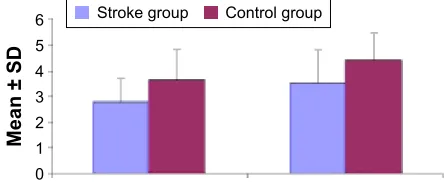
A significant worsening in the instrumental scores was detected among patients with stroke within the first month following the incident as compared to controls (P,0.01) (Figure 1). There were no significant changes from base-line in MMSE and forward counting in study groups (P.0.05), while significantly lower scores were obtained in backward counting and calculation skills in patients with stroke (P,0.01) (Figure 2). Although the two groups were comparable in terms of word list recall and word list recogni-tion skills (P.0.05), an assessment of word list memory and overall scores showed significant worsening among patients with stroke than controls (P,0.01) (Figure 3). Abstraction skills were significantly reduced in patients with stroke (P,0.01) (Figure 4). There were no differences in praxis measurements in either group. Clock drawing test results
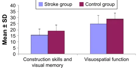
were similar across the groups (P.0.05), while significantly lower scores in construction skills, visual memory, and visu-ospatial functions overall were detected among patients with stroke (P,0.01) (Figure 5). Boston Naming Test results did not differ significantly between the two groups (P.0.05), while FAB was significantly lower at baseline in patients with stroke (P,0.01) (Figure 6).
With a similar level of education between the two groups, patients with stroke exhibited significant worsening in the majority of neuropsychological tests and experienced impairment in many cognitive domains after minor stroke as compared to control group.
A significant improvement from baseline (month 0) to 6 months and from baseline to 12 months was observed in the results of neuropsychological tests. Although no significant improvement occurred in other tests, there was also no worsening in any of these test batteries except for the calculation skills (Figure 7). The majority of the patients had a stable clinical course.
risk factors
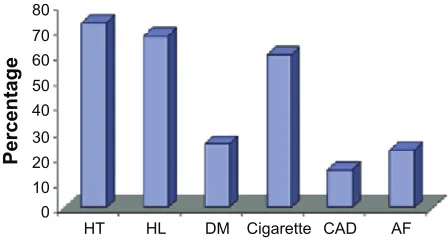
Risk factors most frequently detected in our patients with minor stroke include HT (72.5%), HL, and smoking. HT, HL, diabetes mellitus, smoking, CAD, and AF were presented in 72.5% (n=29), 67.5% (n=27), 25% (n=10), 60% (n=24), 15% (n=6), and 22.5% (n=9) of the patients, respectively (Figure 8).
Table 1 Demographic characteristics
Demographic values Patients with stroke (n=40)
Control subjects (n=40)
P-value
age, years (mean ± sD) 61.03±10.75 61.03±10.75 1.000 sex, n (%)
Female 15 (37.5) 15 (37.5) 1.000
Male 25 (62.5) 25 (62.5)
Abbreviation: sD, standard deviation.
6WURNHJURXS
0HDQ6'
&RQWUROJURXS ,QVWUXPHQWDO
Figure 1 instrumental test results at baseline in study groups.
Abbreviation: sD, standard deviation.
0HDQ6'
6WURNHJURXS &RQWUROJURXS
Figure 2 Backward counting and calculation test results in study groups at baseline (attention).
Abbreviation: sD, standard deviation.
Neuropsychiatric Disease and Treatment downloaded from https://www.dovepress.com/ by 118.70.13.36 on 25-Aug-2020
Dovepress
Deniz et al
Most severe cognitive impairment (ie, in four cognitive tests) was seen among those with CAD and AF, followed by patients with .50% stenosis in Doppler examination who had relatively less severe impairment (ie, in three cognitive tests), suggesting that the frequency of risk factors for stroke does not correlate with the frequency of risk factors associ-ated with cognitive impairment.
Of our patient population, 70% had internal carotid artery stenosis of ,50% and 30% had a stenosis .50%. As expected, those with an internal carotid artery stenosis .50% had greater impairment.
Although cigarette smoking was a risk factor, it was associated with only a minimal increase in the occurrence of cognitive deficits as compared to nonsmokers.
Upon evaluation of impairment ratios in cognitive tests as per risk factors, an improvement occurred in at least one of the cognitive tests.
Except for AF, an improvement was observed in back-ward counting in all risk factor strata. Also, independent of the risk factors, a significant improvement occurred at 6 and 12 months as compared to baseline among the study patients. Among these patients, it was observed that patients with AF and coronary artery disease (CAH) were not included in the group in which significant improvement occurred (Table 2).
The cognitive functions that were most frequently affected among hypertensive patients included word list
memory, FAB test, and backward counting. Hypertensive individuals did not suffer from significant impairment of executive functions as shown by the FAB test.
Radiological findings in study patients included periven-tricular leukoaraiosis in seven, global atrophy in 14, and temporal atrophy in one patient (Table 3). The three tests that were most frequently affected among patients with periven-tricular leukoaraiosis included the FAB test, measuring the frontal executive functions, as well as construction skills and word list memory.
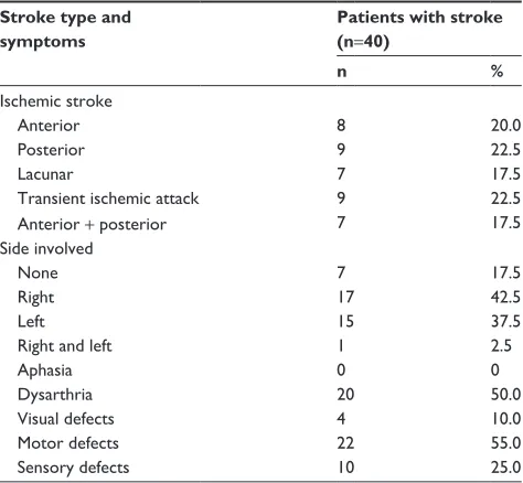
Eight patients had anterior stroke, nine had posterior, and seven had lacunar strokes; also, nine patients had tran-sient ischemic episode, and seven patients had anterior and posterior stroke. For study purposes, thalamic infarcts were classified in the category of lacunar infarcts, which were associated with impairment in all tests, except for word list recall and word list recognition. The most frequently affected tests in patients with lacunar infarcts included the word list memory, backward counting, calculation, and FAB.
There were 17 and 15 patients with right or left hemi-spheric lesions, respectively; a single patient had both right and left hemispheric lesions, while seven patients had no signs of lateralization. Patients with left hemispheric lesions had higher frequency of impairment in all tests as compared
6WURNHJURXS
0HDQ6'
&RQWUROJURXS $EVWUDFWLRQ
Figure 4 abstraction test results in study groups at baseline.
Abbreviation: sD, standard deviation.
0HDQ6'
&RQVWUXFWLRQVNLOOVDQG
YLVXDOPHPRU\ 9LVXRVSDWLDOIXQFWLRQ 6WURNHJURXS &RQWUROJURXS
Figure 5 construction skills and visual memory and visuospatial function results in study groups at baseline.
Abbreviation: sD, standard deviation.
&RQWUROJURXS 6WURNHJURXS
0HDQ6'
)$%WHVW
Figure 6 FAB test results in study groups at baseline.
Abbreviations: FAB, Frontal Assessment Battery; SD, standard deviation.
0HDQ6'
:RUGOLVWPHPRU\ 9HUEDOPHPRU\ 6WURNHJURXS &RQWUROJURXS
Figure 3 Word list memory and verbal memory test results in study groups at baseline.
Abbreviation: sD, standard deviation.
Neuropsychiatric Disease and Treatment downloaded from https://www.dovepress.com/ by 118.70.13.36 on 25-Aug-2020
Dovepress evaluation and follow-up of cognitive functions
to those with right hemispheric lesions. Also, from a clinical viewpoint, patients with left hemispheric lesions had minimal impairment in abstraction, while those with right hemispheric lesions had minimal impairment in executive functions as documented by clock drawing and FAB tests.
A minor stroke occurring over 70 years of age was associated with a more marked cognitive impairment in all test categories as compared to those ,70 years of age. This difference was statistically significant (Table 4).
Discussion
Cerebrovascular diseases may lead to significant reductions in the quality of life, not only due to the associated neurologic deficits but also due to the development of subsequent cogni-tive dysfunction. Identification and prevention of cerebro-vascular events and the associated risk factors carry even a greater significance than the cognitive deficits resulting in decreased functionality.
The conclusions of the current study examining the identification and subsequent course of cognitive functions in patients with minor stroke and transient ischemic attack are described later. An assessment of the association between
cognitive decline and risk factors following a minor stroke showed comparable and highest rates of cognitive ment in patients with CAD and AF (highest cognitive impair-ment in four of the overall cognitive test battery). Patients over 70 years of age had more marked cognitive impairment in all cognitive tests as compared to those ,70 years of age. Also, patients with left hemispheric lesions had more marked impairment in all tests as compared to those with right hemispheric lesions. In comparison with educationally matched controls, patients had significantly more marked impairment in the majority of the neuropsychological tests, and assessment of the results of the neuropsychological tests at early phase and 6 months showed a significant improve-ment only in the backward counting. Despite the absence of a significant improvement in other tests, there was also no worsening in any of the other tests except for the calculation tests. Most patients had a clinically stable course.
cognitive test results according to risk
factors
The most common risk factors in our study population were HT (72.5%), HL, and cigarette smoking. However, an assess-ment of the association between risk factors and cognitive impairment following minor strokes showed highest and similar rates of cognitive dysfunction in those with CAD and AF (most severe impairment in four of all cognitive tests). These were followed by the group of patients who had .40% stenosis in Doppler examination (most severe impairment in three of all cognitive tests). These observations suggest the absence of a correlation between the frequency of risk factors for stroke and risk factors for cognitive dysfunction. Also, when the etiological role of AF in the development of stroke is considered, it seems that examination of the cog-nitive deficits may bear clinical significance in those with cardioembolic stroke.
HT is an important risk factor for cerebrovascular diseases, also with significant roles in the development of vascular cognitive dysfunction and vascular dementia.16
The association among HT, low blood flow rates, and vascular dementia has been clearly established.17,18 Studies
suggest that blood pressure should be maintained below 140/90 mmHg to provide protection against cardiovascular and cerebrovascular diseases and certain types of dementia, as well as for the maintenance adequate cognition in the long-term.18 Longitudinal studies suggested that HT in the
middle age may be associated with cognitive dysfunction developing in later stages of life.17 Kuo et al19 in their 2004
study claimed that high blood pressure in otherwise healthy
)RUZDUGFRXQW WHVW
0HDQ6'
%DFNZDUGFRXQW
WHVW &DOFXODWLRQ PRQWK PRQWKV PRQWKV
Figure 7 Forward counting, backward counting, and calculation test results in the stroke group.
Abbreviation: sD, standard deviation.
3HUFHQWDJH
+7 +/ '0 &LJDUHWWH &$' $)
Figure 8 Distribution of risk factors in the stroke group (n=40).
Abbreviations: AF, atrial fibrillation; CAD, coronary artery disease; DM, diabetes mellitus; HL, hyperlipidemia; HT, hypertension.
Neuropsychiatric Disease and Treatment downloaded from https://www.dovepress.com/ by 118.70.13.36 on 25-Aug-2020
Dovepress
Deniz et al
individuals could be associated with selective impairment of executive functions.
In a study by Khedr et al,14 AF was more common among
patients with PSD, although no significant association with PSD could be established. On the other hand, in 2000, Barba et al1 found correlations between cognitive decline
and AF. AF not only may result in thromboembolic events but also leads to reduced cardiac output, which is more marked in higher ventricular rates, potentially leading to cerebral hypoperfusion. This may also represent an additional mechanism contributing to the development of brain injury and cognitive decline.20
In our study, 70% of the patients had a carotid stenosis of ,50%, while stenosis was $50% in the remaining 30%, with the latter group having more frequent occurrence of cognitive impairment as documented by the cognitive tests. Carotid stenosis, one of the risk factors for such conditions, is a common occurrence in patients with PSD, although no
significant associations could be established between the severity of stenosis and cognitive decline.3 In some recent
studies, common carotid artery intima-media thickness was identified as a risk factor for deficits in memory and cognition, particularly among the elderly females.21 Talelli
et al22 failed to observe a significant association between
common carotid artery intima-media thickness and cognitive decline 12 months after an ischemic stroke when other risk factors were adjusted. Lee and Yeh23 observed a better level
of cognition among patients with lower common carotid artery intima-media thickness, as assessed by Cognitive Abilities Screening Instrument. In 2009, Wendell et al24
found impaired cognitive functions in healthy subjects with increased carotid media thickness. In line with the observations by Wendell et al,24 this result suggests that
the severity of cognitive dysfunction increases with increasing stenosis.
In the present study, a more marked cognitive impairment was found in all tests among patients over 70 years of age as compared to those ,70 years of age, consistent with previ-ous observations suggesting that advanced age represents a significant risk factor for cognitive deficits.
Also, more patients with left hemispheric lesions had cognitive impairment in all cognitive assessment tools than patients with right hemispheric lesions. Also, bilateral infarcts and more extensive involvement of the dominant hemisphere were found to increase the risk of dementia in patients with
Table 3 The distribution of site and clinical signs in patients with stroke
Stroke type and symptoms
Patients with stroke (n=40)
n %
ischemic stroke
anterior 8 20.0
Posterior 9 22.5
lacunar 7 17.5
Transient ischemic attack 9 22.5
anterior + posterior 7 17.5
side involved
None 7 17.5
right 17 42.5
left 15 37.5
right and left 1 2.5
aphasia 0 0
Dysarthria 20 50.0
Visual defects 4 10.0
Motor defects 22 55.0
sensory defects 10 25.0
Table 4 cognitive disturbance according to age group
Time Number of defective cognitive
items
P-values
#70 years age .70 years age
Mean ± SD Mean ± SD
0 month 5.86±2.44 (5) 7.91±2.70 (9) 0.042* 6 months 5.86±2.23 (5) 8.00±2.54 (8) 0.026* 12 months 5.86±2.24 (5) 7.90±2.55 (8) 0.027*
Note: *P,0.05.
Abbreviation: sD, standard deviation. Table 2 risk factors affecting the backward counting skill
Number of patients who had disturbance of backward counting skill
Initial (%) 6 months (%) 12 months (%) P-values
hT (N=29) 25 (82.2) 23 (79.3) 23 (79.3) 0.028
DM (N=10) 9 (90) 8 (80) 8 (80) 0.134
smoker (N=24) 18 (75) 17 (70.8) 17 (70.8) 0.059
hypercholesterolemia (N=27) 22 (81.5) 19 (70.4) 19 (70.4) 0.031
coronary artery disease (N=6) 6 (100) 6 (100) 6 (100) 0.363
Atrial fibrillation (N=9) 7 (77.8) 7 (77.8) 7 (77.8) 0.390
Abbreviations: DM, diabetes mellitus; HT, hypertension.
Neuropsychiatric Disease and Treatment downloaded from https://www.dovepress.com/ by 118.70.13.36 on 25-Aug-2020
Dovepress evaluation and follow-up of cognitive functions
stroke patients who were diagnosed with vascular dementia both radiologically and pathologically.25
Detailed assessment of the cognitive tests
Comparison of patients and controls in our study during the early phase revealed significant impairment in attention (backward counting), visuospatial functions (construction skills and visual memory), language (word list memory), cal-culation, and abstraction, as well as in all executive function domains in the patient group. Srikanth et al26 utilized similarneuropsychological tests, similar timing of tests as well as a similar stroke classification, while in contrast with our study, they also involved patients with intracerebral hemorrhage. In this study, the patient group had significant impairment in attention, visuospatial functions, language, and executive functions as compared to controls, while the impairment in memory was less severe.
All patients with minor stroke or transient ischemia in our study had impaired FAB test results for frontal executive functions. Although verbal memory scores were low among the overall group of patients with vascular cognitive impair-ment and vascular deimpair-mentia, their performance in word list recall and recognition was not significantly different from that in controls.27 Again, Sachdev et al28 showed that impaired
frontal executive functions and psychomotor retardation represent two prominent areas of involvement in vascular cognitive impairment and vascular dementia. Also, dominant and nondominant parietal lobe functions are affected.
In our group of hypertensive patients, the most frequently affected cognitive functions were word list memory, FAB (frontal executive functions), backward counting, and attention. Thus, executive dysfunction was also a marked characteristic in these hypertensive individuals.
The three tests that were most frequently affected among patients with periventricular leukoaraiosis included the FAB test, measuring the frontal executive functions, as well as construction skills and word list memory. Foci of white mat-ter in the periventricular and subcortical regions have been associated with memory and executive functions.29–31
Our patients with posterior infarcts had, in the decreasing order of severity, impairment in memory, executive func-tions, and attention. Cerebellar pathological conditions are generally associated with less severe cognitive dysfunction. In a case report by Paulus et al,32 impairment in attention,
executive functions, and memory were observed in a patient with a large cerebellar infarct. This patient without motor dysfunction was diagnosed as having a pure cerebellar cognitive affective syndrome.
In the current study, thalamic infarcts were categorized within the class of lacunar infarcts, which were found to be associated with impairments in all tests except for word list recall and word list recognition. In other words, these patients had impairment in attention, concentration, and verbal and visual memory. The tests that were most frequently impaired included word list memory, backward counting, calculation, and FAB. While the word list memory was the most frequently involved area of impairment, no finding of disturbance in word list recall and recognition suggested that the cognitive decline in thalamic (lacunar) infarcts leads to a learning difficulty rather than a recall problem.
Thalamic infarcts also represent a significant cause of cognitive deficits, and amnesia developing in association with thalamic infarcts leads to learning difficulties rather than recall problems. Ischemic lesions involving the mam-millothalamic tract in the tuberothalamic artery zone cause learning and short-term memory deficits. Aphatia and abulia generally occur in those with anterior thalamic pedincle.33
Szirmai et al34 studied 19 patients with thalamic infarction
and demonstrated that there were deficits in attention, con-centration, visual and verbal deficits in patients with left or bilateral thalamic infarction. Paramedian lesions of the thalamus may lead to naming difficulty or perseverations, although cortical aphasia does not occur.35 Since the medial
dorsal nucleus is connected with Broca’s and Wernicke’s areas, a variety of language problems may emerge. Szirmai et al34 found an increased incidence of semantic and verbal
disturbances when thalamic lesions occurred in association with striatocapsular infarcts.
an assessment of the prognosis during
follow-up
In the present study, 22.5% of the patients had impairment in a maximum of five test categories at baseline, and similarly at 6 months, 22.5% of the patients had impairment in five tests, while at 12 months, 17.5% had impairment in five tests. The number of tests with impairment decreased from baseline to 12 months, although the difference was not significant. The number of cognitive tests (13 in total) with impairment did not alter significantly during the follow-up period, that is, from 0 to 6, 0 to 12, and 6 to 12 months, suggesting a stable clinical course in the majority of the patients.
Tham et al13 assessed attention, language, verbal memory,
visual memory, visual construction, and visual-motor velocity at baseline and at 6 months in patients enduring transient ischemic attack without permanent disability and found that 56% of the patients had normal cognition
Neuropsychiatric Disease and Treatment downloaded from https://www.dovepress.com/ by 118.70.13.36 on 25-Aug-2020
Dovepress
Deniz et al
at baseline, while 40% had cognitive impairment without dementia, and 4% had dementia. At 1 year, cognitive disorders in 31% of patients had improved, but cognitive impairment occurred in 10% of patients with initial normal cognitive performance, and dementia occurred in 11% of patients who had mild cognitive impairments. These results point out to a long-term variability between patients in terms of the results of cognitive performance tests. In spite of the reports suggesting considerable cognitive dysfunction (ie, 18%–26%) 3 months after stroke, Rasquin et al36 in
their study assessing poststroke memory, mental speed, and executive functions reported that 12% of the patients developed dementia within 1 year.37–39 Again in the same
study, an improvement rather than worsening in cognitive performance was noted between 1 and 6 months.
An examination of the rate of improvement according to risk factor in our study showed that there was an improvement in at least one cognitive test. Except for patients with AF, backward counting improved in all other risk groups. Also, a statistically significant improvement in backward counting occurred from baseline (month 0) to 6 and 12 months in the patient group, regardless of the risk factors. Patients with AF, however, did not belong to the group in which significant improvement occurred.
Neuropsychological tests administered at baseline and 6 months showed a significant improvement only in back-ward counting test between 0 and 6 and 0 and 12 months. Despite the absence of significant improvement in other tests, no significant worsening occurred also, except for the calculation test.
Also, there was an improvement in MMSE scores between 0 and 6 months, although this was not significant. In a study from Israel, patients with lacunar infarcts had an average decline of 1.1 point in MMSE scores.40 In another
study involving elderly patients with vascular dementia, a 1.77 point decrease occurred in MMSE scores at the end of 1 year of follow-up.41
In a study by Ballard et al,42 patients $75 years of age
were examined 3 and 15 months poststroke. A $2 point increase in MMSE was considered to indicate cognitive improvement, while development of dementia according to
Diagnostic and Statistical Manual of Mental Disorders – 4th
Edition was considered cognitive decline; patients without
such changes were considered stable. Among these patients, those with diabetes were less likely to be represented in the group with improvement in cognitive functions.
Accordingly, Srikanth et al43 found no increase in
progressive dementia risk in mild to moderately severe
stroke patients without aphasia, provided that stroke does not recur.
study limitations
Several studies found an association between the severity of stroke and increased risk of dementia,1,3,37,44–47 with
sug-gestion of a higher risk for extensive dominant hemispheric lesions.3,37,45 In our study, patients with severe strokes,
particularly those with total anterior circulation infarcts, were excluded. As inclusion criteria in the study were using the NIHS score, infarct volume in cranial imaging was not measured.
In our study, all patients with minor stroke or transient ischemic episode had impairment in at least two of the neuropsychological tests administered. Possible explanations of the higher rate of impairment compared to previous reports include a detailed examination of the results of a variety of cognitive tests, exclusion of memory disturbance as a study criteria, and small sample size.
Cognitive tests proposed by Turkuaz Alzheimer Work-ing group were used in our study. Addition of a depression scale to those already utilized could have provided a more comprehensive patient assessment.
Further prospective studies examining the total brain volume or infarct volume in larger patient populations and the use of appropriate neuropsychological assessments will prove valuable in the follow-up of these patients.
Conclusion
In patients with vascular dementia, disturbance in other cognitive domains far precedes the disturbance in memory. Recognition of cognitive disturbance may allow earlier diag-nosis and treatment of vascular dementia before advanced vascular dementia develops. This, in turn, may help offset the limitations of preventive therapeutic strategies.
Disclosure
The authors report no conflicts of interest in this work.
References
1. Barba R, Martinez ES, Rodriguez GE, Pondal M, Vivancos J, Del Ser T. Poststroke dementia: clinical features and risk factors. Stroke.2000;31:1494–1501.
2. De Haan RJ, Limburg M, Van Der Meulen JH, Jacobs HM, Aaronson NK. Quality of life after stroke. Impact of stroke type and lesion location. Stroke. 1995;26:402–408.
3. Pohjasvaara T, Erkinjuntti T, Yilkoski R, Hietanen M, Vataja R, Kaste M. Clinical determinants of poststroke dementia. Stroke. 1998;29:75–81. 4. Tatemichi TK, Paik M, Bagiella E, et al. Risk of dementia after stroke in
a hospitalized cohort: results of a longitudinal study. Neurology. 1994; 44:1885–1891.
Neuropsychiatric Disease and Treatment downloaded from https://www.dovepress.com/ by 118.70.13.36 on 25-Aug-2020
Dovepress evaluation and follow-up of cognitive functions
5. Leys D, Henon H, Mackowiak-Cordoliani MA, Pasquier F. Poststroke dementia. Lancet Neurol. 2005;4:752–759.
6. O’Brien JH, Erkinjuntti T, Reisberg B, et al. Vascular cognitive impair-ment. Lancet Neurol.2003;2:89–98.
7. Frisoni GB, Galluzzi S, Bresciani L, Zanetti O, Geroldi C. Mild cognitive impairment with subcortical vascular features. Clinical characteristics and outcome. J Neurol. 2002;249:1423–1432. 8. Ebly EM, Hogan DB, Parhad IM. Cognitive impairment in the
nonde-mented elderly. Results from the Canadian Study of Health and Aging. Arch Neurol. 1995;52:612–619.
9. Ingles JL, Wentzel C, Fisk JD, Rockwood K. Neuropsychological predictors of incident dementia in patients with vascular cognitive impairment, without dementia. Stroke. 2002;33:1999–2002. 10. Wentzel C, Rockwood K, MacKnight C, et al. Progression of
impair-ment in patients with vascular cognitive impairimpair-ment without deimpair-mentia. Neurology. 2001;57:714–716.
11. Petersen RC, Smith GE, Waring SC, Ivnik RJ, Tangalos EG, Kokmen E. Mild cognitive impairment: clinical characterization and outcome. Arch Neurol.1999;56:303–308.
12. Desmond DW, Moroney JT, Sano M, Stern Y. Recovery of cognitive function after stroke. Stroke. 1996;27:1798–1803.
13. Tham W, Auchus AP, Thong M, et al. Progression of cognitive impairment after stroke: one year results from a longitudinal study of Singaporean stroke patients. J Neurol Sci. 2002;203–204:49–52. 14. Khedr EM, Hamed SA, El-Shereef HK, et al. Cognitive impairment
after cerebrovascular stroke: relationship to vascular risk factors. Neu-ropsychiatr Dis Treat.2009;5:103–116.
15. Madureira S, Guerreiro M, Ferro JM. Dementia and cognitive impair-ment three months after stroke. Eur J Neurol. 2001;8:621–627. 16. In’t Veld BA, Ruitenberg A, Hofman A, Stricker BA, Breteler MMB.
Antihypertensive drugs and incidence of dementia: the Rotterdam Study. Neurobiol Aging.2001;22:4071.
17. Harrington F, Saxby BK, McKeith IG, Wesnes K, Ford GA. Cognitive performance in hypertensive and normotensive older subjects. Hypertension.2000;36:1079–1082.
18. Birkenhager WH, Forette F, Seux ML, Wang JG, Staessen JA. Blood pressure, cognitive functions, and prevention of dementias in older patients with hypertension. Arch Intern Med. 2001;161:152–156. 19. Kuo H-K, Sorond F, Iloputaife I, Gagnon M, Milberg W, Lipsitz LA.
Effect of blood pressure on cognitive functions in elderly persons. J GerontolA Biol Sci Med Sci.2004;59(11):1191–1194.
20. Ott A, Breteler MM, de Bruyne MC, van Harskamp F, Grobbee DE, Hofman A. Atrial fibrillation and dementia in a population-based study. The Rotterdam Study. Stroke.1997;28:316–321.
21. Komulainen P, Kivipelto M, Lakka TA, et al. Carotid intima-media thickness and cognitive function in elderly women: a population-based study. Neuroepidemiology.2007;28:207–213.
22. Talelli P, Ellul J, Terzis G, et al. Common carotid artery intima media thickness and post-stroke cognitive impairment. J Neurol Sci. 2004;223: 129–134.
23. Lee YH, Yeh SJ. Correlation of common carotid artery intima media thickness, intracranial arterial stenosis and post-stroke cognitive impair-ment. Acta Neurol Taiwan.2007;16:207–213.
24. Wendell CR, Zonderman AB, Metter EJ, Najjar SS, Waldstein SR. Carotid intimal medial thickness predicts cognitive decline among adults without clinical vascular disease. Stroke. 2009;40(10):3180–3185. 25. Jayakumar PN, Taly AB, Shanmugam V. Multi-infarct dementia:
a computed tomographic study. Acta Neurol Scand. 1989;73:292–295. 26. Srikanth VK, Thrift AG, Saling MM, et al; Community-Based
Prospective Study of Nonaphasic English-Speaking Survivors. Increased risk of cognitive impairment 3 months after mild to moderate first-ever stroke: a community-based prospective study of nonaphasic English-speaking survivors. Stroke.2003;34:1136–1143.
27. Grober E, Kawas C. Learning and retention in preclinical and early Alzheimer’s disease. Psychol Aging.1997;12:183–188.
28. Sachdev PS, Brodaty H, Valenzuela MJ, et al. The neuropsychological profile of vascular cognitive impairment in stroke and TIA patients. Neurology. 2004;62:912–919.
29. De Groot JC, De Leeuw FE, Oudkerk M, Hofman A, Jolles J, Breteler MM. Cerebral white matter lesions and depressive symptoms in elderly adults. Arch Gen Psychiatry.2000;57:1071–1076. 30. Gunning-Dixon FM, Raz N. The cognitive correlates of white matter
abnormalities in normal aging: a quantitative review. Neuropsychology. 2000;14:224–232.
31. Kramer-Ginsberg E, Greenwald BS, Krishnan KR, et al. Neuropsy-chological functioning and MRI signal hyperintensities in geriatric depression. Am J Psychiatry. 1999;156:438.
32. Paulus KS, Magnano I, Conti M, et al. Pure post-stroke cerebellar cogni-tive affeccogni-tive syndrome: a case report. Neurol Sci. 2004;25:220–224. 33. Bhatia KP, Marsden CD. The behavioral and motor consequences of
focal lesions of the basal ganglia in man. Brain.1994;117:859–876. 34. Szirmai I, Vastagh I, Szombathelyi E, Kamondi A. Strategic infarcts of
the thalamus in vascular dementia. Neurol Sci. 2002;203–204:91–97. 35. Stuss DT, Guberman A, Nelson R, Larochelle S. The neuropsychology
of paramedian thalamic infarction. Brain Cogn. 1988;8:348–378. 36. Rasquin SM, van Oosstenbruge RJ, Verhey FR, Lodder J. Vascular
mild cognitive impairment is highly prevalant after lacunar stroke but does not increase over time: a 2 year follow up study. Dement Geriatr Cogn Disorder. 2007;24:396–401.
37. Desmond DW, Moroney JT, Paik MC, et al. Frequency and clinical determinants of dementia after ischemic stroke. Neurology. 2000;54: 1124–1131.
38. Pohjasvaara T, Erkinjuntti T, Vataja R, Kaste M. Dementia three months after stroke: baseline frequency and effect of different definitions of dementia in the Helsinki Stroke Aging Memory Study (SAM) cohort. Stroke.1997;28:785–792.
39. Tatemichi TK, Desmond DW, Mayeux R, et al. Dementia after stroke: baseline frequency, risks and clinical features in a hospitalized cohort. Neurology.1992;42:1185–1193.
40. Aharon-Peretz J, Daskovski E, Mashiach T, Tomer R. Natural history of dementia associated with lacunar infarctions. J Neurol Sci. 2002;203–204: 53–55.
41. Segal-Gidan FI, Lyness SA, Chui HC. A comparison of mental status decline in ischemic vascular dementia and Alzheimer’s disease. Neurology.1999;52(suppl 2):486–487.
42. Ballard C, Stephens S, Kenny R, Kalaria R, Tovee M, O’Brien J. Profile of neuropsychological deficits in older stroke survivors without dementia. Dement Geriatr Cogn Disord.2003;16:52–56.
43. Srikanth VK, Anderson JFI, Donnan GA, et al. Progressive dementia after first ever stroke: a community-based follow-up study. Neurology. 2004;63:785–792.
44. Censori B, Manara O, Agostinis C, et al. Dementia after first stroke. Stroke. 1996;27:1205–1210.
45. Tatemichi TK, Desmond DW, Paik M, et al. Clinical determinants of dementia related to stroke. Ann Neurol. 1993;33:568–575.
46. Henon H, Durieu I, Guerouaou D, Lebert F, Pasquier F, Leys D. Poststroke dementia: incidence and relationship to prestroke cognitive decline. Neurology.2001;57:1216–1222.
47. Inzitari D, Di Carlo A, Pracucci G, et al. Incidence and determinants of poststroke dementia as defined by an informant interview method in a hospital-based stroke registry. Stroke. 1998;29:2087–2093.
Neuropsychiatric Disease and Treatment downloaded from https://www.dovepress.com/ by 118.70.13.36 on 25-Aug-2020
Neuropsychiatric Disease and Treatment
Publish your work in this journal
Submit your manuscript here: http://www.dovepress.com/neuropsychiatric-disease-and-treatment-journal
Neuropsychiatric Disease and Treatment is an international, peer-reviewed journal of clinical therapeutics and pharmacology focusing on concise rapid reporting of clinical or pre-clinical studies on a range of neuropsychiatric and neurological disorders. This journal is indexed on PubMed Central, the ‘PsycINFO’ database and CAS,
and is the official journal of The International Neuropsychiatric Association (INA). The manuscript management system is completely online and includes a very quick and fair peer-review system, which is all easy to use. Visit http://www.dovepress.com/testimonials.php to read real quotes from published authors.
Dovepress
Dove
press
Deniz et al
Neuropsychiatric Disease and Treatment downloaded from https://www.dovepress.com/ by 118.70.13.36 on 25-Aug-2020