University of Pennsylvania
ScholarlyCommons
Publicly Accessible Penn Dissertations
1-1-2012
Conceptual Flexibility: Behavioral and Neural
Variations in Semantic Memory Retrieval
Nina Hsu
University of Pennsylvania, [email protected]
Follow this and additional works at:http://repository.upenn.edu/edissertations
Part of theCognitive Psychology Commons, and theNeuroscience and Neurobiology Commons
Recommended Citation
Hsu, Nina, "Conceptual Flexibility: Behavioral and Neural Variations in Semantic Memory Retrieval" (2012).Publicly Accessible Penn Dissertations. 521.
Conceptual Flexibility: Behavioral and Neural Variations in Semantic
Memory Retrieval
Abstract
Understanding the neural organization of semantic memory - our shared general knowledge - has the potential to uncover the neural mechanisms by which we give meaning to the endless array of objects that we encounter in the world. One prominent set of theories posits a distributed organization of semantic memory -remembering object features activates brain regions overlapping with or adjacent to regions involved in perceiving and acting on those features (e.g., Allport, 1985). Despite accumulated evidence favoring these theories, it is important to understand both the commonalities in the mapping between perception and memory, as well as meaningful variability across sources such as contexts, people, and use. In three studies, we found evidence that while conceptual knowledge is grounded in neural substrates, several factors contribute to variations in semantic memory retrieval. In Chapter 2, we used the logic often used in neuroimaging studies of semantic memory by demonstrating overlapping chromaticity effects (e.g., greater response to colored than grayscale stimuli) in the left lingual gyrus for both color perception and color knowledge. Chapter 3 investigated whether the mapping between perception and memory varied across contexts and participants. Whereas context (here, fidelity of color information as manipulated through task demand) varied the extent to which the left fusiform gyrus was active during a color similarity judgment, individual differences in cognitive style predicted activity in the left lingual gyrus. We replicated these results in a second experiment that controlled for stimulus modality and anticipatory strategies. In Chapter 4, we used a training paradigm to investigate the role of feature diagnosticity (i.e., features that best distinguish between two otherwise similar categories) in semantic representations. Whereas subjects had knowledge of feature importance in novel object categorization, whether they used this information affected neural representations. Ventral temporal brain regions were more active during a separate retrieval task for subjects who learned and used the diagnostic feature for object categorization. Additionally, behavioral ratings of similarity predicted multivariate neural similarity. Collectively, this work suggests that semantic representations, integral to a memory system often thought of as free of contextual constraints, contain meaningful variations across contexts, people, and use.
Degree Type Dissertation
Degree Name
Doctor of Philosophy (PhD)
Graduate Group Neuroscience
First Advisor
Sharon L. Thompson-Schill
Keywords
Subject Categories
CONCEPTUAL FLEXIBILITY: BEHAVIORAL AND NEURAL VARIATIONS IN
SEMANTIC MEMORY RETRIEVAL
Nina Shen Hsu
A DISSERTATION
in
Neuroscience
Presented to the Faculties of the University of Pennsylvania
in
Partial Fulfillment of the Requirements for the
Degree of Doctor of Philosophy
2012
Supervisor of Dissertation
________________________
Sharon L. Thompson-Schill
Professor, Psychology
Graduate Group Chairperson
________________________
Joshua I. Gold, Associate Professor, Neuroscience
Dissertation Committee:
Geoffrey K. Aguirre, Assistant Professor, Neurology David H. Brainard, Professor, Psychology
FORMATTING NOTE: Margins must remain set to: left-hand margin: 1.5 inches,
right-hand margin: 1 inch, top and bottom margins: 1 inch. Please double-space your
dissertation for the sake of readability (except for footnotes, long quoted passages, and
lists of tables and figures, which are to be spaced). You may choose to
single-space the dissertation manuscript. If you use foot notes, it is recommended that you move
the pagination to the top. Pagination must remain within the margin requirements.
NOTHING MAY BE WRITTEN IN THE MARGINS.
Copyright page is optional, use Creative Commons License Deed if using Creative
CONCEPTUAL FLEXIBILITY: BEHAVIORAL AND NEURAL VARIATIONS IN
SEMANTIC MEMORY RETRIEVAL
COPYRIGHT
2012
Nina Shen Hsu
DEDICATION
ACKNOWLEDGEMENTS
I am indebted to a number of invaluable resources in the form of friends, family,
and colleagues, who have helped me in many more ways than they realize.
First, I was supported by the Systems and Integrative Biology Training Grant
during my first two years at Penn (T32-GM007517). After that, I was fortunate to obtain
a pre-doctoral NRSA (F31-AG034743) from the National Institute on Aging under the
National Institutes of Health. Much additional support came from R01-MH070850 to my
advisor, Sharon Thompson-Schill.
Sharon is the kind of advisor and mentor that I aspire to be someday, and I have
learned so much from her over the past six years. She has been supportive, generous, and
kind for all aspects of graduate school. Moreover, she has shown me the importance of
bearing in mind both the nitty-gritty of experimental details, as well as taking a step back
in order to understand the bigger questions being asked. Through it all, she has believed
in and cheerleaded this work passionately, even when I regularly walked into her office
with "good news and bad news." As these projects evolved over the years, she provided a
masterful balance of both guidance and freedom. I am very grateful to have had the
opportunity to work with her.
Next, my thesis committee members – Geoff Aguirre, David Brainard, Joe Kable,
and Kim Curby have my thanks and gratitude for offering helpful suggestions, raising
important theoretical and methodological points, and being a supportive and interactive
The members of the Thompson-Schill lab – and the Center for Cognitive
Neuroscience as a whole – make for a warm and intellectually stimulating community.
Though they all contributed in more ways than they know, I’d like to mention a few in
particular. Steven Frankland and Andrea Houghtling were wonderful office-mates for my
first few years. Steven was also a collaborator for the experiments described in Chapter 2.
In addition to helping me toss around ideas and get their input on my work, both of them
got me to listen to hipper music. David Kraemer was a collaborator for the experiments
in Chapter 3 – his intellectual contributions to the work improved it tremendously, plus
he has mad poker skills to boot. Ranjani Prabhakaran showed me the ropes of fMRI data
analysis when I first arrived, and served as a role model throughout my time in the lab.
It’s quite possible that we discussed general linear modeling and Friday Night Lights in
equal proportion. Irene Kan and Robyn Oliver were always generous with their time,
willing to help me understand theories and several iterations of experimental designs.
Lila Chrysikou, in addition to being a terrific officemate, showed me the utility of
white-boarding my ideas in order to sketch them out (both literally and figuratively). I am
grateful to both Lila and Eiling Yee for their patience, compassion, and generosity in
helping me to talk about ideas, theoretical issues, experimental design, and pretty much
anything else on my mind. Finally, Meg Schlichting was instrumental to the design of the
training study described in Chapter 4. Through her, I was introduced to the intricacies of
Blender and PsyScope, and for better or worse, the music of Ke$ha.
My classmates in the Neuroscience Graduate Group are a terrific bunch, and
poring over Cell 600 textbooks in Barchi as first-years, to the annual pre-Thanksgiving
Thanksgiving Potluck, they are all ridiculously smart, and I’m so glad to have the
pleasure of knowing them.
I was also fortunate to have an incredible group of friends during graduate school.
I’m particularly indebted to Allison Lesher, with whom I’m still convinced we share
parallel lives. Jennifer Gilbert, Maiana Hanshaw, and Vy Hoang have been constant
sources of support and terrific friends to boot. And to the quizzo team – thanks for giving
me a weekly outlet for all of the non-neuroscience ramblings in my head. See you all on
Wednesday.
Chris Gowlland, Lila Chrysikou, and Vanessa Troiani have my thanks for
providing fresh eyes and thoughts as they read through early drafts of this document.
There are simply not enough words to express how grateful I am to Kevin Byron,
who has been a pillar of strength for me, particularly during the last stretch. Whether he is
giving me invaluable support and fresh perspective, sending me daily jokes to make sure
I’m still laughing, or patiently staying up with me when it’s 2:30 AM and I’m obsessing
over my data – love, thank you.
Finally, my family – my parents, Yi-Ming and Pi-Lan, and brother, Carey. My
father has shown me the rewards of a successful academic career, and my mother
instilled in me a diligent work ethic and high standards for myself. My brother provided a
concrete example of surviving (and enjoying!) graduate school, and was a wonderful
mentor as I encountered my own personal and mental obstacles throughout my graduate
ABSTRACT
CONCEPTUAL FLEXIBILITY: BEHAVIORAL AND NEURAL VARIATIONS IN
SEMANTIC MEMORY RETRIEVAL
Nina Shen Hsu
Sharon L. Thompson-Schill
Understanding the neural organization of semantic memory – our shared general
knowledge – has the potential to uncover the neural mechanisms by which we give
meaning to the endless array of objects that we encounter in the world. One prominent set
of theories posits a distributed organization of semantic memory – remembering object
features activates brain regions overlapping with or adjacent to regions involved in
perceiving and acting on those features (e.g., Allport, 1985). Despite accumulated
evidence favoring these theories, it is important to understand both the commonalities in
the mapping between perception and memory, as well as meaningful variability across
sources such as contexts, people, and use. In three studies, we found evidence that while
conceptual knowledge is grounded in neural substrates, several factors contribute to
variations in semantic memory retrieval. In Chapter 2, we used the logic often used in
neuroimaging studies of semantic memory by demonstrating overlapping chromaticity
effects (e.g., greater response to colored than grayscale stimuli) in the left lingual gyrus
for both color perception and color knowledge. Chapter 3 investigated whether the
mapping between perception and memory varied across contexts and participants.
varied the extent to which the left fusiform gyrus was active during a color similarity
judgment, individual differences in cognitive style predicted activity in the left lingual
gyrus. We replicated these results in a second experiment that controlled for stimulus
modality and anticipatory strategies. In Chapter 4, we used a training paradigm to
investigate the role of feature diagnosticity (i.e., features that best distinguish between
two otherwise similar categories) in semantic representations. Whereas subjects had
knowledge of feature importance in novel object categorization, whether they used this
information affected neural representations. Ventral temporal brain regions were more
active during a separate retrieval task for subjects who learned and used the diagnostic
feature for object categorization. Additionally, behavioral ratings of similarity predicted
multivariate neural similarity. Collectively, this work suggests that semantic
representations, integral to a memory system often thought of as free of contextual
TABLE OF CONTENTS
ABSTRACT ... VII
TABLE OF CONTENTS ... IX
LIST OF TABLES ... XI
LIST OF ILLUSTRATIONS ... XII
CHAPTER 1: INTRODUCTION ... 1
CHAPTER 2: CHROMATICITY OF COLOR PERCEPTION AND OBJECT COLOR KNOWLEDGE ... 16
Abstract ... 16
Introduction ... 17
Method ... 19
Results ... 23
Discussion ... 28
CHAPTER 3: COLOR, CONTEXT, AND COGNITIVE STYLE: VARIATIONS IN COLOR KNOWLEDGE RETRIEVAL AS A FUNCTION OF TASK AND SUBJECT VARIABLES ... 45
Abstract ... 45
Introduction ... 47
Experiment 1: Methods ... 51
Experiment 1: Results ... 58
Experiment 1: Discussion ... 61
Experiment 2: Results ... 65
Experiment 2: Discussion ... 67
General Discussion ... 69
CHAPTER 4: FEATURE DIAGNOSTICITY AFFECTS SEMANTIC REPRESENTATIONS OF NOVEL OBJECTS ... 84
Abstract ... 84
Introduction ... 86
Methods ... 91
Results ... 99
Discussion ... 106
CHAPTER 5: GENERAL DISCUSSION AND FUTURE DIRECTIONS ... 126
BIBLIOGRAPHY ... 137
LIST OF TABLES
Table 2.1. Mean and standard deviation of stimuli characteristics used in chromaticity task.………... 34
Table 2.2. List of stimuli used in the chromaticity task………....…… 35
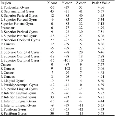
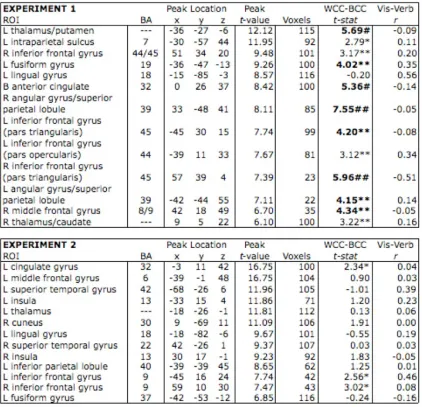
Table 2.3. Regions identified from the 27 maxima in the exploratory, whole-brain analysis………..….. 38
Table 3.1. Clusters identified in the secondary exploratory, whole-brain analysis… 76
Table 4.1. Subject training schedule, in which the specific combination of tasks is indicated for each session of the experiment……… 113
LIST OF ILLUSTRATIONS
Figure 2.1. Examples of chromaticity stimuli from each task ……… 41
Figure 2.2. Co-localization of color perception and color knowledge ……….. 42
Figure 2.3. Exploratory analyses ………... 43
Figure 2.4. Signal change trends in ventral but not dorsal regions……….. 44
Figure 3.1. Design of Experiment 1……… 78
Figure 3.2. Design of Experiment 2……… 79
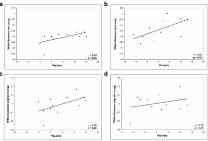
Figure 3.3. Task accuracy and left lingual gyrus activity correlate with cognitive style preference ……….. 80
Figure 3.4. Areas of visual cortex show differences in activity when retrieving color knowledge at differing levels of detail ………. 81
Figure 3.5. Color perception and color knowledge retrieval activate overlapping brain regions……….. 82
Figure 3.6. Color knowledge retrieval recruits overlapping brain regions in both Experiment 1 and Experiment 2……… 83
Figure 4.1. Exemplars from the color+shape object set……….. 117
Figure 4.2. Exemplars from the shape object set………. 118
Figure 4.3. Naming accuracy by session……… 119
Figure 4.4. Differences in knowing versus naming colors……… 120
Figure 4.5. Similarity judgments vary by group………. 121
Figure 4.6. Group differences in activation of a color perception region during a shape retrieval task……….……… 122
Figure 4.7. Task activity in the left inferior temporal gyrus ………. 123
CHAPTER 1: Introduction
What color are fire engines? What sounds do cows make? These are questions
that you can easily answer, but can you remember where or when you first acquired this
information? Cognitive psychologists and neuroscientists often refer to semantic memory
as shared knowledge about the world that gives meaning to objects and concepts. In his
seminal article, “Episodic and semantic memory,” Endel Tulving borrowed the term
semantics from linguists in order to describe a memory system for “words and other
verbal symbols, their meaning and referents, about relations among them, and about
rules, formulas, and algorithms for the manipulation of these symbols, concepts, and
relations” (Tulving, 1972).
Semantic memory refers to shared general knowledge concerning objects, words,
facts, and beliefs. Psychologists distinguish it from episodic memory, which is another
major category of declarative memory, by referring to our ability to retrieve semantic
information independent of specific experiences. In other words, semantic knowledge can
be retrieved without reference to the original circumstances under which it was first
acquired. For example, remembering the color of a fire engine constitutes semantic
knowledge, whereas remembering where you were when you last heard a fire engine
would be episodic knowledge. In a taxonomy of long-term memory (e.g., Squire, 1987),
episodic memory and semantic memory are characterized as being distinct parts of the
associated with events, whereas semantic memory is associated with facts. Tulving
suggests that "the semantic system may be quite independent of the episodic memory" —
but in order to have semantic knowledge, there must have been some episode where the
information was learned (i.e., a specific time and place). Cognitive psychologists and
cognitive neuroscientists have been investigating how we acquire semantic knowledge,
and what neural mechanisms underlie this acquisition and information storage, so that we
can make better sense of the endless number of things in the world around us.
A glimpse of semantic memory through neuropsychological studies
Neuropsychological studies of patients with amnesia due to medial temporal lobe
atrophy often exhibited impairments in both semantic and episodic memory (Gabrieli,
Cohen, & Corkin, 1988; Stefanacci, Buffalo, Schmolck, & Squire, 2000), but other
patient work indicates that impaired episodic memory can be accompanied by relatively
intact semantic memory (Gabrieli et al., 1988; Gardiner, Brandt, Baddeley,
Vargha-Khadem, & Mishkin, 2008; O’Kane, Kensinger, & Corkin, 2004; Vargha-Khadem et al.,
1997). This work suggests that these two systems are at least partially independent. Other
studies have demonstrated that in some cases, semantic knowledge can be selectively
lost. Elizabeth Warrington first noted in 1975 that patients with semantic dementia — a
temporal variant of frontotemporal dementia — demonstrated a “failure to recognize or
identify common objects” that could not be accounted for by intellectual impairment,
sensory or perceptual deficits, or language disorders (Warrington, 1975). Semantic
wherein patients with otherwise intact speech fluency exhibit marked deficits in
identifying objects, concepts and people (Snowden, Griffiths, & Neary, 1999).
Importantly, these patients retain episodic knowledge of recent autobiographical events
(Graham, Lambon, & Hodges, 1997; Snowden, Griffiths, & Neary, 1996).
Some patients exhibit even more selective deficits in semantic knowledge, in
which specific categories of knowledge are lost. Some patients with category-specific
deficits had difficulty naming living things (e.g., parrot, snail) but not nonliving things
(e.g., briefcase, compass), whereas others have presented with the opposite problem
(Warrington & McCarthy, 1983; Warrington & Shallice, 1984). In thinking about ways in
which semantic memory might be organized, one proposal is that semantic knowledge
divides along categorical, or domain-specific, boundaries (Caramazza & Shelton, 1998).
On the other hand, early functional neuroimaging studies showed anatomical distinctions
between objects according to features (sometimes called attributes or properties), even
within a single category (Thompson-Schill, Aguirre, D’Esposito, & Farah, 1999). In one
early study, Martin and colleagues demonstrated that retrieving color information about
an object ("yellow" for pencil) activated ventral temporal cortex, whereas retrieving
motor information about the same object ("write" for pencil) activated middle temporal
and frontal cortex (Martin, Haxby, Lalonde, Wiggs, & Ungerleider, 1995). Further,
distinct but overlapping regions were activated when presenting the objects as words or
as pictures, suggesting that different stimulus modalities tapped into common neural
substrates of color or action information. Subsequent work has demonstrated that in line
of use within motor properties (e.g., Boronat et al., 2005), or color, size, and form within
visual properties (e.g., Thompson-Schill, 2003).
Theories on the organization of semantic memory
There have been a number of theories developed to explain how our semantic
knowledge is represented in the brain. Early models assumed an amodal structure to a
conceptual representation — each concept was represented in a single node that was part
of a larger, unitary semantic network (Collins & Quillian, 1969), and connections to other
nodes allowed us to make meaningful associations between concepts. These and other
models (e.g., Fodor, 1975; Pylyshyn, 1984) posit an arbitrary relationship between
perception and knowledge representation — our semantic knowledge is organized
abstractly and is fundamentally amodal. Though not explicitly addressed, these models
assumed situational invariance — under all contexts and circumstances, the connections
between semantic representations (as well as the representations themselves) remained
stable and fixed. That is, no matter how a particular concept is activated or accessed, that
evoked concept would always be the same each time.
A different explanation for the evident neural dissociation of semantic knowledge
categories is that distinct brain regions may be responsible for different categories of
knowledge — for example, one region for living things and a different region for
non-living things. In this domain-specific model of semantic memory (see Caramazza &
Shelton, 1998), distinct brain regions are innately tuned to represent those categories
and non-overlapping representations of these categories would be stored in corresponding
non-overlapping brain regions that have adapted to specialization for these categories.
Elaborations of this model (Mahon & Caramazza, 2003, 2011) posit that these
representations may be organized by property (i.e., sensorimotor-based), but that within
each modality, the specific categories remain distinct, perhaps constrained by anatomical
or functional connectivity. Domain-specific theories depart from the notion of a unitary
semantic network, but nevertheless maintain that conceptual representations — while
organized by domain or category — remain situationally invariant and fixed.
An alternative to domain-specific theories is sensory-functional theory, which
assumes that semantic memory is organized according to the sensory and functional
properties of objects. Different representations might rely on sensory and functional
information to varying extents (e.g., Farah & McClelland, 1991; Warrington &
McCarthy, 1987), and category-specific deficits could arise from an organization of
semantic knowledge that would not necessarily require explicit categorical boundaries.
Noting that this binary sensory-functional divide might be overly simplistic, Allport
(1985) proposed that sensory information could be further subdivided into multiple
attributes (e.g., color, form, sound), so that "the same neural elements that are involved in
coding the sensory attributes of a (possibly unknown) object presented to eye or hand or
ear also make up the elements of the auto-associated activity-patterns that represent
familiar object-concepts in ‘semantic memory.'" Thus, in sensorimotor- or property-based
theories of semantic memory (Allport, 1985; Barsalou, 1999; Gallese & Lakoff, 2005;
distributed, modality-specific fashion, and is stored in overlapping or adjacent brain
regions that are involved in perceiving and acting on those objects. Importantly, these
theories permit the notion of conceptual flexibility: features can be dynamically recruited
depending on the circumstances. That is, the degree to which a feature contributes to the
concept can vary, depending on both the importance of the feature (relative to other
features) and the context under which the concept is evoked.
A third set of theories considers the correlation of certain features that tend to
occur for the majority of members within a category (Devlin, Gonnerman, Andersen, &
Seidenberg, 1998; Gonnerman, Andersen, Devlin, Kempler, & Seidenberg, 1997; Tyler
& Moss, 2001). Here, features can consist not only of sensory information about an
object, but also of experience-based or encyclopedic knowledge concerning features of
the object. Classes of objects are determined by the differential extent to which particular
features co-occur with other members of the category (e.g., the general category of
“living things” might be predominantly composed of members that "have four legs" and
"have fur"). Importantly, because members within a category can share a large number of
features, these models propose that distinctive or diagnostic features are necessary in
order to identify individual members of the set. In addition to considering the differential
importance of features for a concept, these theories also posit experience-dependence.
Over the long term, contextual constraints that are relevant during learning contribute to
the overall concept.
In sum, these theories generally assume that distinct neural regions are
our ability to perform higher-level abstraction, convergence, and manipulation of this
information? Semantic memory models that incorporate "convergence zones" (Damasio,
1989) posit that distinct brain regions are involved in integrating and processing general
semantic information, as opposed to modality-specific semantic information, while
maintaining category-like topography (Simmons & Barsalou, 2003). There is no general
consensus on the specific regions that are responsible for this higher-level convergence.
Some researchers have suggested that the anterior temporal poles serve a critical role in
semantic integration (Lambon Ralph, Lowe, & Rogers, 2007; Patterson, Nestor, &
Rogers, 2007; Rogers et al., 2004), whereas others posit that integration occurs in an
interactive hierarchy throughout much of the ventral temporal cortex and in the inferior
parietal lobe (Binder & Desai, 2011).
Functional neuroimaging studies of semantic memory
As described above, much research on semantic memory has relied on studies of
patients with different types of problems in semantic knowledge. More recently, the
development of functional neuroimaging techniques, such as positron emission
technology (PET) and functional magnetic resonance imaging (fMRI), has allowed
cognitive neuroscientists to explore various hypotheses regarding the neurobiology of
semantic memory in healthy individuals.
Some early neuroimaging studies sought to examine whether semantic knowledge
could be organized along categorical boundaries, with distinct neural substrates for
and nonliving things, or between animals and tools. When naming living things, subjects
tended to activate medial temporal cortex, whereas naming nonliving things activated the
left medial occipital cortex (Mummery, Patterson, Hodges, & Wise, 1996). Spitzer and
colleagues (Spitzer et al., 1998; Spitzer, Kwong, Kennedy, Rosen, & Belliveau, 1995)
also found category-specific activation on frontal and temporo-parietal regions. Naming
of animals resulted in activation in the medial temporo-occipital cortex, including
fusiform gyrus (Damasio, Grabowski, Tranel, Hichwa, & Damasio, 1996; Grabowski,
Damasio, & Damasio, 1998; Grossman et al., 2002; Martin, Wiggs, Ungerleider, &
Haxby, 1996; Okada et al., 2000; Perani et al., 1995), whereas naming of tools activated
the posterior temporal cortex (Damasio et al., 1996; Grabowski et al., 1998; Grossman et
al., 2002; Martin et al., 1996; Okada et al., 2000; Perani et al., 1995), inferior parietal
regions (Okada et al., 2000), and premotor cortex (Damasio et al., 1996; Grabowski et al.,
1998). One study also found that lateral fusiform activity was specific to animal naming,
whereas medial fusiform activity was specific to tool naming (Chao, Haxby, & Martin,
1999), though a subsequent study did not find category-specific activation in the fusiform
gyrus (Tyler et al., 2003).
Taken together, these results would suggest that semantic memory is organized
categorically, in that distinct anatomical regions are differentially activated when
retrieving information about different object categories. However, as briefly outlined
earlier, sensory-functional and sensorimotor theories can also account for putatively
category-specific activation. These theories posit differential weighting of attributes (e.g.,
not explicitly organized by category. Neuroimaging studies also provide corroboration
for such theories — in an early example, naming of both animals and tools activated the
ventral temporal cortex. Importantly, naming animals selectively activated left medial
occipital areas, whereas naming tools selectively activated left premotor areas (Martin et
al., 1996).
The idea that different features carry differing amounts of importance for a given
object raises several additional areas for study, both on the contextual issues surrounding
the representations, and more generally on how object representations allow us to make
sense of constant variation in the world. Context (i.e., a source of variation) can be
important at different timescales in semantic memory. It may play an important role
initially, in that when we acquire features about an object, contextual variation may
interact with construction of the semantic representation as a whole, over the long term.
Context may also be relevant for short-term demands, in the sense that circumstances
such as the immediate task or the specific type of stimulus might influence differential
access to the semantic representation. As one example, Martin and colleagues (1995)
found that distinct neural regions were involved in remembering color or action
information about objects. They found overlapping but distinct regions when presenting
the objects as words or pictures. The overlap was interpreted as consistency across
stimulus modality, but what about the areas activated for words but not pictures (and vice
versa)? How might contextual factors such as stimulus modality tap into conceptual
representations?
illustrated in at least one neuroimaging study (Thompson-Schill et al., 1999). For
category, if semantic knowledge is distributed by feature, then retrieving different object
features (e.g., visual or functional) about different object categories (e.g., living or
living things) should activate distinct brain regions. For task, specifically, retrieving
non-visual information about living things (e.g., whether zebras live in Africa) would activate
regions involved in visual knowledge, because visual information is strongly tied to the
object representation. Accessing a weaker part of the object representation (i.e.,
non-visual knowledge) would nevertheless be sufficient to activate the stronger part of the
representation (here, visual knowledge). Furthermore, retrieving semantic knowledge
about the visual features of non-living things should activate regions involved in visual
knowledge, even if functional information may be more important overall.
Thompson-Schill and colleagues (1999) found evidence for exactly this type of
dissociation. They observed activity in the left fusiform gyrus, a region associated with
visual knowledge, during retrieval of both visual and non-visual information about living
things. They also observed activity in this same region during retrieval of non-living
things, but only when asking about visual features of such objects. Phillips and colleagues
had complementary findings, in which action and non-action tasks activated the left
middle temporal cortex for tools, whereas only action tasks activated the same region for
fruits (Phillips, Humphreys, Noppeney, & Price, 2002). Together, these findings (and
others) suggest that putatively category-specific activations may instead reflect the
different levels of importance associated with features in different object categories. They
memory retrieval.
The evidence thus far suggests that neuroimaging studies tend to support
feature-based theories of semantic memory. These theories make an additional prediction: the
same neural regions involved in perceiving and acting on objects should also be activated
when retrieving knowledge about those objects. Neuroimaging studies have also provided
some evidence in support of this prediction, by demonstrating overlapping or adjacent
neural substrates for visual knowledge (Chao & Martin, 1999; Kosslyn, Thompson, Kim,
& Alpert, 1995; Martin et al., 1995; Simmons et al., 2007), hearing (Hughes et al., 2001;
Kraemer, Macrae, Green, & Kelley, 2005; Wheeler, Petersen, & Buckner, 2000; Yoo,
Lee, & Choi, 2001), and action (Chao & Martin, 2000; Hauk, Johnsrude, & Pulvermüller,
2004; Kellenbach, Brett, & Patterson, 2003; Oliver, Geiger, Lewandowski, &
Thompson-Schill, 2009; Yee, Drucker, & Thompson-Thompson-Schill, 2010).
Feature-based models predict that there may be considerable overlap of brain
regions involved in both perception and memory of object features. Consequently, we can
propose adding another theoretical principle of information processing. Functional and
anatomical dissection of the brain into multiple visual areas (e.g., V1, V2, etc.), based on
the observation that visual areas respond preferentially to different types of stimuli, has
primarily been associated with explanatory theories focusing on hierarchy and parallel
processing (see Grill-Spector & Malach, 2004 for review). Hierarchical visual processing
suggests that we first locally process visual information in high resolution, with a high
degree of similarity to how that information was initially perceived. After this initial local
moves through visual processing streams. Further, we can conceptually divide this visual
information into two main components: a ventral “what” stream important for object
recognition and form, and a dorsal “where” stream responsible for processing spatial and
motor information about the stimulus. We can consider these distinctions to be roughly
analogous to the divisions described in sensorimotor models of semantic memory.
Moreover, we can consider these “streams” to be pathways along which neural
representations might vary along multiple dimensions, wherein multiple visual areas
underlie multiple semantic representations.
Motivation for the current studies
For the experiments described in Chapters 2-4, we investigated semantic memory
through the domain of object color. Research focusing on object color is convenient for
several reasons. First, as a feature that is often critical for object identification, color
allows research into behavioral and neural measures of semantic memory retrieval, as
well as possible correlations between these attributes. Second, color is a feature that is
solely experienced through the visual modality, unlike features such as shape or size.
Third, several previous studies in neuroimaging have already examined color perception
in the context of semantic memory, which helps to provide a foundation of prior research
for the current set of studies, as well as some a priori predictions which can be tested.
The existing literature on semantic memory (both psychological and neural) has
generally sought to investigate semantic representations that are, as Allport wrote,
words, semantic memory has often been treated as if it were essentially void of context.
Episodic memory carries an autobiographical tag — contextual information comprised of
some degree of spatial and/or temporal association. But, even though semantic memory
may be defined as having some detachment from autobiographical content, there are
reported variabilities, despite the “general” consensus shown by neuroimaging. As one
example, color studies in semantic memory often cite ventral temporal regions as being
involved in both color perception and memory retrieval. Chao & Martin (1999) failed to
find overlap in brain regions involved in both processes, but Simmons and colleagues
(2007) did find overlap in the left fusiform gyrus. Our understanding of the neurobiology
of semantic memory would be enhanced if we can better understand this variability, as it
would help to clarify the extent to which semantic memory may be less stable and fixed
than was previously believed. That is, given this observed variability, are neural
representations stable and fixed, or are they linked to contextual constraints that may
occur at differing timescales?
The goal of this dissertation was to examine contextual factors that contribute to
variability in semantic memory retrieval. The research has two main goals: first, to
investigate systematic factors in memory retrieval; and second, to provide a framework
which would incorporate the varying results which have emerged from prior research.
We begin, in Chapter 2, by first using the logic behind most neuroimaging studies of
semantic memory. Through chromaticity (i.e., a greater response to colored than to
grayscale stimuli), we investigated whether overlapping brain regions are involved in
and psychophysics experiments (Beauchamp, Haxby, Jennings, & DeYoe, 1999;
Simmons et al., 2007). Here, in accordance with sensorimotor theory, we sought to
examine whether memory retrieval would parallel the chromaticity effect in color
perception. If we could demonstrate activation in a given brain region during both color
perception and knowledge retrieval, then this would provide evidence that similar neural
substrates are involved in both processes.
Having established the theoretical paradigm in Chapter 2, we turn to factors that
contribute to variability in semantic memory under a number of different circumstances.
First, in Chapter 3, we examine context and cognitive style, these being two factors that
can modulate differences in brain activity observed during color knowledge retrieval. In
order to examine the role of context, which is a task factor, we manipulated the level of
detail that subjects retrieved about object color. In order to examine the role of cognitive
style, which is a subject factor, we asked subjects to self-report their preference for words
or pictures. By using this approach, we can investigate whether semantic memory — as
expressed in terms of knowledge of object color — would likely be the same under all
conditions (i.e., short-term context within the demands of the current trial), and for all
people (i.e, generalizing to the population as a whole).
In Chapter 4, we consider the notion of how different features might take on
varying levels of importance in a given object representation, and apply this concept to a
novel object training paradigm. More specifically, Chapter 4 uses this training paradigm
to examine the role of feature diagnosticity — those features that permit distinctions
manipulate object stimulus properties, tasks, and object environments. As noted
previously, in Chapter 3 we treated context as a short-term task factor. By contrast, in
Chapter 4 we vary context as a long-term, learned factor, and consequently are able to
test whether semantic memory retrieval will be the same under all conditions (i.e.,
long-term context). The training paradigm laid out in Chapter 4 allows us to explicitly
manipulate long-term context in terms of visual experience and semantic memory, and
thus to study subsequent psychological and neural measures of feature diagnosticity in
newly learned object representations.
Semantic memory allows us to give meaning to, and make sense of, the constant
variation that we experience in the world on a daily basis. In order to better understand
semantic memory, we must investigate sources of these semantic memory variations —
in different contexts, for different people, and in different utilities. Taken together, the
work presented in Chapters 2-4 helps to shed light on some of the sources of variability in
CHAPTER 2: Chromaticity of color perception and object color knowledge
Hsu, N.S., Frankland, S.M., & Thompson-Schill, S.L. (2012). Chromaticity of color perception and object color knowledge. Neuropsychologia, 50(2), 327-333.
Abstract
Sensorimotor theories of semantic memory require overlap between conceptual and
perceptual representations. One source of evidence for such overlap comes from
neuroimaging reports of co-activation during memory retrieval and perception. For
example, regions involved in color perception (i.e., regions that respond more to colored
than grayscale stimuli) are activated by retrieval of object color. One unanswered
question from these studies is whether the distinctions that are observed during
perception are also observed during memory retrieval. That is, are regions defined by a
chromaticity effect in perception similarly modulated by the chromaticity of remembered
objects (e.g., lemons more than coal)? Subjects performed color perception and color
retrieval tasks while undergoing fMRI. We observed increased activation during both
perception and memory retrieval of chromatic compared to achromatic stimuli in
overlapping areas of the left lingual gyrus, but not in dorsal or anterior regions activated
during color perception. These results provide evidence in support of sensorimotor
Introduction
According to sensorimotor theories of semantic memory, object knowledge is
organized in a modality-specific fashion, and distributed in or near the brain regions
responsible for perceiving and acting on objects (Allport, 1985; Barsalou, 1999;
Warrington & McCarthy, 1987). Numerous behavioral, neuroimaging, and
neuropsychological studies have provided evidence supporting these theories, and
neuroimaging studies in particular have demonstrated that retrieval of knowledge about
object features will recruit the brain regions which would be involved in perceiving those
features (Martin, 2007; Thompson-Schill, 2003). For this study, we investigated color in
the visual modality. Color has several features, which are useful for our purposes. It is
especially important for object recognition, and also is perceived solely through the
visual modality, unlike other features of object appearance such as shape or size.
Consequently, sensorimotor theories offer clear predictions about how color information
is represented in semantic memory.
Cortical regions involved in color perception are typically defined as those
responding more to viewing of colored stimuli than grayscale stimuli. Previously,
subjects have passively viewed colored and grayscale Mondrians (Chao & Martin, 1999;
Howard et al., 1998), actively viewed these stimuli by detecting characters in the displays
(Beauchamp et al., 1999), or actively made luminance judgments on the
Farnsworth-Munsell 100 Hue stimuli (Beauchamp et al., 1999; Simmons et al., 2007). There is some
variability with respect to the brain regions activated during these tasks, but they all tend
investigated brain regions involved in color knowledge retrieval, using different tasks
including judgments of color similarity (Howard et al., 1998), object color naming (Chao
& Martin, 1999; Martin et al., 1995), as well as property verification (Simmons et al.,
2007). In particular, two studies found activation of the left fusiform gyrus during both a
color perception task and color knowledge retrieval task, suggesting that areas involved
in perceiving color are also involved when retrieving object color (Hsu, Kraemer, Oliver,
Schlichting, & Thompson-Schill, 2011; Simmons et al., 2007).
This prior research has not resolved two interesting questions. First, tasks
involving color perception tend to result in activation of several brain areas, but
neuroimaging studies found overlap only in one anterior region. Why were posterior
regions not activated during color knowledge retrieval? Second, previous studies did not
use a color knowledge retrieval task that is directly analogous to the color perception
tasks. That is, if brain regions involved in color perception are identified as those
responding more to colored than grayscale stimuli, then we would expect a similar
chromaticity effect in color knowledge (i.e., thinking about the colors of lemons versus
coal). Specifically, we would expect differential recruitment of color perception regions,
when retrieving knowledge about chromatic versus achromatic object colors.
To address these issues, we conducted the current investigation, in which subjects
retrieved color knowledge by comparing luminance of named object pairs while
undergoing functional magnetic resonance imaging (fMRI). For the conditions of interest,
these object pairs were of two chromatic objects, or two achromatic objects. Subjects also
grayscale visual displays. We found overlapping brain regions involved in both
perception and knowledge retrieval in a posterior region, this being the left lingual gyrus.
Further, we found that in ventral but not dorsal regions involved in the color perception
task, there was more activity when retrieving chromatic versus achromatic color
knowledge. Our findings are the first to demonstrate that chromaticity distinctions in
color perception extend to color knowledge, and thus provide further support to
sensorimotor theories of semantic memory.
Method
Participants
Eighteen right-handed, native English speakers with no history of neurological
disorders participated in this study (8 males; average age: 23.3). All subjects provided
informed consent and practiced both tasks prior to scanning. The University of
Pennsylvania IRB approved all experimental procedures. Participants received monetary
compensation for their participation.
Task — color knowledge retrieval
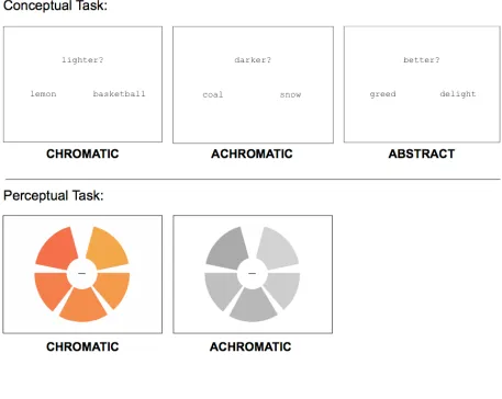
Subjects made a luminance judgment on a named pair of objects, indicating which
object was lighter or darker. The conditions of interest named chromatic (e.g., LEMON
and BASKETBALL) or achromatic (e.g. COAL and SNOW) objects. The 206 objects
(102 achromatic, 104 chromatic) used in the experiment were rated for color agreement
study sample. The conditions did not differ from one another in terms of frequency, word
length, concreteness, familiarity, imageability, or percent color/chromatic agreement (see
Table 2.1 for characteristics of word stimuli). There were 98 trials for each condition. As
a baseline task, subjects judged which of a pair of abstract (e.g., GREED and DELIGHT)
concepts was better or worse. For a list of stimuli used in the color knowledge task, see
Table 2.2.
At the beginning of each trial, a pair of words and a prompt, randomly assigned to
display “lighter?” or “darker?” (“better?” or “worse?” for the abstract condition)
simultaneously appeared on the screen for 2700 msec (see Figure 2.1). During this time
window, the subjects’ task was to decide which named object was lighter or darker,
indicating their response via button press. At the end of the trial, a central fixation cross
appeared for 300 msec, for a total trial duration of 3000 msec.
We blocked conditions as follows: 7 trials of one condition (21 seconds) followed
by 12 seconds of passive fixation, 7 trials of the second condition followed by 12 seconds
of passive fixation, then 7 trials of the third condition, and so on. We permuted condition
order across subjects, but there was always a fixed stimulus onset asynchrony (SOA) for
the 7 trials in each 21 second block. We used E-Prime software (Psychology Software
Tools, Inc.) to present stimuli and to collect response data.
Task — color perception
After subjects completed the color retrieval task, we administered a functional
the Farnsworth-Munsell 100 hue stimuli, in which they judged whether the wedges
making up colored or grayscale wheels were sequentially ordered from lightest to
darkest. The methods and stimuli for this task have been used previously to identify brain
regions involved in color perception (Beauchamp et al., 1999; Hsu et al., 2011; W.
Simmons et al., 2007).
Scanning Procedure
We acquired imaging data using a 3T Siemens Trio system with a standard
8-channel head coil and foam padding to secure the head position. After we acquired
T1-weighted anatomical images (TR = 1620 msec, TE = 3 msec, TI = 950 msec, voxel size =
0.9766 mm x 0.9766 mm x 1.0 mm), each subject performed four runs of the color
knowledge retrieval task, followed by two runs of the color perception task, while
undergoing blood oxygen dependent (BOLD) imaging (Ogawa et al., 1993). We collected
774 sets of 42 slices using interleaved, gradient echo, echoplanar imaging (TR = 3000
msec, TE = 30 msec, FOV = 19.2 cm x 19.2 cm, voxel size = 3.0 mm x 3.0 mm x 3.0
mm). Nine seconds of “dummy” gradient and radio frequency pulses preceded each
functional scan to allow for steady-state magnetization; during this initial time period, we
did not present stimuli or collect fMRI data.
Image Processing
We analyzed the data using VoxBo (www.voxbo.org) and SPM2
FMRIB Software Library (FSL) toolkit (http://www.fmrib.ox.ac.uk/fsl) to correct for
spatial inhomogeneities and to perform non-linear noise reduction. Functional data were
sinc interpolated in time to correct for the slice acquisition sequence; motion corrected
with a six-parameter, least squares, rigid body realignment routine using the first
functional image as a reference; and normalized in SPM2 to a standard template in
Montreal Neurological Institute (MNI) space. Data were smoothed using a 9mm
full-width half-max Gaussian smoothing kernel. Following preprocessing for each subject, a
power spectrum for one functional run was fit with a 1/frequency function, and this
model was used to estimate the intrinsic temporal autocorrelation of the functional data
(Zarahn, Aguirre, & D’Esposito, 1997).
We fit a modified general linear model (Worsley & Friston, 1995) to each
subject’s data to the four runs of the color retrieval task, in which the conditions of
interest (chromatic, achromatic, abstract) were each modeled as a 21-second block and
convolved with a standard hemodynamic response function. Several covariates of no
interest (global signal, scan effects, movement, spikes) were also included in the model.
An adjusted response latency for each trial for all conditions (i.e., a mean centered log
transformation of each subject’s RT) was also entered as a continuous covariate of no
interest, to address any difficulty or “time on task” confounds. From this model, we
computed parameter estimates for each condition (compared to fixation baseline) at each
voxel, and these estimates were included in the group-level random effects analyses
described below. Independently, we fit a second modified GLM to each subject’s data
(colored versus grayscale stimuli) were modeled as blocks in the same manner as
described above. Aside from this difference in the conditions of interest, the two models
were constructed identically.
Results
Behavioral Results
Color knowledge retrieval task: There was a significant RT difference across
conditions, F(2, 51) = 5.78, p = 0.005 (chromatic: M = 1873 ms, SD = 173 ms;
achromatic: M = 1782 ms, SD = 145 ms; abstract: M = 1694 ms, SD = 155 ms). We found
this RT difference substantial enough to warrant entering the RT for each trial as a
continuous covariate of no interest in the GLM, such that any differences reported below
describe condition differences, rather than RT differences. Note that the inclusion of this
covariate has the effect of underestimating the chromaticity effect on the BOLD
response.
Color perception task: There were no RT differences between chromatic and
achromatic perceptual judgments [(chromatic: M = 1473 ms, SD = 204 ms; achromatic:
M = 1439 ms, SD = 230 ms), t(17) = 1.43, p = 0.17)], though participants were
significantly worse at chromatic judgments [(chromatic: M = 73%, SD = 5.6%;
achromatic: M = 79%, SD = 5.9%), t(17) = 3.00, p = 0.008].
Functional Region of Interest Analyses: Left Lingual Gyrus
effects of chromaticity on color knowledge retrieval, we first performed a group-level
random effects analysis on the color perception data, comparing brain activity of colored
stimuli to that of grayscale stimuli as in prior studies. No regions responded more to
grayscale than colored stimuli. We corrected for multiple comparisons (at α = 0.05) by
performing 1000 Monte Carlo permutations of the data, deriving a critical threshold of t =
6.16 (Nichols & Holmes, 2002). When examining those regions that responded more to
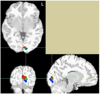
colored than grayscale stimuli, only one fROI (17 voxels) in the left lingual gyrus
(Talairach coordinates: -9, -87, 4) surpassed the corrected threshold. Within this region,
we calculated parameter estimates for each subject, for each condition of the color
retrieval task, on the spatially-averaged time series. We assessed an effect of concrete
versus abstract concepts by testing a comparison of both types of conceptual knowledge
to abstract knowledge. The chromaticity effect was assessed by using a paired t-test of
the difference between the chromatic and achromatic parameters. In the left lingual gyrus,
there was significantly greater activity when retrieving concrete versus abstract
knowledge ([Chromatic + Achromatic] — Abstract; t(17) = 2.85, p = 0.01). Critically,
there was significantly greater activity when retrieving chromatic versus achromatic
knowledge ([Chromatic – Achromatic]; t(17) = 2.29, p = 0.04]).
The preceding analyses establish that a chromaticity effect during color retrieval
is observed in a region that exhibits a chromaticity effect during color perception. This
effect, while a direct test of the hypothesis of interest, is a narrow way to address the
extent and location of overlap during perception and memory processing. Towards this
similar to the group-level random effects analysis performed on the color perception data,
we performed a similar analysis on the color knowledge retrieval data, again from all 18
subjects. For this dataset, we compared brain activity in the chromatic condition to that in
the achromatic condition. Next, within occipital brain regions, we identified the top 50
voxels that were most active during each task, irrespective of threshold. For the color
perception task, Talairach coordinates for the peak voxel were -9, -87, 4. For the color
knowledge task, Talairach coordinates for the peak voxel were -9, -93, 2. As seen in
Figure 2.2, we found co-localization of seven voxels in the left lingual gyrus. This result
suggests not only that the lingual gyrus is involved in both color perception and
knowledge retrieval, but that it is recruited more for the chromatic condition in both
processes (more so than the achromatic condition of both processes).
Exploratory Whole Brain Analyses: Ventral and Dorsal Distinctions
In order to ascertain the specificity of this effect, we examined brain activity at a
less stringent threshold to determine whether other brain regions were active for either
task. For this exploratory analysis, we examined the group color perception data at an
uncorrected threshold of p < 0.001(t = 3.97), which yielded 27 distinct clusters, as shown
in Figure 2.3. We then created 27 fROIs of comparable size by identifying each
individual local maximum and any of the 26 surrounding voxels that also surpassed the
uncorrected threshold (see Table 2.3 for the coordinates of these local maxima). In order
to assess the chromaticity effect during memory retrieval within these regions, we
on the spatially-averaged time series across voxels within each fROI.
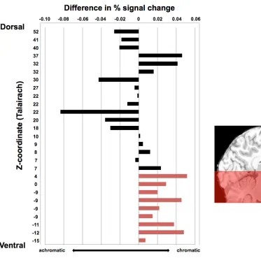
Upon doing this, we observed an unanticipated pattern, which is evident in the
order we have arranged the clusters in Figure 2.4: The difference in response to
chromatic versus achromatic memory retrieval was larger and more consistent in the
ventral fROIs than in the dorsal fROIs. Although the chromaticity effects did not reach
significance in individual fROIs, the reliability of the pattern can be established with a
post-hoc binomial test. In the nine most ventral fROIs (z < 5), all nine of the fROIs
showed numerically greater activation for chromatic compared to achromatic stimuli (p =
0.004). In contrast, the pattern was not consistent across the 18 dorsal fROIs (p = 0.48).
Although these results should be interpreted with caution, they suggest that the effect
reported above in the left lingual gyrus may be a more widespread pattern in ventral but
not dorsal regions of the visual system. In the following section, motivated by previous
work, we take a closer look at one such region, the left fusiform gyrus.
Secondary Region of Interest Analyses: Fusiform Gyrus
Previous research has shown that the fusiform gyrus is involved in both color
knowledge retrieval and color imagery and perception (Howard et al., 1998; Simmons et
al., 2007). In all analyses thus far, ROIs were functionally defined, and the fusiform
gyrus did not emerge as an active region when contrasting the chromatic to achromatic
conditions in either task (i.e., activity did not surpass either the uncorrected or permuted
threshold). However, for our color knowledge task, the left fusiform gyrus was robustly
pairs (t[17] = 4.63, p < .001). The Talairach coordinates for the peak active voxel for this
contrast were -33, -36, -13 ; this is almost identical to the peak voxel in left fusiform
gyrus as reported by Simmons and colleagues for their color knowledge task ( 33, 36,
-16). The proximity of these peak voxel coordinates across studies suggests that our
memory task, and other tasks which have been used in prior research, are both tapping
into color knowledge retrieval processes, though again, there was no main effect of
chromaticity (t[17] = 0.38, p = 0.71).
Several prior studies have reported that this region of the fusiform gyrus is active
during color perception (that is, more active during perception of chromatic compared to
achromatic stimuli) in addition to during retrieval of color (Simmons et al., 2007). We
did not observe this effect in the analyses we reported above; although we used their
same procedure and stimuli to manipulate color perception, there were some differences
in our analyses. Subsequently, we repeated the analysis of the perception data without
including global signal as a covariate in the model (following from Simmons et al.,
2007), and this new analysis does show a chromaticity effect in color perception in the
fusiform gyrus. In this functionally-defined fusiform region, there was a trend towards a
chromaticity effect on the knowledge retrieval task, though it did not reach statistical
significance (t(17) = 1.82, p = 0.09). Finally, in applying a similar approach as described
earlier — in which we identified the top voxels in ventral temporal regions that were
involved in both tasks, irrespective of threshold — in analyses without global signal in
the models, we observed near (but not direct) voxel overlap between task activation (in
In sum, these additional analyses suggest a role for the left fusiform gyrus in color
perception and memory. However, they also show that the pattern of activation in this
region is different in a number of ways from that observed in the left lingual region,
being more correlated with global signal, and less sensitive to chromaticity in memory.
We will return to these differences and their possible implications below.
Discussion
The results of this study demonstrate that a chromaticity effect (namely, greater
activation to colored than grayscale stimuli), which has already been documented in color
perception, is paralleled in memory retrieval. This tests an important prediction of
sensorimotor models of memory, which propose that the same processes invoked during
perception of a sensory property will also contribute to memory of that property.
Therefore, under these models, if there are different neural patterns associated with the
perception of chromatic versus achromatic stimuli, this difference should emerge during
memory retrieval. Our data confirm this prediction. This experimental strategy could be
applied to many other sensorimotor properties, in order to test for similarities between
perception (or action) and memory processes.
Somewhat to our surprise, the chromaticity effect on memory was strongest in a
very posterior region, namely the left lingual gyrus. This was the one region where we
found direct overlap of voxels activated by both perception and memory tasks. Ventral
temporal regions, and in particular the left fusiform gyrus, showed robust task activation
from the peak voxel reported by Simmons and colleagues (2007). However, the
chromaticity effect in the left fusiform gyrus associated with the memory task was weak
at best, and evident only as a marginally significant difference in an analysis without
global signal in the model. These findings suggest that future research might fruitfully
focus on possible differences between posterior (i.e., lingual) and more anterior (i.e.,
fusiform) regions of the visual system.
As noted, the decision to include global signal in the model did not affect the
detection of the perceptual chromaticity effect in the left lingual region, but did have an
impact in the left fusiform region. Often, considerations about inclusion of the global
signal in a model are based on the correlation between the global signal covariate and the
task covariate — because when the two are highly correlated, teasing apart the effects of
each becomes nearly impossible (see Aguirre, Zarahn, & D’Esposito (1998) for further
discussion). The correlation between global signal and the chromaticity covariate in the
perceptual task ranged from 0.11-0.31 across subjects, though of course the co-linearity
across predictors in the model is the same for the two regions in question here.
What could differ, however, is the correlation between the global signal and the
regionally-specific signal. In other words, if activation in the fusiform region is more
strongly correlated with the global signal than is activation in the lingual region, then the
inclusion of global signal in the model could differentially affect the fusiform findings.
This was indeed the pattern that we observed in these two regions (p < 0.01). The fact
that global signal explains more variance in the fusiform (average t = 10.8) than in the
differential effects in these two regions.
We find this idea interesting in light of an argument made in Beauchamp et al.
(1999), namely that fusiform activation may depend on the attentional demands of the
task. For example, previous work has demonstrated that the fusiform gyrus is activated
during an attentionally demanding color perception task, but not during passive viewing
tasks (see Beauchamp et al., 1999). In other research, we have shown that activation in
this region during color retrieval is modulated by specificity of the color information
required in the task (Hsu et al., 2011). If activation in the fusiform gyrus does reflect a
more general attentional process, then it is conceivable that activity there would be
correlated with a spatially-distributed attentional network, which could in turn lead to a
higher correlation with global signal — because the global signal is the average of every
voxel’s response at a given time point. More generally, this discussion highlights the
potential value of comparing models that include or omit covariates which are generally
considered to have “no interest.” As shown above, global signal might indeed be of
interest.
Turning to the memory data, we found a strong effect of chromaticity in the
lingual gyrus which is co-localized with the perceptual chromaticity effect, but a weak
effect, at best, in the fusiform gyrus. We find the absence of a chromaticity effect in the
fusiform gyrus particularly interesting, given that the memory task resulted in robust
activation in this region. How might this area be contributing to the process of memory
retrieval, particularly in light of its association with color perception as reviewed above?
that the interaction between chromaticity and region did not reach significance (p = 0.19),
so we do not have evidence that the patterns we observed in fusiform and lingual regions
are reliably different. For this reason, these ideas should be understood as suggestions for
future research aimed at distinguishing the response properties of these regions.
Firstly, echoing a suggestion in Simmons et al. (2007), the posterior lingual area
might be generating a purely sensory response to any colored stimuli, regardless of task,
whereas the more anterior fusiform area may be more directly involved in the process of
color categorization. The luminance judgment for our color knowledge task did not
explicitly require subjects to categorize color stimuli, which may explain the lack of a
robust chromaticity effect in fusiform gyrus. Hence, the two areas can be understood as
supporting different processes that are invoked to varying degrees during different
perceptual and memory tasks. This account leads to the testable prediction that
chromaticity effects would be observed during a memory retrieval task that required
greater attention to color categories.
A second possibility concerns the nature of the representations of different colors
in these two regions. An area could be said to represent color, if the response in that area
varies as a function of variation in color. One such variation would be that chromatic
stimuli, as a group, tend to elicit a different pattern of activity (in this case, a higher
magnitude response) than do achromatic stimuli. Observation of a chromaticity effect
requires that within-class differences (that is, variation between different colors, among
the chromatic stimuli, or between different shades of gray, among the achromatic stimuli)