INTRODUCTION
Chronic, nonhealing wounds arise from diverse etiologies such as diabetes, venous stasis and pressure. Wounds occur in 12–25% of diabetic patients and contribute to a high incidence of limb loss and death (1). Despite improve-ments in glycemic control, antibiotics, and wound care adjuvants, nonhealing wounds continue to present a formidable problem. The underlying molecular mechanisms of impaired wound healing are poorly understood despite extensive
knowledge of the normal wound repair process. Efficient wound repair involves many cytokines, chemokines and growth factors that regulate the inflammatory and regenerative phases of wound heal-ing (2). Unfortunately, novel therapies targeting these factors, such as topical platelet-derived growth factor, have had limited success (3).
Inflammation is a necessary response to tissue injury, leading to the production of nitric oxide (NO) and reactive oxygen species (ROS) and reactive nitrogen
species (RNS) that mediate oxidative stress (4). Recent evidence demonstrates that physiologic oxidative stress is essen-tial for normal wound healing (5,6). Redox signaling has been reported to play an important role in antibiosis, hemosta-sis, inflammation, re-epithelialization, an-giogenesis and growth factor modulation (7,8). However, an overabundance of in-flammatory by-products has deleterious effects on the wound (4). For example, ROS contributed to the failure of wound healing in a rodent ischemic flap model (9) and high doses of topical hydrogen peroxide (H2O2) delayed wound repair by reducing collagen deposition and angio-genesis (10). Achieving the ideal balance of ROS/RNS in damaged tissues may de-termine wound healing efficiency.
Wound repair also depends on NO. NO synthesis in wounds occurs through the arginine-NO synthase (NOS) path-way (11) and modulates inflammation, chemotaxis, antibacterial defenses, colla-gen production and angiocolla-genesis
Wound Healing
Michael C Madigan,
1,2Ryan M McEnaney,
1,2Ankur J Shukla,
1,2Guiying Hong,
1,2Eric E Kelley,
3Margaret M Tarpey,
1,3Mark Gladwin,
4Brian S Zuckerbraun,
1,2and Edith Tzeng
1,21
Surgery Services, Department of Veterans Affairs Medical Center, Pittsburgh, Pennsylvania, United States of America; Departments of 2Surgery, 3Anesthesia, and 4Medicine, University of Pittsburgh, Pittsburgh, Pennsylvania, United States of America
Chronic, nonhealing wounds result in patient morbidity and disability. Reactive oxygen species (ROS) and nitric oxide (NO) are both required for normal wound repair, and derangements of these result in impaired healing. Xanthine oxidoreductase (XOR) has the unique capacity to produce both ROS and NO. We hypothesize that XOR contributes to normal wound healing. Cutaneous wounds were created in C57Bl6 mice. XOR was inhibited with dietary tungsten or allopurinol. Topical hydrogen peroxide (H2O2, 0.15%) or allopurinol (30 μg) was applied to wounds every other day. Wounds were monitored until closure or collected at d 5 to assess XOR expression and activity, cell proliferation and histology. The effects of XOR, nitrite, H2O2and allopurinol on keratinocyte cell (KC) and endothelial cell (EC) behavior were assessed. We identified XOR expression and activity in the skin and wound edges as well as granulation tissue. Cultured human KCs also expressed XOR. Tungsten significantly inhibited XOR activity and impaired healing with reduced ROS production with reduced angiogenesis and KC proliferation. The expression and activity of other tung-sten-sensitive enzymes were minimal in the wound tissues. Oral allopurinol did not reduce XOR activity or alter wound healing but topical allopurinol significantly reduced XOR activity and delayed healing. Topical H2O2restored wound healing in tungsten-fed
mice. In vitro, nitrite and H2O2both stimulated KC and EC proliferation and EC migration. These studies demonstrate for the first time that XOR is abundant in wounds and participates in normal wound healing through effects on ROS production.
Online address: http://www.molmed.org doi: 10.2119/molmed.2014.00191
Address correspondence toEdith Tzeng, Chief of Vascular Surgery, VA Pittsburgh Health-care System, Professor of Surgery, Department of Surgery, University of Pittsburgh, A1010 PUH, 200 Lothrop Street, Pittsburgh, PA 15213. Phone: 412-802-3025; Fax: 412-291-1669; E-mail: [email protected].
(12–14). NO deficiency has been impli-cated in the delayed wound healing in diabetic rodent models (15,16), where NOS was downregulated. In addition, arginase I uses L-arginine to produce polyamines and proline, both essential for collagen synthesis and cell prolifera-tion, and is induced and competes for the NOS substrate (16–18). Inducible NOS (iNOS) or endothelial NOS (eNOS) deficiency both delayed wound closure in normal mice, whereas restoration of NOS activity in these mice improved healing (19,20), supporting an essential role of NO in wound repair.
Xanthine oxidoreductase (XOR) is a ho-modimeric enzyme that metabolizes xan-thine and generates ROS. It is also a po-tent nitrite reductase, converting nitrite back to NO (21,22). This pathway is espe-cially efficient in hypoxia, a condition that reduces NOS-dependent NO produc-tion. We and others have reported that ni-trite can serve as a source of NO through XOR in models of vascular injury and pulmonary hypertension (23,24). XOR has been detected in skin, where it is in-volved in ROS production in response to lipopolysaccharide (LPS) (25) and ultravi-olet irradiation (26). Thus, we hypothe-size that XOR participates in normal wound healing through ROS/RNS and NO production. We used a murine exci-sional wound-healing model and manip-ulated XOR activity (27,28) to examine its role in normal wound repair.
MATERIALS AND METHODS
Excisional Wound Healing Model
All procedures conformed to the Guide for the Care and Use of Laboratory Animals (29) and the policies of the Institutional Animal Use and Care Committee of the University of Pittsburgh (protocol #1104675A). Male C57BL/6 mice (8–12 wks old; The Jackson Laboratory, Bar Harbor, ME, USA) were anesthetized with Nembutal (70 mg/kg, Abbott Labs, Chicago, IL, USA) and isoflurane. After shaving, a 1.5 × 1.5–cm excisional wound was created on the back of each mouse and then covered with bio-occlusive
dressings (Systagenix, Quincy, MA, USA). Wound area was measured by ac-etate tracings every other day until wound closure. The areas were calcu-lated using MetaMorph® (Version 7.7.5.0; Molecular Devices, Inc., Sunny-vale, CA, USA). Wounds were also col-lected at earlier time points for protein and immunohistochemical analyses.
Dietary and Topical Wound Treatments
Tungsten-enriched diet (#960350; MP Biomedicals, Irvine, CA, USA) was started 2 wks before wounding to opti-mize molybdenum replacement in XOR and maintained thereafter. Allopurinol (100 mg/kg/day; Sigma-Aldrich, St. Louis, MO, USA) in drinking water, sodium nitrite (300 mg/L in deionized water; Sigma-Aldrich) or nitrite-free diet (Harlan Teklad amino acid diet, TD 99366; Harlan, Indianapolis, IN, USA) was initiated 1 wk before wounding and continued.
Topical H2O2was applied to the wound as a 0.15% H2O2solution (Thermo Fisher Scientific Inc., Waltham, MA, USA) in normal saline, and the wound was covered. Topical allopurinol (30 μg/wound) was similarly applied to each wound. Treatment was initiated im-mediately after wounding and continued every other day.
Western Blot Analysis
Wound samples were collected and di-vided into the granulation tissue and the wound edge. Skin adjacent to the wound was also collected. Samples were homog-enized in lysis buffer (Cell Signaling Technology, Danvers, MA, USA) and quantified using a Pierce® BCA Protein assay (Thermo Fisher Scientific). Western blot analysis for XOR (rabbit mono-clonal, 1:5,000; ab109235; Abcam, Cam-bridge, MA, USA), iNOS (rabbit poly-clonal, 1:200; ab15323; Abcam) or arginase I (mouse monoclonal, 1:2,000; BD Biosciences, San Jose, CA, USA) was performed using horseradish peroxi-dase–linked goat rabbit or anti-mouse secondary antibody (1:10,000;
Thermo Fisher Scientific). The mem-branes were developed by using Super-Signal® West Pico Chemiluminescent #34080 (Thermo Fisher Scientific).
Wound Immunohistochemistry
Wounds were collected on d 7 or at wound closure and fixed in 2% parafor -maldehyde, cryoprotected in 30% sucrose, embedded in OCT (Tissue Tek®; Sakura Finetek, Torrance, CA, USA) and sectioned (7 μm). Sections were treated with rabbit polyclonal anti-XOR (1:100; Santa Cruz Biotechnology, Santa Cruz, CA, USA), rabbit polyclonal anti- collagen I (1:200; Abcam), monoclonal anti-Ki67 (1:200; Abcam) or rat monoclonal anti-CD31 (1:50; BD Biosciences) antibody followed by goat anti-rabbit 488 or goat anti-rat Cy5 at 1:1,000 (Invitrogen [Thermo Fisher Scientific]). Nuclei were counterstained with Hoechst 33325 (2 μg/mL, Sigma-Aldrich). Images were collected using the Fluoview® FV1000 confocal microscope (Olympus, Center Valley, PA, USA).
Wound Angiogenesis
Wound sections were stained with CD31, and two confocal images of the wound granulation tissue were obtained for each section. Wound angiogenesis was calculated as the number of CD31-stained lumens with ImageJ (version 1.45s; National Institutes of Health, Be-thesda, MD, USA) and as the percent area of CD31 staining using MetaMorph®.
XOR and Aldehyde Oxidase Activity
ac-tivity was measured by incubating tissue homogenates with the AO substrate 4-(dimethylamino)cinnamaldehyde (DMAC) (25 μmol/L in potassium phos-phate [KPi], pH 7.8, and at 25°C) and monitored for a decrease in absorbance at 398 nm.
Wound ROS/RNS Measurement
Total ROS/RNS was quantified by using the OxiSelect™ In Vitro ROS/RNS Assay Kit (Cell Biolabs, Inc., San Diego, CA, USA) per instructions. Homoge-nized wound tissues (50 μg) were as-sayed for ROS/RNS by the conversion of dichlorodihydrofluorescein to 2′,7′ -dichlorodihydrofluorescein diacetate.
In Vitro Studies in Keratinocyte Cells and Endothelial Cells
Human epidermal keratinocyte cells (KCs) (PCS-200-011; ATCC, Manassas, VA, USA) were cultured in Dermal Cell Basal Media (PCS-200-030, ATCC) plus Keratinocyte Growth Kit (PCS-200-040, ATCC). KCs were differentiated with 1.2 mmol/L of calcium. Whole cell lysates were used for Western blot analysis for XOR (1:100; Santa Cruz). For immunocy-tochemistry, KCs were cultured on glass slides, fixed with 2% paraformaldehyde, blocked in 2% BSA in PBS and then incu-bated with rabbit polyclonal anti-XOR (1:100; Santa Cruz) and mouse mono-clonal anti-K10 (1:100; Abcam) followed by secondary goat rabbit or anti-mouse antibody (1:1,000). Nuclei were counterstained with Hoechst 33325 (2 μg/mL) (Sigma-Aldrich). Images were acquired on the Fluoview® FV1000 con-focal microscope (Olympus).
Human dermal microvascular endothe-lial cells (ECs) (VEC Technologies; Rensse-laer, NY, USA) were cultured as described (30) and low passage was used. Briefly, cells were grown in a 1:1 mix of MCDB131 (VEC Technologies) and Dulbecco modi-fied Eagle medium (DMEM) with 5% fetal bovine serum (FBS). In proliferation assays, ECs were cultured in DMEM with 1% FBS overnight. Proliferation was mea-sured by using 3H-thymidine as described (31) in 1% FBS medium with nitrite,
H2O2, catalase or allopurinol (Sigma-Aldrich). Migration was measured using the scratch assay at 6 h (32) and quanti-fied with ImageJ. For in vitro angiogene-sis, ECs were cultured on Matrigel™ (BD Biosciences) as described (30) with and without nitrite, allopurinol, H2O2or catalase. Tube formation was examined at 6 h, quantifying boxes and number of long tubes (>110 pixels in length) as a marker of healthy tubes.
Statistical Analysis
Data are presented as mean ± standard error of the mean (SEM). Statistical anal-ysis was performed with a Student ttest or analysis of variance by using the Sig-maStat 11.0 software (Systat Software Inc., San Jose, CA), and significance was determined at P< 0.05. All pairwise mul-tiple comparisons were performed by using the Holm-Sidak method.
RESULTS
XOR, iNOS and Arginase I Expression in Wound Tissue
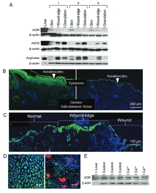
C57BL/6 mouse skin had easily detect-able XOR expression by Western blot (Figure 1A). XOR expression was not al-tered by allopurinol, tungsten and nitrite diets (data not shown). The wound edge had less XOR expression, whereas the granulation tissue had the lowest. In con-trast, iNOS expression was minimal in the skin but was upregulated in the wound edge and granulation tissue (Fig-ure 1A). Arginase I had a similar expres-sion pattern to iNOS (Figure 1A). Im-munostaining of wounds at d 7 and after healing at d 14 revealed that the epider-mal KCs expressed high levels of XOR (Figure 1B). Much less XOR was detected in the dermis or subcutaneous tissues. XOR expression was most pronounced in the thick, proliferating layer of KCs sur-rounding the healing wound as well as in the thinner layer of KCs in normal skin. At 2 d after wounding (Figure 1C), the upregulation of XOR expression in the wound edge was evident compared with the surrounding skin. In addition, there were cells in the peri-wound
der-mis and subcutaneous tissues that stained for XOR and are consistent with neutrophils by the appearance of the nu-clear morphology. This finding indicates that XOR is highly expressed in the wound throughout the healing process and early inflammatory infiltrates also express XOR. Cultured KCs also ex-pressed XOR by immunostaining and Western blot (Figures 1D, E). Differentia-tion of the KCs with calcium, as indi-cated by the expression of keratin-10, did not affect XOR expression.
Tungsten-Inhibited XOR Activity and Delayed Wound Healing in C57BL/6 Mice
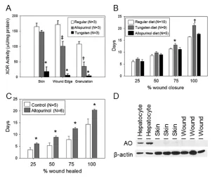
Tungsten substitutes for the molybde-num (Mb) in XOR, inhibiting it irre-versibly. Tungsten diet significantly in-hibited skin XOR activity compared with mice fed allopurinol (18.5 ± 15.2 versus 147.6 ± 4.8 μU/mg protein, P< 0.001) or a chow diet (165.3 ± 10.7 μU/mg protein, P< 0.001) (Figure 2A). XOR activity was similar between allopurinol and control mice. XOR activity at the wound edge was moderately reduced in allopurinol-treated versus control mice (100.9 ± 15.5 versus 178.7 ± 12.1 μU/mg protein, P< 0.001) but tungsten-treated mice had near complete inhibition of XOR activity (6.6 ± 4.1 μU/mg protein, P< 0.001 ver-sus control or allopurinol). Both allopuri-nol and tungsten-treated mice (35.3 ± 14.4 and 4.6 ± 2.9 μU/mg protein, re-spectively) had significantly less XOR ac-tivity in granulation tissue than control mice (107.6 ± 8.3 μU/mg protein, P< 0.01 versus tungsten or allopurinol). Wound healing was significantly delayed in mice receiving the tungsten-enriched diet versus mice on chow (Figure 2B; 21.2 ± 0.7 versus 16.4 ± 0.2 d, P< 0.001). Dietary allopurinol did not delay wound healing (Figure 2B).
compared with wounds from control mice (Figure 2C). Measurement of XOR activity showed that allopurinol applied to the wounds markedly reduced XOR activity by over 70% compared with con-trol wound tissues (156.2 ± 90.5 μU/mg versus 547.4 ± 15.9, respectively; n = 3/ group; P= 0.013).
Minimal Expression of Other Mb-Containing Oxidases Was Detected in Wound Tissue
AO and sulfite oxidase (SO) both con-tain Mb and can be inhibited by tungsten. They can also function as nitrite reduc-tases. Very low levels of AO were detected in the skin and wound tissues by Western
blot (Figure 2D). SO, which is primarily mitochondrial, was not detectible in the wounds as well (data not shown). Assay of wound tissues for AO activity showed no detectable oxidation of DMAC (n = 3), supporting the absence of AO activity in wounds. These findings support that the predominant effect of tungsten on wound healing is mediated through XOR.
XOR Inhibition with Tungsten Reduced ROS/RNS in Granulation Tissue
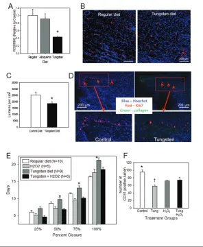
To determine the effect of dietary tung-sten or allopurinol on wound levels of ROS/RNS, granulation tissue was col-lected at d 5. By using the OxiSelect In Vitro ROS/RNS Assay, granulation tissue from control mice had similar levels of ROS/RNS as wounds from mice treated with dietary allopurinol (Figure 3A). However, tungsten reduced wound ROS/ RNS by ~60%, confirming the efficient in-hibition of XOR by tungsten but not di-etary allopurinol (n = 7/group; P< 0.01 versus control, P< 0.05 versus allopuri-nol). In a separate experiment, topical al-lopurinol did significantly reduce wound ROS/RNS production compared with un-treated wounds (n = 3/ group, 0.55 ± 0.10 versus 1.00 ± 0.09-fold change; P= 0.029).
XOR Inhibition with Tungsten Reduced Wound Angiogenesis and KC
Proliferation
Tissue sections from healed wounds were stained for CD31 as a marker for ECs (Figure 3B). Wounds from tungsten-treated mice had 25.3% fewer CD31-positive lumi-nal structures versus wounds from control mice (Figure 3C; P< 0.05; n = 6). Wound proliferation was measured by Ki67 stain-ing at d 7. Ki67 was most prominent in the wound edge basilar KCs in control mice (30.7 ± 5.3%, Figure 3D). However, tung-sten-treated mice (Figure 3D) had signifi-cantly fewer proliferating basilar KCs (9.1 ± 4.8%, P< 0.05).
Topical H2O2Reversed Tungsten-Induced Delay in Wound Healing and Improved Angiogenesis
To determine if the role of XOR in wound healing was mediated through
Figure 1.Examination of XOR expression in wounds and keratinocytes. (A) Western blot anal-ysis for XOR, iNOS and arginase I expression in mouse skin, wound edge and granulation tis-sue is shown. Wound samples at d 7 from three separate animals are represented. β-Actin was used as a loading control. (B) XOR expression in healing wounds at d 14 (left) and nor-mal skin (right) was examined by immunostaining (green = XOR, blue = nuclei, red = colla-gen; magnification of 100×). The area of the healing wound that is shown is the healed area adjacent to the wound bed. (C) XOR expression in early wounds (d 2) is demonstrated on a panoramic view showing normal skin, wound edge and the wound (green = XOR, blue = nu-clei). (D) Immunostaining of cultured undifferentiated (left) and differentiated (right) human keratinocytes (200× magnification; green = XOR, blue = nuclei, red = keratin; representative photomicrographs, n = 3/group). (D) Western blot analysis for XOR expression in cultured human keratinocytes. Treatment with Ca2+differentiated the cells to mature keratinocytes.
H2O2production, wounds were treated topically with 0.15% H2O2or saline every other day until complete wound closure. H2O2did not alter wound healing rates in control mice (Figure 3E), but it signifi-cantly improved healing in tungsten-fed mice versus tungsten diet alone (18.5 ± 0.7 versus 21.0 ± 0.7 d; P< 0.01), ap-proaching the rates in control mice. H2O2 treatment of wounds in tungsten-treated mice showed a trend toward improved angiogenesis (Figure 3F; P= 0.067).
Dietary Nitrite Manipulations Did Not Alter Systemic Levels of Nitrite or Wound Healing
Mice receiving supplemental sodium nitrite showed no improvement in wound healing compared with controls (17.4 ± 0.3 versus 16.7 ± 0.3 d, P= nonsignificant [NS], n = 5). Similarly, mice on a nitrite-free diet showed no change in wound
healing versus controls (17.2 ± 0.3 versus 16.7 ± 0.3 d, respectively; P= NS). How-ever, serum nitrite levels remained un-changed in mice receiving nitrite supple-mentation or depletion compared with regular chow (282.5 ± 35.5 versus 254.8 ± 64.0 versus 199.3 ± 53.9 nmol/L, respec-tively; P= 0.54, n = 4). The inability to change serum nitrite levels with diet may explain the lack of effect observed.
XOR Mediates KC and EC Function
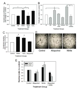
KCs are the predominant cell type in the wounds expressing XOR and tung-sten inhibition of XOR reduced basilar KC proliferation. Thus, we evaluated the effect of XOR activity on KCs in vitro. In-hibition of XOR with allopurinol did not affect proliferation in cultured human KCs (Figure 4A). Nitrite treatment signif-icantly increased KC proliferation, which was reversed by allopurinol, indicating
nitrite functioned through XOR to pro-mote proliferation. H2O2also increased KC proliferation.
Because tungsten also reduced wound angiogenesis, the role of XOR in EC func-tion was also examined. By Western blot, cultured human ECs express XOR at baseline (data not shown). There was a trend toward reduced proliferation when ECs were treated with allopurinol (P= 0.064) and catalase (P= 0.073) (Figure 4B), suggesting XOR and H2O2contribute to baseline EC proliferation. Nitrite sig-nificantly increased EC proliferation, and this was reversed with allopurinol, indi-cating that nitrite effects were mediated by XOR. Treatment with H2O2also in-creased proliferation. Similarly, nitrite and H2O2both increased cell migration (Figure 4C). Allopurinol did not alter mi-gration but nitrite could enhance XOR-mediated NO production to increase mi-gration, suggesting that XOR did not regulate baseline migration but could be used to improve migration with nitrite. EC tubing, a measure of angiogenic activ-ity, was inhibited by allopurinol, which reduced box formation and reduced the number of long tubes (Figures 4D, E). Ni-trite significantly increased tubing com-plexity and tube lengths in an allopurinol reversible fashion.
DISCUSSION
Our investigations identified a novel role for XOR in the normal wound heal-ing process. High levels of XOR expres-sion were detected in the skin and wound edge of normal mice, with most of the expression located within the basi-lar KCs. This expression was upregu-lated shortly after wounding and likely coincides with the proliferative activity of the KCs at the wound edge. This en-hanced XOR expression is observed throughout the healing process and likely subsides as the KCs stop prolifer-ating and the epidermis matures. We also observed XOR expression in the early in-flammatory infiltrates in the wound. XOR inhibition with tungsten signifi-cantly delayed wound closure. Local wound inhibition of XOR with topical
al-Figure 2.Effect of XOR inhibition on excisional wound-healing rates. (A) XOR activity was measured in the skin, wound edge and granulation tissue in control, tungsten-fed or allop-urinol-fed mice (n = 3 mice/group; *P < 0.05 versus control and allopurinol groups, ‡P < 0.05
versus control and tungsten groups). (B) Wound healing rates in control, tungsten-fed and allopurinol-fed mice as reported as days to reach 25%, 50%, 75% and 100% wound closure (*P < 0.05 and †P < 0.001 versus regular or allopurinol diet). (C) Effect of topical allopurinol
lopurinol also significantly delayed wound healing. Both of these findings support an important role for XOR in wound repair. The ability to reverse ef-fect of tungsten with topical H2O2 sug-gests that XOR contributes to wound H2O2production and is required for nor-mal healing. In vitrostudies demon-strated the ability of XOR to mediate angiogenic functions in ECs and pro-liferation in KCs. These findings together strongly support the contribution of XOR in the normal wound healing process.
XOR represents a mix of xanthine oxi-dase (XO) and dehydrogenase (XDH) (both are involved in purine metabolism [21]) and is clinically notable for its role in gout, where metabolism of xanthine to uric acid (UA) results in joint deposition and inflammation. This pathway also yields H2O2and superoxide, both of which are associated with tissue injury during ischemia/reperfusion (I/R) (33,34). Inhibition of XOR reduces ROS-mediated I/R injury (28). In the setting of ischemia and hypoxia, XO predomi-nates and more efficiently generates su-peroxide, although XDH also possesses partial oxidase activity (35). While detri-mental in excess, physiologic levels of ROS are essential in cell signaling (36) and tissue homeostasis and repair (6,7). Most tissue production of ROS has been attributed to nicotinamide adenine dinu-cleotide phosphate (NADPH) oxidase, and the role of XOR in the generation of physiologic ROS has been underappreci-ated. More recently, XOR gained atten-tion for its ability to convert nitrite to NO to mediate cytoprotection (23,37). We and others have shown that XOR is re-quired for the beneficial actions of sup-plemental nitrite in the inhibition of inti-mal hyperplasia after vascular injury (23) and in reversing pulmonary hyperten-sion (24). XOR expreshyperten-sion in skin has been previously reported, linked to the pathogenesis of sunburn and skin in-flammation (24,25). Beyond that, little is known about the role of XOR in the skin.
XOR inhibition with dietary tungsten significantly delayed wound closure. Tungsten replaces the Mb within XOR,
Figure 3.Effect of XOR inhibition on wound angiogenesis and proliferation. (A) Effect of tung-sten and allopurinol diet on wound ROS/RNS production. Wound tissue was collected from mice at 5 d after wounding. ROS/RNS production was measured by using the OxiSelect In Vitro ROS/RNS Assay Kit, which detects H2O2, peroxyl radical, NO and peroxynitrite anion. Levels of
ROS/RNS are normalized to the levels measured in mice fed regular diets (n = 5 mice/group, *P = 0.006 versus regular diet and P = 0.016 versus allopurinol diet). (B) Effect of XOR inhibition on wound angiogenesis, as indicated by CD31-positive structures, was examined in wound tis-sues collected from control mice and tungsten fed mice immediately upon wound closure (CD31 for ECs, red; Hoechst 33325 for nuclei, blue; collagen, green). Representative photomi-crographs of wounds from mice fed regular and tungsten diets are shown (100× magnifica-tion). (C) Quantification of wound angiogenesis was performed by counting luminal structures (two sections per wound, n = 6/group; *P < 0.05 versus control diet). (D) Effect of XOR inhibition with tungsten on wound keratinocyte proliferation. Wound tissue was collected from control and tungsten diet–fed mice at d 7 after wounding. Tissues were stained for cell proliferation with Ki67 (red) and for cell nuclei with Hoechst 33325 (blue). Representative photomicrographs (100× magnification) are shown. Red arrows indicate proliferating basilar KCs at the wound edge. Magnified views of the red box areas are provided. (E) Effect of H2O2on wound healing in tungsten-treated mice. C57BL/6 mice were fed either regular diet or a tungsten-enriched diet starting 2 wks before wounding and then maintained. Wounds were treated with either topical H2O2(0.15%) or saline application every other day. Wounds were measured every
other day until complete closure. The time required to achieve 25%, 50%, 75% and 100% closure for each treatment group is presented graphically (*P < 0.01 versus H2O2and tungsten + H2O2).
(F) Quantification of wound angiogenesis was performed in wounds treated with and without H2O2. Data are presented as number of CD31-positive lumen structures per high-power field
irreversibly inhibiting its function (21). However, Mb is also essential for other oxidoreductases such as AO and SO. We found low levels of these enzymes in
wound tissues. The other inhibitor of XOR that we used was allopurinol, a drug used to clinically treat gout and as an investigative tool in other
experi-ments (23,24). In our studies, dietary al-lopurinol did not alter wound healing rates and raised concerns that tungsten effects may be mediated by actions on AO or SO. However, we identified essen-tially no AO or SO expression and no measurable AO activity as well in un-treated wounds. We did find that tung-sten dramatically inhibited skin and wound XOR activity, while dietary allop-urinol only reduced it modestly in the wound edge and granulation tissue. Al-lopurinol has been used to block XOR ac-tivity in other tissues, such as in carotid and pulmonary arteries, but was much less effective in our model. The reduced ability of allopurinol to inhibit XOR in wounds may be due to poor biodistribu-tion in the relatively hypoperfused wound bed and skin. Its bioavailability may also be diminished by renal clear-ance and the requirement for conversion to its active form, oxypurinol (38,39). In addition, the binding of XOR to endothe-lial glycosaminoglycans results in resist-ance to allopurinol (38,39). In contrast, topical allopurinol significantly delayed wound closure and reduced XOR activity and wound ROS/RNS levels. These find-ings confirm the role of wound XOR function in mediating normal wound re-pair. They also illustrate the potential ability to target wound XOR by topical treatments.
NO is required for normal tissue repair (12,14), and its deficiency contributes to delayed wound healing, whereas NOS gene transfer restores healing rates to normal (19). Diabetic wounds have re-duced NOS expression and NO produc-tion (16,18), resulting in delayed wound healing. Arginase I is also upregulated during wound healing (16) for the pro-duction of polyamines that are essential for cell proliferation and collagen synthe-sis but competes with NOS for arginine and can reduce NO production (17). Di-etary nitrite can serve as an alternate source of NO through XOR (21,22). This pathway has been shown to be biologi-cally important in the setting of vascular injury and pulmonary hypertension where dietary nitrite reduced the adverse
Figure 4.Effect of XOR function on keratinocyte and endothelial cell behavior in vitro. (A) Human KCs were cultured in 1% serum media with allopurinol (allo; 100 μmol/L), nitrite (100 μmol/L), nitrite + allopurinol and H2O2(250 μmol/L) for 24 h. Proliferation was measured by 3H-thymidine uptake, and results are expressed as fold-change over control (mean ±
SEM of four experiments; *P < 0.05 nitrite versus control or nitrite + allopurinol). (B) ECs were grown in 1% serum media with the addition of allopurinol (allo; 100 μmol/L), nitrite (100
μmol/L), nitrite + allopurinol, H2O2(250 μmol/L), catalase (1,000 U) or catalase + H2O2. EC proliferation was quantified by 3H-thymidine incorporation at 24 h (mean ± SEM of 6–11
ex-periments; *P < 0.05 versus control and nitrite + allopurinol; †P < 0.001 versus H
2O2+
vascular remodeling (23,24,37). On the basis of these reports and the abundance of XOR in skin, we proposed that wound NO production may be increased through XOR-mediated nitrite conver-sion to NO. Harnessing this local nitrite reductase activity may be an attractive mechanism to increase wound NO pro-duction in settings where NOS activity is reduced, such as diabetic wounds (15–18). In our study, neither dietary ni-trite depletion nor supplementation al-tered healing rates. Measurements of serum nitrite levels revealed that these dietary manipulations did not change serum nitrite levels. A potential explana-tion for these findings is that systemic ni-trite production may contribute to signif-icant serum stores, making it difficult to reduce or elevate systemic levels with diet alone. The poor vascularity of the wounds may also reduce tissue nitrite bioavailability. Thus, no conclusion can yet be drawn about the ability to manip-ulate XOR-mediated NO production in wound healing. In addition, normal wound healing is efficient and difficult to improve. Future studies will examine XOR in models of impaired wound heal-ing such as diabetes.
Other products of XOR that may con-tribute to wound healing are ROS. While high levels of ROS in the skin are linked to injury and disease (40,41), physiologic levels of ROS, particularly H2O2, are es-sential for wound repair (6,8). Overex-pression of catalase, which breaks down H2O2, delayed wound healing, whereas physiologic levels of H2O2applied to a wound-enhanced healing (6). In ze-brafish, H2O2production is upregulated immediately after wounding and pro-motes the inflammatory phase by rapid leukocyte recruitment (8). The decompo-sition of H2O2stalled vascular endothe-lial growth factor (VEGF)–VEGF receptor signaling and impaired angiogenesis and wound healing (42). In all of these stud-ies, NADPH oxidase was determined to be the source of the H2O2and other ROS. In the tungsten-fed mice and in topical allopurinol-treated wounds, ROS/RNS levels were significantly reduced,
sug-gesting that a deficiency in oxidants con-tributes to poor wound healing. In a hy-poxic environment, such as the wound bed, the predominant ROS generated by XOR is H2O2(43). Thus, we applied di-lute H2O2topically on wounds in tung-sten-fed mice and restored healing to near normal and improved wound an-giogenesis. These findings support that XOR production of ROS/RNS, likely H2O2, mediates normal wound healing that was previously attributed to NADPH oxidase. Our studies do not quantify or discount the role of NADPH oxidase–derived ROS/H2O2in wound healing but strongly support the impor-tant contribution of XOR in the produc-tion of oxidants necessary for normal bi-ologic processes. Interestingly, Nam et al. (44) showed that NADPH oxidase inhibi-tion reduced KC H2O2production at early time points (30 min) but did not alter production at 20 h, supporting an alternate source of H2O2.
Another product of XOR function is UA. UA has been associated with inflam-mation and has been detected in wound fluids, where its concentrations correlated with degree of impaired healing (45). UA may have direct injurious actions inde-pendent of XOR-derived ROS, but this has yet to be determined. We hypothesize that, in the setting of abnormal wound re-pair, such as in diabetes, XOR may be up-regulated and leads to the increased ox-idative stress that results in protracted inflammation and poor healing. NO and H2O2have been implicated in KC prolif-eration and in the modulation of epider-mal growth factor receptor in lung and human foreskin KC (46,47). Similarly, they have also been implicated in wound angiogenesis (5). We found prominent proliferation in the basilar KC lining the wound edge, and tungsten reduced the number of proliferating KCs in the wound. We also observed reduced angio-genesis in wounds from tungsten-treated mice, supporting a role for XOR in wound angiogenesis. Our in vitrostudies confirm an important role for XOR in me-diating EC proliferation, migration and angiogenic activity. Allopurinol blocked
proliferation and tube formation in vitro, indicating that endogenous XOR activity contributes to these key EC behaviors. The ability of nitrite and H2O2to enhance EC proliferation and migration suggests that both NO and H2O2production likely mediate the wound-healing properties of XOR in vivo. Kou et al.(48) reported the role of endogenous XO in maintaining VEGF- induced EC survival. It was also reported that NO derived from XOR reg-ulates hypoxia-inducible factor 1-α (HIF1α)- and VEGF-dependent angiogen-esis (49). The impaired angiogenangiogen-esis re-sulting from XOR inhibition may be due to loss of H2O2-mediated induction of an-giogenic VEGF (42). It was also shown that VEGF downstream signaling de-pends on ROS production (42), again pre-sumably through NADPH oxidase. Fu-ture studies will isolate the contribution of XOR-derived ROS from that of NADPH oxidase.
Isolated XOR deficiency in humans is rare and is only documented in a few case reports (50,51). However, Mb cofactor de-ficiency has been described and is associ-ated with severe refractory seizure activ-ity with childhood fatalactiv-ity (reviewed in ref. [52]). Global XOR deficiency is fatal, and, thus, no XOR-deficient mouse is cur-rently available to assist in the investiga-tion of XOR funcinvestiga-tion in wound healing. The generation of tissue-specific or condi-tional XOR knockouts will be extremely helpful and is currently underway.
CONCLUSION
NO production, possibly achieved through topical routes that are extremely attractive in the management of patients with difficult wounds.
ACKNOWLEDGMENTS
We gratefully acknowledge the excel-lent technical assistance provided by NJ Hundley, SI Zharikov and N Cantu-Medellin. This material is based on work supported in part by the Department of Veterans Affairs, Veterans Health Admin-istration and Office of Biomedical Labo-ratory Research and Development (ET), through funding from the National Insti-tutes of Health (NIH) (NIH T32 HL098036 to MC Madigan, RM McEnaney and AJ Shukla and NIH R01 HL058115 to MM Tarpey). M Gladwin received re-search support from NIH grants R01HL098032, R01HL096973 and PO1HL103455 as well as from the Insti-tute for Transfusion Medicine and the Hemophilia Center of Western Pennsylvania.
The contents of this manuscript do not represent the views of the Department of Veterans Affairs or the United States Government.
DISCLOSURE
The authors declare that they have no competing interests as defined by Molec-ular Medicine, or other interests that might be perceived to influence the re-sults and discussion reported in this paper.
REFERENCES
1. Singh N, Armstrong DG, Lipsky BA. (2005) Pre-venting foot ulcers in patients with diabetes.
JAMA. 293:217–28.
2. Barrientos S, Stojadinovic O, Golinko MS, Brem H, Tomic-Canic M. (2008) Growth factors and cy-tokines in wound healing. Wound Repair Regen.
16:585–601.
3. Smiell JM, et al.(1999) Efficacy and safety of be-caplermin (recombinant human platelet-derived growth factor-BB) in patients with non-healing, lower extremity diabetic ulcers: a combined anal-ysis of four randomized studies. Wound Repair Regen.7:335–46.
4. Soneja A, Drews M, Malinski T. (2005) Role of ni-tric oxide, nitroxidative and oxidative stress in wound healing. Pharmacol. Rep. 57 Suppl:108–19. 5. Schafer M, Werner S. (2008) Oxidative stress in
normal and impaired wound repair. Pharmacol. Res. 58:165–71.
6. Roy S, Khanna S, Nallu K, Hunt TK, Sen CK. (2006) Dermal wound healing is subject to redox control. Mol. Ther.13:211–20.
7. Sen CK, Roy S. (2008) Redox signals in wound healing. Biochim. Biophys. Acta. 1780:1348–61. 8. Niethammer P, Grabher C, Look AT, Mitchison
TJ. (2009) A tissue-scale gradient of hydrogen peroxide mediates rapid wound detection in ze-brafish. Nature. 459:996–9.
9. Senel O, Cetinkale O, Ozbay G, Ahcioglu F, Bulan R. (1997) Oxygen free radicals impair wound healing in ischemic rat skin. Ann. Plast. Surg.39:516–23.
10. Loo AE, et al.(2012) Effects of hydrogen peroxide on wound healing in mice in relation to oxida-tive damage. PLoS One.7:e49215.
11. Lee RH, Efron D, Tantry U, Barbul A. (2001) Ni-tric oxide in the healing wound: a time-course study. J. Surg. Res.1001:104–8.
12. Fukumura D, et al.(2001) Predominant role of endothelial nitric oxide synthase in vascular en-dothelial growth factor-induced angiogenesis and vascular permeability. Proc. Natl. Acad. Sci. U. S. A. 98:2604–9.
13. Schäffer MR, et al.(1997) Nitric oxide, and au-tocrine regulator of wound fibroblast synthetic function. J. Immunol. 158:2375–81.
14. Ziche M, et al.(1994) Nitric oxide mediates an-giogenesis in vivo and endothelial cell growth and migration in vitro promoted by Substance P.
J. Clin. Invest. 94:2036–44.
15. Schäffer MR, et al.(1997) Diabetes-impaired heal-ing and reduced wound nitric oxide synthesis: a possible pathophysiologic correlation. Surgery.
121:513–9.
16. Kampfer H, Pfeilschifter J, Frank S. (2003) Ex-pression and activity of arginase isoenzymes during normal and diabetes-impaired skin repair.
J. Invest. Dermatol.121:1544–51.
17. Albina JE, Mills CD, Henry WL, Caldwell MD. (1990) Temporal expression of different pathways of L-arginine metabolism in healing wounds. J. Im-munol.144:3877–80.
18. Stallmeyer B, et al.(2002) Regulation of eNOS in normal and diabetes-impaired skin repair: impli-cations for tissue regeneration. Nitric Oxide.
6:168–77.
19. Yamasaki K, et al.(1998) Reversal of impaired wound repair in iNOS-deficient mice by topical adenoviral-mediated iNOS gene transfer. J. Clin. Invest. 101:967–71.
20. Lee PC, et al.(1999) Impaired wound healing and angiogenesis in eNOS-deficient mice. Am. J. Physiol.277:H1600–8.
21. Brondino CD, Romao MJ, Moura I, Moura JJ. (2006) Molybdenum and tungsten enzymes: the xanthine oxidase family. Curr. Opin. Chem. Biol.
10:109–14.
22. Li H, Cui H, Kundu TK, Alzawahra W, Zweier JL. (2008) Nitric oxide production from nitrite oc-curs primarily in tissues not in the blood. J. Biol.
Chem. 283:17855–63.
23. Alef MJ, et al.(2011) Nitrite-generated NO cir-cumvents dysregulated arginine/NOS signaling to protect against intimal hyperplasia in Sprague-Dawley rats. J. Clin. Invest.121:1646–56. 24. Zuckerbraun BS, et al.(2010) Nitrite potently
in-hibits hypoxic and inflammatory artery hyper-tension and smooth muscle proliferation via xan-thine oxidoreductase-dependent nitric oxide generation. Circulation. 121:98–109. 25. Nakai K, Kadiiska MB, Jiang JJ, Stadler K,
Mason RP. (2006) Free radical production re-quires both inducible nitric oxide synthase and xanthine oxidase in LPS-treated skin. Proc. Natl. Acad. Sci. U. S. A. 103:4616–21.
26. Portugal-Cohen M, et al.(2011) Skin organ cul-ture as a model to study oxidative stress, inflam-mation and structural alterations associated with UVB-induced photodamage. Exp. Dermatol.
20:749–55.
27. Johnson JL, Rajagopalan KV, Cohen HJ. (1974) Molecular basis of the biological function of molybdenum. J. Biol. Chem.249:859–66. 28. Pacher P, Nivorozhkin A, Szabo A. (2006)
Thera-peutic effects of xanthine oxidase inhibitors: ren-aissance half a century after the discovery of al-lopurinol. Pharmacol. Rev.58:87–114.
29. Committee for the Update of the Guide for the Care and Use of Laboratory Animals, Institute for Laboratory Animal Research, Division on Earth and Life Studies, National Research Coun-cil of the National Academies. (2011) Guide for the Care and Use of Laboratory Animals. 8th edition. Washington (DC): National Academies Press. 30. Sachdev U, et al.(2012) High mobility group box 1 promotes endothelial cell angiogenic be-havior in vitro and improves muscle perfusion in vivo in response to ischemic injury. J. Vasc.
Surg.55:180–91.
31. Kibbe MR, et al.(2000) Inducible nitric oxide synthase (iNOS) expression upregulates p21 and inhibits vascular smooth muscle cell prolifera-tion through p42/44 mitogen-activated protein kinase activation and independent of p53 and cyclic guanosine monophosphate. J. Vasc. Surg.
31:1214–28.
32. Liang CC, Park AY, Guan JL. (2007) In vitro scratch assay: a convenient and inexpensive method for analysis of cell migration in vitro.
Nat. Protoc.2:329–33.
33. Adkins WK, Taylor AE. (1990) Role of xanthine oxidase and neutrophils in ischemia-reperfusion injury in rabbit lung. J. Appl. Physiol.69:2012–8. 34. White CR, et al.(1996) Circulating plasma xan-thine oxidase contributes to vascular dysfunction in hypercholesterolemic rabbits. Proc. Natl. Acad. Sci. U. S. A. 93:8745–9.
35. Linas SL, Whittenburg D, Repine JE. (1990) Role of xanthine oxidase is ischemia/reprofusion in-jury. Am. J. Physiol.258:F711–6.
36. Sen CK. (2003) The general case for redox con-trol and wound repair. Wound Repair Regen.
11:431–8.
endothelial nitric oxide synthase and xanthine oxidoreductase. Circulation. 125:2922–32. 38. Malik UZ, et al.(2011) Febuxostat inhibition of
endothelial-bound XO: implications for targeting vascular ROS production. Free Radic. Biol. Med.
51:179–84.
39. Kelley EE, et al.(2004) Binding of xanthine oxi-dase to glycosaminoglycans limits inhibition by oxypurinol. J. Biol. Chem.279:37231–4. 40. Pence BC, Reiners JJ Jr. (1987) Murine epidermal
xanthine oxidase activity: correlation with degree of hyperplasia induced by tumor promoters.
Cancer Res.47:6388–92.
41. Vermeij WP, Backendorf C. (2010) Skin cornifica-tion proteins provide global link between ROS detoxification and cell migration during wound healing. PLoS One.5:e11957.
42. Roy S, Khanna S, Sen CK. (2008) Redox regula-tion of the VEGF signaling path and tissue vas-cularization: hydrogen peroxide, the common link between physical exercise and cutaneous wound healing. Free Radic. Biol. Med.44:180–92. 43. Kelley EE, et al.(2010) Hydrogen peroxide is the
major oxidant product of xanthine oxidase. Free Radic. Biol. Med. 48:493–8.
44. Nam HJ, Park YY, Yoon G, Cho H, Lee JH. (2010) Co-treatment with hepatocyte growth factor and TGF-β1 enhance migration of HaCaT cells through NADPH oxidase-dependent ROS gener-ation. Exp. Mol. Med.42:270–9.
45. Fernandez ML, Upton Z, Shooter GK. (2014) Uric acid and xanthine oxidoreductase in wound healing. Curr. Rheumatol. Rep.16:396.
46. Goldkorn T, et al.(1998) EGF-receptor phosphory-lation and signaling are targeted by H2O2 redox stress. Am. J. Respir. Cell Mol. Biol.19:786–98. 47. Peus D, et al.(1998) H2O2is an important
media-tor of UVB-induced EGF-recepmedia-tor phosphoryla-tion in cultured keratinocytes. J. Invest. Dermatol.
110:966–71.
48. Kou B, Ni J, Vatish M, Singer DR. (2008) Xanthine oxidase interaction with vascular endothelial growth factor in human endothelial cell angiogen-esis. Microcirculation.15:251–67.
49. Bir SC, et al.(2012) Hydrogen sulfide stimulates ischemic vascular remodeling through nitric oxide synthase and nitrite reduction activity reg-ulating hypoxia-inducible factor-1a and vascular endothelial growth factor-dependent angiogene-sis. J. Am. Heart Assoc.1:e004093.
50. Ichida, K, et al.(1997) Identification of two muta-tions in human xanthine dehydrogenase gene re-sponsible for classical type I xanthinuria. J. Clin. Invest.99:2391–7.
51. Mateos FA, Puig JG, Jimenez ML, Fox IH. (1987) Hereditary xanthinuria: evidence for enhanced hypozanthine salvage. J. Clin. Invest.79:847–52. 52. Reiss J, Johnson JL. (2003) Mutations in the
molybdenum cofactor biosynthetic genes MOCS1, MOCS2, and GEPH. Hum. Mutat.
21:569–76.