PRICKLE1 MUTATION CAUSES PLANAR CELL POLARITY AND DIRECTIONAL CELL MIGRATION DEFECTS ASSOCIATED WITH CARDIAC OUTFLOW TRACT
ANOMALIES AND OTHER STRUCTURAL BIRTH DEFECTS
Brian C. Gibbs
A dissertation submitted to the faculty of the University of North Carolina at Chapel Hill in partial fulfillment of the requirements for the degree of Doctor of Philosophy in the
Department Cell Biology and Physiology.
Chapel Hill 2015
Approved by:
Cecilia Lo
Patrick Brennwald
Deborah O’Brien
Larry Ostrowski
ABSTRACT
Brian C. Gibbs: Prickle1 mutation causes planar cell polarity and directional cell migration defects associated with cardiac outflow tract anomalies
and other structural birth defects (Under the direction of Cecilia Lo)
Planar cell polarity (PCP) is controlled by a highly conserved pathway that
regulates directional cell behavior during development. Here, we show that Bj mutant
mice harboring a mutation in Prickle1 (Pk1), a core PCP component, exhibit a wide spectrum of developmental phenotypes with a common etiology involving cell polarity
defects, including skeletal anomalies, cochlear patterning defects, and congenital
cardiac anomalies. As a result, Bj mutants die at birth with cardiac outflow tract (OFT)
malalignment. This is associated with a shortened OFT due to loss of polarized cell
orientation and failure of second heart field cell intercalation in the dorsal pericardial wall
required for OFT lengthening. OFT myocardialization was also disrupted with
cardiomyocytes failing to align with the direction of cell invasion into the outflow
cushions. The expression of genes mediating canonical and noncanonical Wnt
signaling were altered. Also noted in the Bj mutants were shortened but widened bile
ducts, along with reduced b-catenin expression indicating disruption in canonical Wnt
signaling. Using an in vitro wound closure assay to examine cell migration behavior, we showed Bj mutant mouse embryonic fibroblast (MEF) cells are unable to establish polarized cell morphology or engage in directional cell migration. The actin cytoskeleton
mutants exhibited cilia defects, shown by reduction of primary cilia formation in Bj
mutant MEFs and functional and structural defects associated with motile cilia in the
tracheal epithelia. These findings are intriguing given the phenotypes exhibited by the Bj
mutants are reminiscent of those seen in ciliopathies, suggesting Pk1 may play a role in regulation of cilia. Together these findings show Pk1 plays an essential role in PCP and the regulation of cell polarity and directional cell migration essential for development of
I dedicate this dissertation to my community in Cuba Alabama, my late teacher, coach,
and bus driver Willie James Rumley, Sr., my high school band director James E. Dailey,
my late grandmother Christine A. Nixon, and my daughter Madeleine Elizabeth
Stanley-Gibbs. Words can’t express how thankful I am for all that you have done. Thank you for
ACKNOWLEDGEMENTS
First, I would like to thank UNC Chapel Hill’s Biological and Biomedical Sciences
Program for giving me the opportunity to pursue a Ph.D. I’m beyond grateful for the
opportunity and the continuous support that my mentors, Dr. Vytas Bankaitis and/or Dr.
Cecilia W. Lo, who gave me the opportunity to work in the laboratory. Some of my most
exciting times in the Bankaitis and Lo lab have been conversing about the literature,
experimental design, and career goals. Both taught me how critical it is to be aware of
the current literature and to always push to conduct clean and thoughtful science, and
most importantly to never sell myself short and to always be my own self advocate.
I would like to thank my family and extended family, Martha and John Gibbs,
Annie and Arthur Collins, Gus Haywood, Chris Richardson, Georgia Bush, Darlene
Gibbs, Brenda Brown, Kathy and Dr. Theo Towns, and Carol and Dr. Robert Gagnon for
being extremely supportive, as I have made this journey. Their continuous support and
confidence in me to not only reach for high goals, but also obtain them is something I
will carry with me, inspired others to obtain, and pass on within my family. I would also
like to thank my brothers John and Jamie, for their support and encouragement. I would
like to thank Rev. Gregory Bentley, Carl Montgomery, and Byron Wratee Esq. for a
listening ear when times were tough, but also times of reward and thankfulness.
My opportunity as an undergraduate at Stillman College to work in Dr. Ruth
cardiovascular biology. My time in Dr. Washington’s lab and academic guidance from
Dr. Aggison inspired me to pursue a graduate level education in science.
My opportunity to attend graduate school largely comes from the opportunity
given by Dr. Helena Mishoe and Dr. Cecilia Lo (Biomedical Research Training Program
For Underrepresented Minorities, BRTPUM) at National Heart, Lung, and Blood Institute
(NHLBI, NIH). Dr. Mishoe and Dr. Lo saw potential in me and provided unconditional
mentorship and unlimited resources essential to my success. The confidence and
skill-set engrained into me by excellent mentorship and training plays a pivotal role in why I
am able to share this dissertation.
Finally, I would like to thank my many friends, colleagues, and lab members at
UNC Chapel Hill and University of Pittsburgh. Specifically, I would like to thank Pat
Phelps and Ashalla Freeman of IMSD for creating a group capable of not only career
support, but created an environment where life-long friendship developed. I would like
to give an enormous amount of gratitude to the administrative staff, Janice Warfford,
and to my graduate school committee for providing support when it was needed the
most. I also would like to thank Dr. Sean Bailey, PhD, Dr. Daniel Dominguez, PhD,
Christopher Barnes, Dr. Eric James, PhD, Dr. Elizabeth Phillips, MD, Dr. Unwanaobong
Nseyo, MD, Joshua Lee, PharmD, Riean Norman, JD, Dr. Oladi Bentho, MD, Dr.
Samuel Quaynor MD/PhD, and my best friend Byron Wratee, Esq. for being true friends
and keeping the vision that hard work and commitment makes ones’ goal more
TABLE OF CONTENTS
List of Tables………...xi
List of Figures.………...…...xii
List of Abbreviations………...……..xiii
CHAPTER1: GENERAL INTRODUCTION………16
1.1 Planar Cell Polarity (PCP) Signaling Pathway………16
1.2 Planar Cell Polarity in Vertebrate Development……….19
1.2.1 Neurulation and Neural Tube Defects.………..20
1.2.2 PCP, Oriented Cell Division, and Developmental Patterning...21
1.2.3 PCP and Development of Tubular Organs………...22
1.2.4 PCP in Cochlea Development………23
1.3 Planar Cell Polarity in Cardiac Development………..24
1.3.1 Mammalian Cardiac Development...…….………...24
1.3.2 Role of PCP in Congenital Heart Defects……….25
1.3.3 Congenital Heart Defects Involving Malalignment of the Great Arteries...27
1.4 Cilia and Planar Cell Polarity……….29
1.5 Role of PCP Core Components in Development………31
1.5.1 Van Gogh (Vang1/2)………31
1.5.2 Dishevelled (Dvl)………..32
1.6 Role of Pk1 in Development………...…...…33
1.6.1 Pk1 Structure...34
CHAPTER 2: CHARACTERIZATION OF NOVEL Pk1 MUTANT PHENOTYPES 2.1 Introduction………...36
2.2 Results………...38
2.2.1 ENU mutagenesis breeding scheme and workflow….………...38
2.2.2 Recovery of the Pk1 Mutation………...39
2.2.3 Outflow tract malalignment defects in Bj mutants………...39
2.2.4 Bj mutants have shortened outflow tract and defects on neural crest and second heart derivatives………...……40
2.2.5 Disruption in epithelial integrity in the developing outflow tract of the Bj mutant embryos...………...41
2.2.6 Bj mutants show a reduction in Pk1 expression in the outflow tract………....42
2.2.7 Bj mutants show disruption of noncanonical Wnt signaling...42
2.2.8 Cell polarity and myocardialization defects in OFT of Bj mutant...43
2.2.9 Craniofacial defects in Bj mutants and perturbation of cranial neural crest cells...43
2.2.10 Bj mutants show stereocilia patterning defects……...………44
2.2.11 Bj mutants show biliary duct (BD) hypoplasia...…………...…45
2.2.12 Wound closure assay show defect in cell polarity and polarized cell migration in Bj mutant MEFs………..……...…45
2.2.13 Bj mutants exhibit primary cilia defects…………...………....47
2.2.14 Cilia defects in Bj mutant tracheae………...…………...47
2.3.1 PCP is required for early heart development...49
2.3.2 Pk1 Required for Addition of SFH cells to the OFT and OFT lengthening...50
2.3.3 Pk1 Modulates Cell Migration, Wnt Signaling, and Cochlea Development...52
2.3.4 Pk1 Modulates NCC Migration and Craniofacial Development...54
2.3.5 Pk1 Mutation Causes Shortening of the Biliary Duct...55
2.3.6 Pk1 Modulates Cell Polarity and Directional Cell Migration...55
2.3.7 Role of Pk1 in Ciliogenesis and Cilia Axonemal Integrity...56
2.4 Summary...58
2.5 Figures...59-76 2.6 Supplemental Figures………...77-79 2.7 Materials and Methods………80
LIST OF TABLES
2.1: Cardiovascular Malformation Detected in Bj Neonates………...62
Supplemental Table 1: Genome Scan to identify mutations in in Bj mutants...82
Supplemental Table 2: Exome Sequence Analysis...84
LIST OF FIGURES
1.1. Three Wnt-dependent pathways……….18
1.2. PCP Complex……….19
1.3. Early steps in heart development………25
1.3.2. Adult heart and the structures that are affected by CHD……….26
1.3.3. Double outlet right ventricle heart structure………...28
2.2.1. ENU Mutagenesis breeding scheme and workflow...59
2.2.2. Recovery of the Pk1 Mutation………...60
2.2.3. Outflow tract malalignment in Bj mutants………...61
2.2.4. Bj mutants have shortened outflow tract and defects on neural crest and second heart derivatives…...63
2.2.5. Disruption in epithelial integrity in the developing outflow tract of the Bjmutant embryos………...………...64
2.2.6. Bj mutants show a reduction in Pk1 expression in the outflow tract………...66
2.2.7. Bj mutants show disruption of noncanonical Wnt signaling...67
2.2.8. Cell polarity and myocardialization defects in OFT of Bj mutants...68
2.2.9. Craniofacial defects in Bj mutants and perturbation of cranial neural crest cells………...70
2.2.10. Bj mutants show stereocilia patterning defects………...71
2.2.11. Bj mutants show biliary duct (BD) hypoplasia………...72
2.2.12. Wound closure assay show defect in cell polarity and polarized cell migration in Bj mutant MEFs………...73
2.2.13. Bj mutants exhibit primary cilia defects………...75
LIST OF ABBREVIATIONS AP: anteroposterior
Ao: aorta
AO: aorta override
AB: apical-basal
AVSD: atrioventricular septal defects
BD: biliary duct
Bj: Beetlejuice
CNCCs: cardiac neural crest cells
CBF: cilia beat frequency
CHD: congenital heart defects
Dvl: Dishevelled
D: distal
DPW: dorsal pericardial wall
DV: dorsoventral
DORV: double outlet right ventricle
EFIC: episcopic fluorescence image capture
EMT: epithelial mesenchymal transition
ENU: ethylnitrosourea
FHF: first heart field
GB: gallbladder
GOLGA2: Golgin subfamily A member 2
H: hypertrophic zone
IHC: inner hair cells
JNK: Jun n-terminal kinase
Isl1: Islet1
LV: left ventricle
Mdp: mandibular prominence
Mxp: maxillary prominence
MEFs: mouse embryonic fibroblasts
NCC: neural crest cells
NTDs: neural tube defects
OFT: outflow tract
ODA: outer dynein arms
OHC: outer hair cells
pmVSD: perimembranous ventricular septal defect
PTA: persistent truncus arteriosus
Phal: phalloidin
PCP: planar cell polarity
PH: prehypertrophic zone
Pk1: Prickle1
PZ: proliferative zone
PCK: protein kinase c
PCKζ: protein kinase c zeta
RZ: resting zone
RV: right ventricle
Scrib: Scribble
SEM: scanning electron microscopy
SHF: second heart field
TZ: transition zone
TEM: transmission electron microscopy
UBM: ultrasound biomicroscopy
CHAPTER 1 INTRODUCTION
Embryogenesis is the process by which cells divide and differentiate to form
tissues that organize into distinct organs and organ systems. Drastic changes in
embryo shape and emergence of organs with tissues that serve specialized functions
require collective cell movements, distinct cell alignment, and polarized cell orientation
organized in three dimensions. This is particularly well studied in the zebrafish embryo
given its transparency and rapid development. Studies in zebrafish have revealed
several polarized cell behaviors critical during early embryogenesis, including (i)
mediolateral intercalation (Jessen et al., 2002), (ii) radial intercalation (Yin et al., 2008),
(iii) polarized cell division (Gong et al., 2004), and (iv) anterior directed cell migrations.
Central to this process of morphogenesis is the establishment of polarity in epithelial cell
sheets (Adler, 1992; Nejsum and Nelson, 2009). Cells within the epithelial sheets of the
developing embryo require information about their orientation, both with respect to
apico-basal polarity, and also polarity within the plane (Adler, 1992; Nejsum and Nelson,
2009). The latter is referred to as planar cell polarity (PCP), a process regulated by an
evolutionarily conserved molecular signaling pathway referred to as PCP signaling
1.1 Planar Cell Polarity (PCP) Signaling Pathway
Planar cell polarity is a β-catenin independent Wnt signaling pathway comprised
of two distinct branches, commonly referred to as canonical vs. non-canonical Wnt
β-catenin, which can undergo translocation to the nucleus to regulate transcription.
Noncanonical Wnt pathway, also known as planar cell polarity, is essential for regulating
cell-cell arrangement, cell migration, and coordination of planar polarization during
embryonic development (Logan and Nusse, 2004; Veeman et al., 2003) (Figure.1.1).
PCP signaling was first described from studies in Drosophila examining patterning in the
wing imaginal disk and in development of the compound eye. Using the power of
Drosophila genetics, PCP signaling was identified to involve formation of distinct multi-protein complexes. These studies showed central to PCP are core component multi-proteins
comprising of Frizzled, Dishevelled, Van Gogh/Strabismus, Prickle, Diego, and
Flamingo (Bastock et al., 2003; Jenny et al., 2005). These PCP core proteins can be
membrane localized and during development, show asymmetric accumulation at the
distal (D) vs. proximal (P) end of cells organized within epithelial sheets. This
asymmetric distribution plays an important role in specifying cell-cell alignment, cell
orientation within tissue axes, and polarized cell migration essential for developmental
patterning (Axelrod, 2009). This is mediated through the downstream regulation of the
cytoskeleton through modulation of the small GTPases RhoA, Rho kinase (ROCK),
protein kinase C (PKC), and Jun N-terminal kinase (JNK) (Veeman et al., 2003; Zhou et
al., 2007) (Figure 1.2). This PCP pathway is highly conserved in evolution, with
orthologs identified for all the PCP core components in vertebrates. Moreover,
mutations in these PCP core components have been observed to cause a broad range
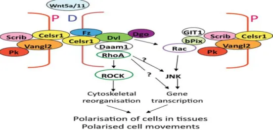
Figure 1.2: PCP Complex. Planar cell polarity is established by the accumulation of core proteins in the proximal and distal regions of plasma membrane. This modification ultimately results in the polarization of cells within epithelia and polarized cell migration (modified from Strutt, 2005; Jenny and Mlodzik, 2006). bPIX, beta PIX; Dgo, Diego; D, distal; Dvl, Disheveled; Fz, frizzled; P, Proximal; Pk, Prickle; Scrib, Scribble; all other acronyms are standard usage. Henderson et al. Birth Defects Research (Part A): Clinical and Molecular Teratology 91:460467 (2011)
1.2 Planar Cell Polarity in Vertebrate Development
Much of the mechanistic insights into the role of PCP signaling in development
were originally obtained from studies in Drosophila. However, the PCP signaling
pathway is highly conserved and more recent studies have shown PCP signaling has
critical roles in vertebrate development. Thus disruption or loss of PCP components
can lead to a wide spectrum of developmental anomalies that can include structural
birth defects such as stereocilia malalignment and mislocalized kinocilia in the cochlear
hair cells, cystic kidneys, delayed or abnormal convergent-extension movement during
gastrulation, craniofacial defects associated with neural crest migration defects, biliary
ductal abnormalities, and neural tube closure defects (Montcouquiol et al., 2003;
Simons et al., 2005; Simons and Mlodzik, 2008; Tada and Smith, 2000; Wallingford and
Harland, 2002). These animal model findings are consistent with clinical studies
atresia, congenital heart defects, and other structural birth defects typically associated
with ciliopathies (Cui et al., 2011b; Juriloff and Harris, 2012). This would further indicate
a role for cilia in PCP signaling, a possibility supported by the observation that
mutations in PCP core components can perturb ciliogenesis (Hildebrandt et al., 2011).
Below, a brief summary is provided on the spectrum of developmental anomalies seen
in conjunction with the disruption of PCP core components in vertebrate development.
1.2.1 Neurulation and Neural Tube Defects
Neurulation is the dynamic process by which the ectoderm gives rise to the
neural plate, followed by formation of the neural tube, the precursor of the brain and
spinal cord. In mice and human, neurulation entails two distinct processes - primary
and secondary neurulation (Greene and Copp, 2009; Rufener et al., 2011). During
primary neurulation, the edges of the neural plate fold and form a midline fusion
resulting in formation of the neural tube anteriorly. Extension of the neural tube
posteriorly involves secondary neurulation mediated by mesenchymal progenitor cells in
the tail bud. These cells actively proliferate and undergo condensation, followed by
cavitation and fusion with the primary neural tube (Cai and Shi, 2014). Perturbation in
the processes that regulate neurulation can lead to neural tube defects (NTDs). While
genetic and non-genetic factors (parental age, maternal nutrition, and chemical
teratogenic agents) (Gurvich et al., 2005; Loeken, 2005; Vieira and Castillo Taucher,
2005) contribute to the etiology of NTD, more than 200 genes have been identified to
play an essential role in neural tube closure, a small set of these genes belong to the
PCP gene family(Harris and Juriloff, 2010). Insights gained from analysis of PCP
NTDs. For example, in mice and humans, mutations in Vangl1 and Vangl2 have been
shown to cause craniorachischisis, the most severe form of NTD in which the neural
tube remains open from the midbrain and extending through the entire length of the
spinal cord (Copp et al., 2003). Also in myelomeningocele, a milder NTD phenotype in
which the spinal canal does not close (Kibar et al., 2007). Interestingly, most mouse
models with mutations in the PCP components and their downstream effectors,
including Vangl2, Celsr1, Dvl2, Scrib, Fzd1/2, and Wnt5a-/-show NTDs of varying
degrees of severity, showing the critical role of PCP in orchestrating neural tube closure
(Greene and Copp, 2009; Wang et al., 2006; Yu et al., 2010).
1.2.2 PCP, Oriented Cell Division, and Developmental Patterning
Cell division orientation plays an integral role in embryo body plan specification,
axis determination and morphogenesis of tissues and organs. In most tissues, cell
division orientation is regulated by the planar cell polarity (PCP) pathway. Such PCP
controlled signaling regulating division orientation has been studied in Drosophila, C.
elegans, and vertebrates; however, the mechanisms regulating division orientation remain to be fully elucidated (Simons and Mlodzik, 2008). The actin cytoskeleton plays
an essential role in orientation of the cleavage plane during mitotic cell division, a
process that can be disrupted by mutation in PCP genes, leading to patterning defects
(McCarthy and Goldstein, 2006). For example in Drosophila Frizzled mutants, the
cuticular hair cells generated by the wing imaginal disk are randomized. In
Strabismus/Vangl2 Drosophila mutants, the photoreceptor cells that are normally stereotypically arranged in the trapezoidal ommatidia become disorganized and fail to
are also necessary for convergent-extensions during embryonic gastrulation and
neurulation. In vertebrate Vangl2 mutants, cells in the embryo failed to converge
towards the midline and instead show abnormal extension along the anterior-posterior
axis (Seifert and Mlodzik, 2007). Oriented cell divisions are also found in association
with formation of renal tubules in the kidneys, with PCP disruption associated with
polycystic kidney disease (Fischer et al., 2006). In Wnt5a/Vangl2 double mouse
mutants, there is disruption of oriented cell division planes required for normal extension
of the fore-stomach, suggesting an essential role for PCP signaling in elongation of the
gastrointestinal tract (Matsuyama et al., 2009).
1.2.3 PCP and Development of Tubular Organs
Epithelial tubes are the functional units of many organ systems, and its proper
development is crucial for organ function. Examples of organs with prominent tubular
architecture include the circulatory system, the outflow tract, the lungs and kidneys, and
the secretory and respiratory organs. In mice and humans, a disruption in proper
development of the tubular networks can cause cardiovascular disease, pulmonary
system defects, gastrointestinal tract defects, and reproductive organ system defects.
In epithelia, cells are polarized along the apical-basal axis. Multicellular animals’
organs are composed of tubular structures to transport essential fluid and gases that
sustain life.
As cited above, inactivation of Wnt5a and Vanlg2 alleles lead to forestomach
defects in mutant mice (Matsuyama et al., 2009). In the kidneys, mice with a
Wnt ligands (Wnt7b, Wnt11, and Wnt9b) also display ureteric bud branching defects
(Karner et al., 2009; Majumdar et al., 2003; Yu et al., 2009). A recent study in Xenopus
showed that morpholino knockdown of Daam1 led to reduced tubular branching in
pronephric proximal segments. Furthermore, daam1 knockdown in zebrafish exhibited
phenotypes with defective convergence extension and convoluted tubules (Miller et al.,
2011). In the uterus and vagina of Vangl2Lp/Lp mice, the morphology is abnormal. These defects include a septated vagina, lack of oviduct coiling, and short uterine horns
(Vandenberg and Sassoon, 2009). Finally, the heart is derived from the anterior
mesoderm that fuses with the midline to form a crescent shaped structure that will give
rise to the cardiac tube derived from the first heart field (FHF) and additional cardiogenic
mesoderm derived from the second heart field (SHF) (Brade et al., 2006). In Vangl2Lp/Lp, Dvl1-/-, Dvl2-/-, and Wnt5a-/- mice, cardiac abnormalities such as shortened outflow tract and abnormal positioning of the aortic arch were observed (Henderson et al., 2006;
Torban et al., 2008).
1.2.4 PCP in Cochlea Development
The inner ear plays a critical role in providing auditory perception and perception
essential for establishing balance. This is mediated by cells in the sensory epithelia that
transmit auditory signals together with nonsensory supporting cells in the organ of Corti.
The sensory epithelia contain mechanosensory hair cells that have stereocilia, which
are actin filament bundles projecting from the luminal surface of the cochlea. These
stereocilia bundles show a chevron organization and are normally all aligned in the
same orientation (Barald and Kelley, 2004). Mutations in several PCP components can
Thus in the PCP mutant Celsr1Crash, the outer sensory hair cells in the organ of Corti are
misaligned (Curtin et al., 2003). In the Vangl2LP/LP mutant, the hair cell orientation is
randomized with a loss of Fzd3 in the cell membrane (Montcouquiol et al., 2003). These
are classic PCP defect phenotypes and can result in disruption of the normal
asymmetric membrane localization of PCP core components. PCP mutants also can
exhibit shorter cochlear tubes in conjunction with the misalignment of cochlear
mechanosensory receptors in the stereociliary bundles of the organ of Corti
(Montcouquiol et al., 2003).
1.3. PCP in Cardiac Development
1.3.1 Mammalian Cardiac Development:
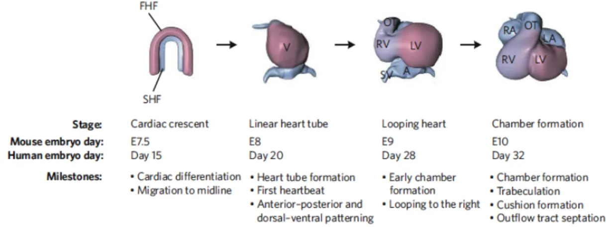
The heart is one of first organs to form and is orchestrated through multiple steps
that include the early specification of cardiac progenitors, followed by cell migration and
morphogenetic movements to form the linear heart tube. This is followed by distinct
dextral looping of the heart tube, then formation and septation to form the four distinct
cardiac chambers with two separate outflows comprising the aorta and pulmonary artery
(Buckingham et al., 2005; Gessert and Kuhl, 2010). This four-chamber anatomy with
two outflows observed in mammals play an essential role in maintaining separate
systemic vs. pulmonary circulation required for efficient oxygenation of blood in air
breathing animals. Hence, the most lethal congenital heart defects involve disruption of
this four chambers and two outflow cardiac anatomy.
The cardiac precursor cells are derived from the primitive streak, and entail
migration of cells in an anterior-lateral direction to positions under the head-folds,
1997). These cells migrate across the midline and merge to form a crescent-shaped
epithelium called the cardiac crescent or first heart field (FHF). These FHF cells
undergo further morphogenetic movements forming the primitive heart tube, which is
the anlage of the future left ventricle (Buckingham et al., 2005). The primitive linear
heart tube then expands by cell proliferation and also recruits additional cells originating
from the second heart field (SHF), a cardiac progenitor population distinct from the FHF,
and is situated medial and dorsal to the cardiac crescent (Moorman et al., 2007). The
SHF derivatives via distinct cell lineage pools will give rise to the outflow tract (OFT), the
right ventricle, and both atria (Cai et al., 2003) (Buckingham et al., 2005) (Figure1.3).
Significantly, mutations in various PCP core components have been shown to cause
congenital heart defects associated with outflow tract malalignment defects. This has
been observed for mutations in PCP components such as Vangl2, Scribbled, and more
recently Prickle1, showing the importance of PCP signaling in cardiac outflow tract
development and in CHD involving outflow tract malalignment defects (Henderson et al.,
2006; Torban et al., 2008).
pools of cardiac precursors exist. The first heart field (FHF) contributes to the left
ventricle (LV), and the second heart field (SHF) contributes to the right ventricle (RV)
and later to the outflow tract (OT), sinus venosus (SV), and left and right atria (LA and
RA, respectively), V, ventricle. Benoit G. Bruneau Nature|Vol 451|21 February 2008
1.3.2 Role of PCP in Congenital Heart Defects:
Recent studies have indicated an important role for PCP dysfunction in
congenital heart defects (CHD). CHD is the most common birth defect in the human
population, observed in 1% of live newborns. CHD is defined as a defect in the structure
of the heart and great vessels present before birth (Hoffman JIE, 2000) (Bruneau,
2008). Each year, more than 35,000 babies in the United States are born with CHD
[NIH, 2011]. It is an important cause of childhood morbidity and mortality worldwide
(Bruneau, 2008). Despite recent advances in critical care and surgical intervention, the
genetic and developmental etiology of CHD is still not well understood.
Overall, CHD can be classified into three broad categories; cyanotic heart
disease, left-sided obstruction defects and septation defects (Figure 1.3.2)(Bruneau,
2008). The most prevalent CHD are ventricular and atrial septal defects, with outflow
tract anomalies being the most common complex congenital heart defects. The
prognosis, morbidity, and mortality, are dependent on the specific type of CHD defects
and associated anomalies. Of great concern to surgeons are outflow tract defects,
because babies that are diagnosed require urgent and complex surgeries shortly after
birth. Outflow tract malalignment defects are one of the most common complex CHD,
and can involve a spectrum of phenotypes such as double-outlet right ventricle (DORV),
Several mouse models with mutations targeting the planar cell polarity pathway (PCP)
have shown PCP signaling plays an important role in CHD involving outflow tract
malalignment defects.
Figure 1.3.2: Adult heart and the structures that are affected by CHD. The estimated incidence of each CHD per 1,000 live births indicated in parentheses. AC, aortic coarctation; AS, aortic stenosis; ASD, atrial septal defect; AVSD, atrioventricular septal defect; BAV, bicuspid aortic valve; DORV, double outlet right ventricle; Ebstein’s, Ebstein’s anomaly of the tricuspid valve; HLHS, hypoplastic left heart syndrome; HRHS, hypoplastic right heart; IAA, interrupted aortic arch; MA, mitral atresia; MS, mitral
stenosis; PDA, patent ductus arteriosus; PS, pulmonary artery stenosis; PTA, persistent truncus arteriosus; TA, tricuspid atresia; TAPVR, total anomalous pulmonary venous return; TGA, transposition of the great arteries; TOF, tetralogy of Fallot; VSD, ventricular septal defect. Benoit G. Bruneau Nature|Vol 451|21 February 2008
1.3.3 Congenital Heart Defect Involving Malalignment of the Great Arteries: The pulmonary artery and the aorta in the cardiovascular system play critical
roles in mediating efficient oxygenation of blood and are known as the great arteries. In
a normal heart, the pulmonary artery is connected to the right ventricle and allows the
flow of oxygen-poor blood from the right ventricle to the lungs. The aorta is connected to
the body. Mutations disrupting PCP components can cause malalignment of the great
arteries such that the relative position of the aorta and pulmonary artery are shifted to
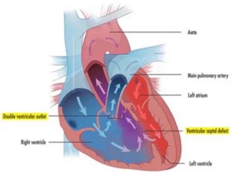
varying degrees, with the most severe form being double outlet right ventricle (DORV)
where both the aorta and pulmonary artery are connected to the right ventricle. DORV is
always accompanied by a ventricular septal defect (VSD), a hole in the ventricular wall
allowing mixing of blood from the two chambers. This type of outflow tract malalignment
defect causes severe cyanosis and is relatively rare, accounting for 1-3% of all
congenital heart defects (Figure 1.3.3).
Less severe and more commonly observed clinically are outflow tract
malalignment defects where the aorta is only partly shifted to the right ventricle, referred
to as an overriding aorta. In the latter, the aorta is situated above the ventricular
septum with a ventricular septal defect, causing aortic blood flow to be partly derived
partly from the right and left ventricle. This condition is often accompanied by additional
narrowing of the pulmonary outflow (pulmonary stenosis) and hypertrophy of the right
ventricle, a congenital heart defect referred to as Tetrology of Fallot (TOF). TOF is
observed in 1 of every 2500 live births (Parker et al., 2010), and is the most common
Figure 1.3.3: Double Outlet Right Ventricle Heart Structure
The two main defects show; ventricular septum defect, aorta originating from the right ventricle. http://www.childrenshospital.org/conditions-and-treatments/conditions/double-outlet-right-ventricle-dorv
1.4 Cilia and Planar Cell Polarity
Cilia have been suggested to play an important role in the modulation of PCP
signaling. Cilia are evolutionarily conserved membrane-bound, microtubule-based
organelles that play important roles in motility and sensory function. These organelles
are conserved through evolution, seen from single cell organisms, such as
Tetrahymena and Chlamydamonas, to mouse and human cells (Satir and Christensen, 2007). Cilia are functionally divided into two types according to their axonemal
arrangement and their ability to engage in active movement, either cilia with motile
function, or nonmotile cilia, also referred to as primary cilia that have sensory and cell
signaling function. Important to note is the fact that many of the same proteins found in
primary cilia also are expressed in motile cilia, indicating there is significant functional
Motile cilia, such as those observed in oviducts, brain ventricles, and respiratory
epithelia, are characterized by a 9+2 microtubule arrangement with nine outer
microtubule doublets, a central pair of microtubules, and interconnecting radial spokes
(Satir and Christensen, 2007). The 9-microtubule doublets have outer dynein arms
(ODA) and inner dynein arms (IDA) that play an essential role in regulating ciliary
motility, which can be quantified with the measurement of ciliary beat frequency and
analysis of the beat waveform. The coordinated action of the ciliary beat mediates fluid
movement that affects mucus clearance in the airway, or mediate cerebral spinal fluid
flow required for development and sustaining metabolic function in the brain. In
contrast, non-motile primary cilia, which are on the surface of most nondividing cells in
the body (e.g. in kidneys and developing heart tissue) are composed of a 9+0
microtubule composition and lack inner/outer dynein arms and radial spokes (Satir and
Christensen, 2007).
Both motile and primary cilia are templated on a modified centriole known as the
basal body. Outgrowth of the ciliary axoneme is mediated by a system of protein
transport known as intraflagellar transport (IFT) and also requires many other
cytoplasmic ciliary assembly factors including proteins in large multiprotein complexes,
known as the BBsome. Together they regulate the assembly, maintenance, and
signaling properties of the cilia. Mutations in genes required for ciliary assembly,
maintenance, motility, and sensory function have led to a series of syndromic disease
and developmental disorders referred to collectively as ciliopathies. These are notable
in exhibiting a wide spectrum of structural birth defects that can include laterality
degeneration, renal and hepatic defects, congenital heart defects, and cystic kidneys
and other renal anomalies (Eggenschwiler and Anderson, 2007; Hildebrandt et al.,
2011; Waters and Beales, 2011). It is significant to note that many of the birth defects
associated with ciliopathies are also observed with disruption of PCP signaling in
mutant mouse models.
Several studies have reported genetic interactions between cilia and the Wnt
signaling pathway. The first connection was demonstrated with analysis of mutation in
Inversin, a basal body protein that also physically interacts with Dishevelled (Simons et
al., 2005). In addition, ciliary proteins BBS1 and BBS4 localize to the basal bodies were
shown to interact with Vangl2 in the stereocilia, with BBS knockout mice observed to
have malaligned stereocilia in the cochlea (Ross et al., 2005). To date, mammalian
core PCP genes include Celsr, Frizzled3 (Fzd3), Fzd6, Vangl1-2, Dvl1-3, and Prickle1-4
are implicated in cilia development and function. Mutations in these genes affect apical
docking and rotational polarity of cilia in the brain ependyma, leading to impaired fluid
flow (Guirao et al., 2010; Tissir et al., 2010). Most recently, Prickle2 mutants were
reported to have decreased ciliary beat frequency, with scanning electron microscopy
showing abnormal blebbing in the ciliary axoneme (Sowers et al., 2014).
1.5 Role of PCP Core Components in Development 1.5.1 Van Gogh (Vangl 1,2)
In Drosophila, mutations in Vangl genes disrupt the organization of various
epithelial structures, causing abnormal wing hair cell patterning and disorganization of
the ommatidia in the Drosophila compound eye (Torban et al., 2004a). Vertebrates have
Gogh/Strabismus (Vang/Stbm). Vangl1 and Vangl2 proteins share ~70% similarity including identical predicted secondary structures. This underlies their conserved
functions, with both Vangl1/2 proteins shown to bind to the three mammalian Dvl1
proteins. Mutation in Vangl2 cytoplasmic domain abrogated interaction of Vangl2 with
Dishevelled (Dvl) (Torban et al., 2004b). Interestingly, Vangl2 encodes four large
transmembrane domains and a large intercellular domain with a PDZ-domain-binding
motif at its carboxyl terminus (Kibar et al., 2011). VANG1 and VANGL2 mutations in
humans can lead to NTDs (Kibar et al., 2011). Vangl2 mutations in mice have been
shown to cause NTDs and cardiac outflow tract anomalies (Torban et al., 2008).
1.5.2 Disheveled (Dvl)
Three Dishevelled (dsh in Drosophila, Dvl in mice) proteins have been identified
in human and mice. They are highly conserved and play a role in both canonical Wnt
and noncanonical Wnt-PCP pathways (Gao and Chen, 2010) (Etheridge et al., 2008).
Dv1 is a scaffold protein than contains three highly conserved domains: DIX
(Dishevelled/Axin), PDZ (PSD-95, DLG, ZO1), and DEP (Dishevelled, EGL-10,
Pleckstrin) domains (Gao and Chen, 2010). Dvl proteins can interact with other adapter
proteins, kinases, and phosphatases (Wallingford and Habas, 2005). The N-terminal
DIX domain of Dvl functions mainly in canonical Wnt signaling, while the central PDZ
domain is required in both pathways via interactions with the cytoplasmic tail of Frizzled
(Fz). The more carboxyl DEP domain has been shown to be critical for PCP signaling
through its role in mediating Dvl cytoplasmic-to-membrane translocation (MacDonald et
al., 2009). Dvl1 and Dvl2 deficiency in mice result in cardiac outflow tract anomalies,
in outflow tract septation with persistent truncus arteriosus (PTA). In addition to these
congenital heart defects, mutants also exhibited randomized stereocilia orientation in
the sensory hair cells of the cochlea, cochlear shortening, and NTDs, phenotypes
thought to reflect the disruption of Dvl1-mediated PCP signaling (Hamblet et al., 2002;
Wang et al., 2006).
1.5.3 Scribble (Scrib)
Scrib is a large cytoplasmic scaffolding protein with four PDZ domains that is a
member of the LASP protein family (Assemat et al., 2008). It can form complexes with
cell-cell adhesion junctions and play a role in regulating cell proliferation, migration,
differentiation, and vesicular trafficking (Qin et al., 2005). Scrib mouse mutants exhibit
heart looping defects and disrupted myocardial organization. They also can exhibit
neural tube closure defects (Murdoch et al., 2001), abnormalities known to be
associated with disruption in PCP signaling and likely to involve Scrib interaction with
Vangl2 (Kallay et al., 2006).
1.6 Role of Pk1 in Development
Pk1 expression is broadly expressed, and is seen in many regions of the embryo
involved in morphogenetic movements, consistent with a conserved role in regulating
planar cell polarity in development (Wallingford et al., 2002). In humans, Pk1 is critical
for human nervous system development. Mutations in Prickle1 are associated with
neural tube defects (NTD) and myoclonus epilepsy in humans (Bassuk et al., 2008;
Bosoi et al., 2011). Pk1 knock out mice are embryonic lethal at E6.5 due to
disorganization of the epiblast. This is associated with abnormal cell shape,
2009). Hypomorphic Pk1 alleles in homozygosity can cause many different types of
birth defects including craniofacial and skeletal defects, cardiac anomalies, and kidneys
and neural tube defects. These defects are suggested to involve disturbance in cell
polarity and may involve disruption of downstream Wnt5a signaling (Liu et al., 2014a).
Interestingly, heterozygous Pk1 mutations in human and mice have been shown to
cause epilepsy (Tao et al., 2011). These findings suggest that Pk1 role in regulating
cell polarity is conserved from flies to humans.
1.6.1 Pk1 Structure
Pk1 is a prenylated (Maurer-Stroh et al., 2007) core PCP component with distinct
N-terminal Prickle, Espinas, and Testin (PET) domains and C-terminal Lin11, Isl-1, and
Mec-3 (LIM) domains (Takeuchi et al., 2003). In humans, there are 4 Prickles. Pk1,
Pk2, and Pk3 have similar domains: a N-terminus PET (Prickle, Espinas, and Testin)
and three LIM domains (Lin11, Isl1, and Mec3) in the C-terminus (Shimojo and Hersh,
2003, and Shimojo and Hersh, 2006). The least known Prickle, Pk4 has a N-terminus
PET domain and two LIM domains (Lin11, and Isl1). These proteins belong to a group
of LIM proteins that interact with actin and various transcription factors, indicating a dual
role in cargo transportation and gene regulation (Kadrmas and Beckerle, 2004; Zheng
and Zhao, 2007). The PET domain and LIM domain are known to be in involved in
protein-protein interaction with other LIM domain proteins (Kadrmas and Beckerle,
2004). In Drosophila, the PET domain and LIM of Pk1 plays a role in membrane
assertion, which localizes disheveled (dsh) to the membrane to help establish the PCP
In addition, downstream of the third LIM domain, Pk1 protein has several
N-glycosylation sites, a cAMP-dependent protein kinase A site, several nuclear
localization signals, and a putative prenylation motif in the C-terminus (Liu et al., 2013;
Shimojo and Hersh, 2003). These structures are consistent with a potential role in
transcription regulation (Bassuk et al., 2008 and Mapp et al., 2011). However, these
structures present in Pk2-4 remain to be elucidated. Therefore, it has not been reported
and unclear how Pk1 is similar to the other Prickles. Given what’s known about Pk1, it
suggests that Pk1 might regulate gene transcription, cytoskeleton rearrangement, and
cellular cargo transport.
We have recovered a novel missense mutation in Pk1 from a large-scale mouse
ENU mutagenesis screen for mutations causing CHD (Li et al., 2015). While this
mutant was recovered based on the finding of outflow tract malalignment defects
causing hemodynamic perturbation observed by fetal echocardiography, our detailed
analysis of this mutant model revealed a plethora of structural birth defects, including
the first description of a mammalian PCP mutant with a biliary atresia spectrum of
structural birth defect phenotype. We showed this mutant has a missense mutation in
Pk1 that is likely a hypomorphic allele. This likely accounts for its viability to term, and suggests this mouse model is more relevant for modeling human disease. Analysis of
this novel Pk1 mutant provided new insights into how PCP disruption may contribute to
structural birth defect phenotypes. These studies also showed a novel role for Pk1 in
the modulation of motile cilia function, suggesting an intimate connection between PCP
CHAPTER 2
Characterization of Novel Phenotypes in the Bj Mouse Model 2.1 Introduction
Planar cell polarity (PCP) is an evolutionarily conserved pathway that plays an
important role in development. It was first described in the context of developmental
patterning in the Drosophila wing imaginal disk and compound eye, and refers to the
apical-basal polarity of cells in an epithelial sheet. Genetic analysis in the Drosophila
system identified a group of interacting PCP core components that included Frizzled,
Dishevelled, Van Gogh/Strabismus, Prickle, Diego, and Flamingo (Bastock et al., 2003;
Jenny et al., 2005). These proteins accumulate asymmetrically in the cell membrane
and are responsible for cell-cell alignment and polarized cell orientation within the
forming tissue axes (Axelrod, 2009). These same proteins are conserved in
vertebrates, with deficiency or mutations in these proteins observed to cause a wide
spectrum of developmental anomalies (Wansleeben et al., 2011). This can involve
defects with malpatterning of hair cells in the cochlea, cystic kidneys, neural tube
defects, brain anomalies, skeletal defects, and congenital heart disease (Cui et al.,
2011).
The requirement for PCP components in development has been shown to involve
the critical regulation of planar cell polarity and convergent-extension movement
required for tissue morphogenesis. Thus PCP has been shown to regulate
Convergent-extension movements also play a critical role in neural tube closure (Wallingford et al.,
2002), with neural tube defects associated with mutations in PCP core components in
human and animal models (Cui et al., 2011b; Juriloff and Harris, 2012). PCP dependent
convergent-extension movement also has been suggested to regulate neural crest cell
migration. This may contribute to the cardiac and craniofacial developmental anomalies
in animal models with mutations in PCP core components (Montcouquiol et al., 2003;
Simons et al., 2005; Simons and Mlodzik, 2008; Tada and Smith, 2000; Wallingford and
Harland, 2002). Many of the mutant mouse models harboring mutations in PCP core
components such as Vangl2, Scrib (Phillips et al., 2007), and Dvl 1, 2, and 3 (Etheridge
et al., 2008; Hamblet et al., 2002; Sinha et al., 2012) exhibit a similar spectrum of
cardiac defect phenotypes involving outflow tract malalignment and septation defects.
These cardiac defects likely involve not only perturbation of CNC cells, which is required
for outflow septation, but also the second heart field (SHF). SHF cells migrate into the
developing heart tube, forming most of the outflow tract, a structure that is the target of
PCP mutations (Verizi et al., 2005) (Cohen et al., 2007) (Schleiffarth et al., 2007).
In this study, we present findings from a mouse mutant, Beetlejuice (Bj), harboring a mutation Prickle1, one of six PCP core proteins that makes up the PCP
signaling complex. Pk1 mutations in patients have been shown to be associated with progressive monoclonic epilepsy-ataxis syndrome (Bassuk et al., 2008). Previous
analysis of a hypomorphic Pk1C251X mutant mouse model have shown homozygous mutants are prenatal lethal with neural tube defects, cleft palate, and kidney defects,
while heterozygote animals are viable and can exhibit epilepsy (Yang et al., 2014) (Liu
pregasturlation embryonic lethality (Tao et al., 2009), with selected Cre deletion of a
floxed allele showing a spectrum of defects that is suggested to phenocopy human
Robinow syndrome via the disruption of Wnt5a signaling (Liu et al., 2014a).
The Bj mutant harbors a missense mutation in the LIM domain of Pk1p.C161F, a region previously shown to harbor pathogenic missense mutation causing myoclonus
epilepsy in patients (Toa et al 2011). We showed homozygous Bj mutants exhibit a wide spectrum of developmental anomalies that include cardiac outflow tract
malalignment defects, skeletal and craniofacial anomalies, and cochlea defects. In
addition, we show for the first time, biliary atresia spectrum of defects in the biliary duct,
suggesting mutations in PCP core components should be investigated in patients with
biliary atresia. Overall, the multiple findings from our study suggest an intrinsic defect in
cell polarity and directional cell migration contributing to the wide spectrum of
developmental anomalies in the Bj mutant. We also obtained evidence indicating an essential role for Pk1 in canonical and noncanonical Wnt signaling, and the regulation of
both primary and motile cilia function.
2.2 Results
2.2.1 ENU Mutagenesis breeding scheme and workflow
To investigate the spectrum of CHD and affiliated organ system defects, a large
ethylnitrosourea (ENU) mouse mutagenesis forward genetic screen was done for
modeling human congenital heart defects (Li et al., 2015). Using noninvasive fetal
ultrasound scanning of 87,355 G3 fetuses, most of the CHD observed in the clinical
setting were recovered in the screen. Fetuses were recovered with structural cardiac
Persistent Truncus Arteriosus (PTA), and Atrioventricular Septal Defects (ASD) to name
a few. The CHD diagnoses were confirmed with micro-computed tomography
(micro-CT) or magnetic resonance imaging (micro-MRI) and episcopic fluorescence image
capture (EFIC)(Rosenthal J, ET AL 2004) (Figure 2.1 A, B).
2.2.2 Recovery of the Pk1 Mutation
To recover the disease causing mutation in Beetlejuice (Bj), the Bj mutant line
was intercrossed into the C57BLB10 background for one generation and conducted a
genome scan in the hybrid mutant offspring using polymorphic B6/B10 markers to map
the CHD causing mutation. This analysis mapped the mutation to a 19Mb interval on
chromosome 15, between position 83,618,701 and the end of the chromosome
(Supplemental Table S1). To identify the pathogenic mutation, we carried out whole
mouse exome sequencing analysis at 50X coverage using DNA from one mutant.
Comparison of the sequencing data obtained from the Bj mutant vs. the C57BL/6J
reference genome identified three homozygous coding mutations (Supplemental Table
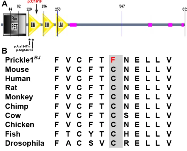
S2). Genotyping analysis of over 20 Bj mutants identified Pk1c.G482T:p.C161F as the pathogenic mutation, as only the Pk1 mutation is consistently homozygous in all the Bj
mutants, (Supplemental Table S2). The mutation in Pk1 is a missense mutation in a
highly conserved amino acid residue, suggesting it is likely to have functional
importance (Figure 2.2 A,B).
2.2.3 Outflow tract malalignment in Pk1 mutants
Beetlejuice (Bj) was recovered by using noninvasive fetal echocardiography (Liu et al., 2014b) to visualized fetuses with structural cardiac defects. Color flow imaging of
malalignment defects encompassing an overriding aorta (OA) or double outlet right
ventricle (DORV) and ventricular septum defect (VSD) (Figure 2.3 G) compared to
wildtype (Figure 2.3 A). Follow up necropsy examination showed the Bj mutant had
parallel positioning of the aorta and pulmonary artery (Figure 2.3 H), while the control
embryo showed normal outflow tract positioning (Figure 2.3 B). Further ECM
histopathology confirmed the mutant heart has a DORV with a perimembranous VSD
(Figure 2.3 I vs. wildtype Figure 2.3 C). Further examination of the cardiac valves
showed the atrioventricular and semilunar valves in the Bj mutant heart were normal
(Figure 2.3 D,L), indistinguishable from that seen in wildtype littermate control (Figure
2.3 E-L), indicating the outflow malalignment defect is not likely related to defects in
valvular morphogenesis. Systematic analysis of a large number of Bj mutants showed
63% of Bj mutants have CHD comprising of a DORV, with 17% exhibiting overriding
aorta. These are closely related phenotypes that differ only by the degree to which the
aorta is shifted to the right ventricle – using the clinical definition; DORV refers to when
the aorta is situated more than 50% over the right ventricle, while OA is when there is
less than 50%. In 20.6% of the Bj mutants, only a simple perimembranous VSD
observed with normally positioned great arteries (Table 2).
2.2.4 Pk1 mutants have shortened outflow tract and defects in neural crest and second heart derivatives
Development of the looped heart tube requires recruitment of second heart field
(SHF) cardiac precursors that contribute to lengthening of the heart tube, eventually
giving rise to much of the right ventricle, atria, and outflow tract (Cai et al., 2003; van
den Berg et al., 2009; Waldo et al., 2005; Waldo et al., 2001). In the E10.5 Bj mutant
p=0.002), suggesting possible defect in the recruitment of the SHF cells. Antibody
staining with Islet1, a SHF marker (Cai et al., 2003), showed the splanchnic mesoderm
in the dorsal posterior wall (DPW) and transition zone (TZ), regions contiguous to the
distal end of the OFT, are comprised of SHF cardiac precursors (Figure 2.4 I, J). While
these Islet1 positive SHF cells exhibited a flat squamous epithelial cell morphology in
the control embryo, a distinct cuboidal cell morphology was observed in the Bj mutant
embryo, suggesting a disruption in planar cell polarity (Figure 2.4 arrowheads K, L).
This suggested a possible convergent-extension defect that may contribute to
shortening of the OFT.
We also examined whether there may be changes in the distribution of cardiac
neural crest cells (cNCC), as cNCC deficiency can also cause OFT malalignment
defects (Cai et al., 2003; Waldo et al., 2001; van den Berg et al., 2009). For this
analysis, we introduced into the Bj mutant line, a Cx43 promoter driven LacZ reporter
transgene previously shown to label neural crest cells (Lo et al. 1997). X-gal staining of
E10.5 wildtype and Bj mutant embryos showed a reduction in cNCC in the OFT of the Bj
mutant embryos, indicating the Pk1 mutation may have disrupted CNC migration into
the OFT (Figure 2.4 F, G).
2.2.5 Disruption of epithelial integrity in the developing outflow tract of the Bj mutant embryos
To examine the possible basis for the altered cell morphology in the SHF cardiac
precursors in the DPW and TZ, we examined the expression of various epithelia cell
markers using confocal microscopy. This analysis showed no change in the cell surface
localization of b-catenin in the DPW and TZ of the E10.5 Bj mutant embryos (Figure 2.5
apically and basally, a pattern observed in both the TZ and DPW (Figure 2.5 A-F). This
contrasts with the observation that in wildtype embryos, laminin is expressed only in the
TZ where it is basally localized (Figure 2.5 A-F). Further examination of PKCζ, an
atypical protein kinase C required for oriented cell division, showed apical cell
localization in the TZ and DPW of wildtype embryos (Figure 2.5 G, I, K), but in the Bj
mutant embryos, PKCζ expression was markedly reduced or absent, with some areas
showing mislocalization (Figure 2.5 H, J, L).
2.2.6 Bj mutants show a reduction in PK1 expression in the outflow tract
Together these findings indicated the disruption of apical-basal cell polarity in in
the SHF cardiac precursors. This was associated with a loss of the pseudostratified
epithelial architecture, and instead, the DPW became multi-cell layered, exhibiting a
disorganized. To examine whether these changes may involve the perturbation of other
PCP core components, we further examined the expression of Vangl2, and Scribbled
(Scrib) in the Bj mutant embryos. Both exhibited the same pattern of apical localization
as in the wildtype embryo (Figure 2.6 A, C ,E). However, PK1 expression was markedly
reduced in the Bj mutant (Figure 2.6 B, D, F).
2.2.7 Bj mutants show disruption of noncanonical Wnt signaling
Given that Wnt signaling is known to play a role in regulating OFT development
(Schleiffarth et al., 2007) (Cohen et al., 2012), we examined whether the OFT defects in
the Bj mutant embryo may involve perturbation in Wnt signaling. For this analysis, we
made use of the canonical Wnt BAT-lacZ reporter, crossing it into the Bj mutant mouse
line and using X-gal staining to assay for canonical Wnt signaling. This analysis
mutant heart, indicating increased canonical Wnt signaling (Figure 2.7 A, B).
Quantitative real-time PCR analysis (Figure 2.7 C) of RNA obtained from the base of
the OFT where BAT-lacZ expression was observed to be elevated showed gene
expression changes that suggested the perturbation of both canonical (Ctnnb1, Apc,
Tcf7, Wnt2b) and noncanonical (Wnt5a, Wnt11, RhoA) Wnt signaling in the Bj mutant heart (Figure 2.7 C, D).
2.2.8 Cell polarity and myocardialization defects in OFT of Bj mutants
PCP has been shown to play a role in modulating cardiomyocyte migration to
muscularize the outflow septum, a process referred to as myocardialization (Cui et al.,
2013). Using MF20 immunostaining, two prongs of myocardial cells can be observed
projecting into the outflow septum in the E13.5 mouse heart (Figure 2.8 A,C). The
direction of cardiomyocyte invasion is aligned with the orientation of actin filaments in
the septal mesenchyme (visualized by phalloidin; Figure 2.8 C). Magnified views show
the myofiament projections in the invading cardiomyocytes are largely aligned with the
direction of cell migration (Figure 2.8 E). In the Bj mutant heart, the myocardial prongs
are not observed, and myofilaments in the outflow cardiomyocytes are not aligned with
the normal direction of myocardialization (Figure 2.8 B, F). Quantitative analysis
showed a significant loss of oriented myofilament alignment (Fig 2.8 G; p=0.0149).
Phalloidin staining also revealed random orientation of actin filaments in the septal
mesenchyme (Figure 2.8 D). These observations suggest Bj mutants may fail to
undergo normal myocardialization of the outflow septum due to a PCP defect that
2.2.9 Craniofacial defects in Bj mutants and perturbation of cranial neural crest cells
Bj mutants exhibited various extracardiac defects that included skeletal anomalies and craniofacial defects (Figure 2.9 B, D). The craniofacial defects included
micrognathia, smaller frontal bones, and short snouts compared to wildtype (Figure 2.9
A, C). Bj mutants also exhibited shortened limbs, confirmed by quantitative analysis of
skeletal preparations, which showed reductions in the length of the long bones and
metacarpals (Supplemental Figure S1). Histological analysis showed chondrocytes in
the growth plate failed to align along the proximal-distal axis, likely contributing to the
reduction in skeletal outgrowth (Supplemental Figure S2). This contrasts with the
well-organized chondrocytes aligned in columns along the long axis of limb outgrowth in
wildtype embryos (Supplemental Figure S2). We also examined the distribution of
cranial neural crest cells in Bj mutants using the Cx43-LacZ transgene, as neural crest
cells play essential roles in craniofacial development (Minoux and Rijli, 2010). Tracking
neural crest cells in the E14.5 embryo via the LacZ reporter revealed a decrease in
neural crest cells in the mandibular and maxillary prominences in the Bj mutant (Figure
2.9 F) vs. wildtype embryo (Figure 2.9 E).
2.2.10 Bj mutants show stereocilia patterning defects
We examined Bj mutants for cochlea defects using phalloidin to visualize the
distribution of stereocilia and Vangl2 to delineate PCP defined cell polarity. Normally,
hair cells in the cochlea are arranged in repeating rows, with the actin based stereocilia
bundles exhibiting an identical polarized stereotypical chevron orientation, a patterning
process that is PCP regulated (Figure 2.10 A) (Kelly and Chen, 2007). Analysis of Bj
Vangl2 expression remained membrane localized in the supporting hair cells, they
pattern of Vangl2 distribution showed they are misaligned relative to the inner hair cell
(IHC) in the Bj mutant cochleae (Figure 2.10 D). Together these findings indicate a
PCP defect in the cochlea.
2.2.11 Bj mutants show biliary duct (BD) hypoplasia
Zebrafish is an established model for studying the hepatobiliary development.
We investigated biliary duct formation in the Bj mutant, as previous work in the zebrafish
had shown antisense morpholino knockdown of pk1-1a and other PCP components can
impair bile duct formation (Cui et al., 2011b). Interestingly, Bj mutant bile ducts
appeared significantly shorter (Figure 2.11 A, B). Quantitative measurements revealed
a decrease in length (p=0.0495) and an increase in width (p=0.0133) of the Bj mutant
bile ducts (C). This was accompanied by a reduction in the number of mucosal folds
(Figure 2.11 D, F; p=0.0093). Given several studies have suggested a role for β-catenin
dependent canonical Wnt signaling in biliary fate determination (Tan et al., 2008), we
conducted confocal immunohistology to examine the expression of both β-catenin and
E-cadherin in the biliary duct. In the wildtype biliary duct, β-catenin and E-cadherin
were highly expressed and show colocalization at the cell surface (Figure 2.11 G-K). In
the Bj mutant, there was no change in the pattern of E-Cadherin expression, but β
-catenin expression was significantly reduced (Figure 2.11 H-L). These findings indicate
a role for Pk1 in biliary duct morphogenesis via a β−catenin dependent process.
2.2.12 Wound closure assay show defect in cell polarity and polarized cell migration in Bj mutant MEFs
Previous studies have shown disruption of PCP components can perturb
potential role of Pk1 in modulating directional cell movement, we generated Bj mutant
and wildtype MEFs for wound scratch assays. MEFS were grown to confluence, and a
wound gap created with a scratch in the monolayer. Wildtype MEFs become aligned to
the direction of wound closure, but the mutant MEFs appeared randomly oriented
(Figure 2.12 A, B). This was demonstrated quantitatively with analysis of Golgi
orientation using a Golgi marker, Golga2 (Figure 2.12 C, D). In migrating cells, Golgi is
expected at the cell’s leading edge, aligned with the direction of cell migration. This was
observed in wildtype MEFs, which exhibited Golgi mostly positioned less than 60oC
relative to the direction of cell migration in the wound gap (Figure 2.12 C, E). In contrast,
in Bj mutant MEFs, this distribution was significantly broadened; indicating Golgi
orientation is randomized (Figure 2.12 D, E). These results indicate Bj mutant MEFs
are unable to establish the cell polarity required for efficient directional cell migration.
To examine further how Prickle1 may regulate directional cell migration, we
conducted time-lapse videomicroscopy over 36 hrs to track the migratory behavior of
individual cells during wound closure. By tracing the migratory paths of individual cells
in the time-lapse videos for 8hrs, we observed a relatively straight migratory path in the
wildtype MEFs (Figure 2.12 F), while a more tortuous migratory path was observed for
the Bj mutant MEFs (Figure 2.12 F). This pattern of cell migration was correlated with a
significant increase in the speed of cell locomotion (p=0.0017), but no net change in the
directionality of cell movement in the Bj mutant MEFS (Figure 2.12 F, G).
To examine for possible cytoskeletal changes that may account for the defect in
polarized cell migration, we carried out phalloidin staining to visualize the actin
direction of cell migration (Figure 2.12 H, J), but in Bj mutant MEFs (Figure 2.12 I, K),
the actin stress fibers were organized in a basket configuration around the cell cortex.
We also carried out immunostaining with a Prickle1 antibody to determine if changes in
Prickle1 distribution may contribute to the defects observed in directional cell migration
in the BJ mutant MEFs. This analysis showed Pk1 is localized in the cytoplasm in
wildtype MEFs, but in the Bj mutant MEFs, it is largely nucleus localized, suggesting a
previously unknown transcriptional role for Prickle1.
2.2.13 Bj mutants exhibit primary cilia defects
Given previous studies indicating a role for cilia in constraining canonical vs.
noncanonical Wnt signaling, we examined ciliogenesis in Bj mutants MEFs. Cilia were
visualized by immunostaining with acetylated and gamma-tubulin after stimulation
serum starvation to stimulate ciliogenesis. Bj mutant MEFs (Figure 2.13 B, C, D)
exhibited a reduction in ciliogenesis and the cilia formed were shorter compared to
wildtype MEFs (Figure 2.13 A, C, D).
2.2.14 Cilia defects in Bjmutant tracheae
To further examine for possible defects in motile cilia, we obtained the tracheal
epithelia of newborn Bj mutants and littermate controls and conducted videomicroscopy
to examine ciliary motion. Surprisingly, the Bj mutant exhibited a significant reduction in
cilia beat frequency (Figure 2.14 A; p<0.0001). Transmission electron microscopy
(TEM) showed normal orientation of the basal feet of cilia in the Bj mutant tracheal
epithelia as observed in littermate controls (Figure 2.14 B, C). Scanning electron
microscopy (SEM) showed abnormal large apical membrane bulges that incorporated
membrane blebs at the surface of ciliary axoneme (Figure 2.14 I’ insets). In contrast,
cilia in wildtype tracheal epithelia were smooth (Figure 2.14 F’ insets). Further analysis
by TEM showed abnormal axonemal microtubules (Figure 2.14 K, L), and compound
axonemes (Figure 2.14 J). Enlarge views (Figure 2.14 M-P) show absent microtubule
doublet in the axoneme (Figure 2.14 N). Taken together, these results suggest Pk1
may play a role in the regulation of primary and motile cilia function.
2.3 Discussion
In this study, we showed Bj, a novel mouse mutant recovered from a prenatal
echocardiography screen of ENU mutagenized mice, has a wide spectrum of
developmental defects. We showed this mutant harbors a C161F point mutation in Pk1,
a core PCP gene. The phenotypes observed in Bj mutants are indeed similar to those
previously seen in other mutant mouse models with mutations in PCP pathway
components (Montcouquiol et al., 2003; Ramsbottom et al., 2014; Simons et al., 2005;
Tada and Smith, 2000). The Pk1 mutation is predicted to be pathogenic, as it is
situated in a highly conserved region comprising the first LIM domain of the protein. In
flies, mice, and human, Pk1 mutations have been shown to cause various defects
including cleft palate, epilepsy, skeletal defects, and neural tube defects (Bosoi et al.,
2011; Tao et al., 2011; Yang et al., 2014).
While previous studies showed complete loss of Pk1 function in null mutation is
embryonic lethal (Tao et al., 2009), the Bj mutants stand out as a Pk1 mutant mouse
model that can survive to term, and yet its phenotypes are consistent with those seen in
other Prickle1 mutant mice. This included skeletal defects, craniofacial defects, and