Address for correspondence Prof. Ijaz Ahmad
Professor of Dermatology
Department of Dermatology, Ziauddin University, Karachi.
Email: [email protected]
Original Article
Clinical presentations of cutaneous tuberculosis
Najam-us Saher*, Zarnaz Wahid*, Ijaz Ahmed**, Sadaf Ahmed Asim*, Farzana Riaz* Department of Dermatology, Civil Hospital Karachi and Dow University of health Sciences, Karachi
** Department of Dermatology, Ziauddin University, Karachi
Abstract
Objective To determine the frequency of different clinical presentations of cutaneous tuberculosis in a tertiary care hospital.Methods This cross-sectional study was carried out in the Department of Dermatology, Civil Hospital, Karachi and Department of Dermatology, Ziauddin University, Karachi, over three years from 5th March 2007 to 4th March 2010. Patients of both sexes and all age groups suffering from histologically-confirmed cutaneous tuberculosis for last 1 month to 10 years were enrolled. A detailed history and examination were recorded on a predesigned proforma. The collected data were computed and analyzed.
Results 57 diagnosed (biopsy proven) cases of cutaneous tuberculosis comprising 35 females (61.4%) and 22 males (38.6%) were enrolled. The age range was 1 up to 80 years. Among these subjects, half the patients were aged between 130 years. 17 patients (29.8%) had the disease for 1-2 years constituting the highest frequency for the duration of illness. Most of the patients i.e. 33 (57.5%) had more than one lesion. The lesions were most commonly seen on limbs in 25 (43.4%) patients, followed by face and neck, trunk and genitalia. Chronic discharging sinuses and plaques were the most common presentations. Scrofuloderma was the most common tuberculosis cutis seen in 35 (62%) patients followed by lupus vulgaris, warty tuberculosis and tuberculids. Overall frequency was higher in females. However, some of the types were more frequent in females while others in males. Mean age of presentation for scrofuloderma was 25.7±16.9 years, warty tuberculosis 25.7±15.9 years, lupus vulgaris 29.4±16.5 years and tuberculids 30±11.9 years (p=0.197).
Conclusion Scrofuloderma is the most common clinical presentation of tuberculosis cutis seen in our setting followed by lupus vulgaris, tuberculosis verrucosa cutis and tuberculids.
Key words
Cutaneous tuberculosis, scrofuloderma, lupus vulgaris, tuberculosis verrucosa cutis, tuberculids.
Introduction
More than 2 billion people (about one-third of the world population) are estimated to be infected with Mycobacterium tuberculosis.1
Cutaneous tuberculosis has re-emerged in the last 15 years together with a higher incidence of
pulmonary tuberculosis and multidrug resistance.2,3 Cutaneous tuberculosis has a
worldwide distribution with malnourishment and low socioeconomic status being the main predispositions.
M. tuberculosis can induce a spectrum of
infection in an immunologically competent host. Post-primary skin lesion e.g. lupus vulgaris, is a common presentation of cutaneous tuberculosis. Scrofuloderma results from contiguous involvement of skin overlying a tuberculous focus e.g. lymph nodes, bones, joints or testicles. Hematogenous spread can lead to miliary tuberculosis. Orificial, perioral or perianal tuberculosis can occur following ingested mycobacteria. Tuberculids once attributed to cutaneous immunological reactions to tuberculosis elsewhere in the body in an immune host are now known to be true forms of cutaneous tuberculosis.4
Early diagnosis and treatment yields better outcome for the disease and if left untreated or misdiagnosed it can lead to complications like squamous cell or basal cell carcinoma especially in lupus vulgaris (8%).5
The incidence of different forms of cutaneous tuberculosis varies globally. Scrofuloderma has been reported to be the commonest form from UK6 and lupus vulgaris from South Africa.7 In
India, scrofuloderma had the highest frequency in children while lupus vulgaris in adults.8,9 In
Pakistan, scrofuloderma has been reported to be the most common form (64.9%).10 Studies
carried out in our country reveal a limited data regarding the frequency of different clinical presentations of tuberculosis cutis.10
The current study was targeted to determine the frequency of different clinical presentations of cutaneous tuberculosis in our part of the world. The study will be helpful towards planning more strategies for awareness about tuberculosis cutis for health care providers leading to early diagnosis and preventing lethal complications.
Methods
Current, descriptive, cross-sectional study was carried out in the Department of Dermatology, Civil Hospital, Karachi and Department of Dermatology, Ziauddin University, Karachi.The study was completed over a period of three years, spanning from 5th March, 2007 till 4th March, 2010.
Patients were selected by non-probability purposive sampling. After an informed consent, patients of both sexes and all age groups suffering from the disease for 1 month to 10 years were enrolled for the study. All the patients were histologically proven cases of cutaneous tuberculosis. Patients having any other concomitant dermatological or systemic problems were ruled out.
After a detailed history and examination, the findings were recorded on a predesigned proforma. The clinical diagnosis was confirmed by biopsy and histopathology. Any relevant investigations were performed wherever required. These included complete blood count, biochemical profile and urinalysis.
The data were compiled, tabulated and analyzed with the help of SPSS Program version 10.0. Frequency and percentages were calculated for categorical data like gender and clinical type of cutaneous tuberculosis. Meanstandard deviation was calculated for numerical variables like age. Frequency of clinical type of cutaneous tuberculosis was also presented according to gender, duration of illness and age in order to control effect modifiers.
Results
Table 1 Duration of illness (n=57).
Duration N (%)
Less than 6 months 12 (21)
6 months – 1 year 11 (19.3)
1-2 years 17 (29.8)
2-4 years 9 (15.8)
4-8 years 8 (14)
Table 2 Types of lesions (n=57).
Duration (%)
Discharging sinuses 56.5%
Plaques 47.8%
Ulcers 39%
Warty growth 39%
Nodules 30.4%
comprising 35 (61.4%) females and 22 (38.6%) males.
The age range was 1 up to 80 years. Among these subjects, 9 (15.8%) patients aged 1-10 years, while 19 (33.3%) between 11-20 years, 9 (15.8%) ranged from 21-30 years, another 8 (14%) in 31-40 years and remaining 12 (21%) patients were above 40 years. Duration of illness can be appreciated from Table 1. 17 (29.8%) patients had the disease for 1-2 years constituting the highest frequency for the duration of illness.
Most of the patients had more than one lesion 33 (57.5%) and solitary lesions were seen in 24 (42.5%). The lesions were most commonly seen on limbs, 25 (43.4%) patients followed by face and neck 10 (18.6%) each, trunk in 8 (14%) and genitalia were involved in 4 (7%) patients.
Table 2 reveals the morphological types of lesions. Chronic discharging sinuses and plaques
were the most common presenting lesions. Some patients had a combination of different types of lesions as well. Scrofuloderma was the most common tuberculosis cutis seen in 35 (62%) patients, followed by lupus vulgaris 11 (19%), warty tuberculosis 6 (10%) and tuberculids in 5 (9%).
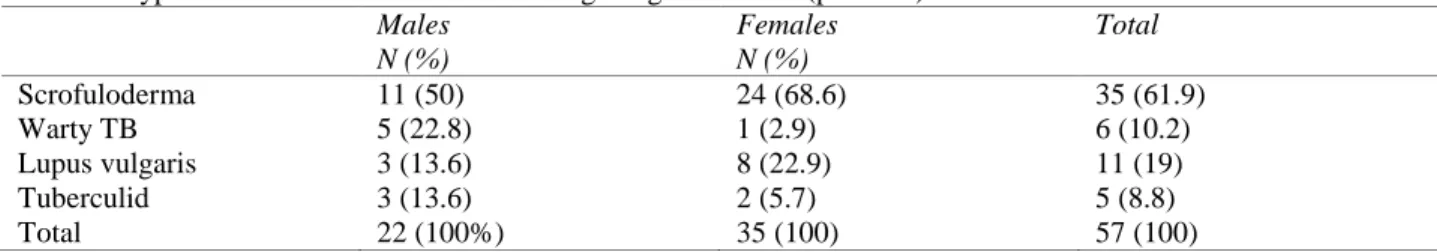
Table 3 reveals gender wise frequency of tuberculosis cutis in our study. Overall frequency was higher in females. However, some of the types were more frequent in females while others in males. Scrofuloderma and lupus vulgaris were more frequent in females while warty tuberculosis and tuberculids had higher frequency in males.
In the current study population, the mean age of presentation with scrofuloderma was 25.7±16.9 years, warty TB 25.7±15.9 years, lupus vulgaris 29.4±16.5 years and tuberculids 30±11.9 years (p=0.197).
Discussion
Tuberculosis has plagued humans and animals from ancient times but little has been said about cutaneous tuberculosis in ancient medical scriptures.11 However, scrofuloderma remains
the earliest reported cutaneous tuberculous lesion by Rene Laennec in 1918. In 1882, Robert Koch not only discovered tubercle bacillus but also found it to be the causative agent of lupus.12This potentially curable illness
is still prevalent in developing countries due to low socioeconomic status, malnutrition and Table 3 Types of cutaneous tuberculosis among the gender n=57 (p=0.033)
Males N (%)
Females N (%)
Total
Scrofuloderma 11 (50) 24 (68.6) 35 (61.9)
Warty TB 5 (22.8) 1 (2.9) 6 (10.2)
Lupus vulgaris 3 (13.6) 8 (22.9) 11 (19)
Tuberculid 3 (13.6) 2 (5.7) 5 (8.8)
overcrowded living. Lack of medical facilities because of poverty or poor health infrastructure has led to longstanding and more extensive tuberculous skin lesions.
It can be elucidated from the current study that cutaneous tuberculosis is still far from extinct and even prevails in cities like Karachi. A rising trend over the years has been observed. The exact reason for this increase in incidence could not be identified as this was not the aim of study. Tuberculosis cutis has also been reported from our neighbor country India.3 Chin et al.13 have
reported a considerable drop in its frequency in Hong Kong where it was a chronic problem. A decrease in the frequency of tuberculosis cutis has been reported in India in another study.14
This relatively rare clinical entity in western countries is still prevalent in the developing world such as Far East.15
A female preponderance was observed in our study. However, some of the types were more frequent in females while others in males. Scrofuloderma and lupus vulgaris were more frequent in females while warty tuberculosis and tuberculids in males. Padmavathy et al.9 have
also reported female preponderance that is comparable to the current study.
No age is immune from cutaneous tuberculosis, as indicated by the age range 1-80 years in our study.16 Approximately 50% of our patients were
aged 11-30 years for an unknown reason. However, low socioeconomic status, malnutrition and overcrowded living may be the contributing factors. Moreover, scrofuloderma was more common in younger children and lupus vulgaris in adolescents. The findings in our study are comparable with international studies.17 Pandhi et al.18 have also reported the
highest frequency of tuberculosis cutis in young subjects from neighbor country, India. In India,
scrofuloderma had the highest frequency in children while lupus vulgaris in adults.8,9 Paul et
al.19 have also reported the disease in an infant
as young as 5 months.
In this study, most of the patients had more than one lesion and solitary lesions were seen less frequently. Padmavathy et al.9 have reported
solitary lesions to be more common in contrast to the current study. However, this could be due to small sample size and a higher frequency of lupus vulgaris.
In our study, lesions were most commonly seen on limbs followed by face, neck, trunk and genitalia. However, the site of involvement varies in accordance with the type of tuberculosis cutis. In warty tuberculosis, lesions occur on areas exposed to trauma e.g. hands, knees, ankles and buttocks. Similarly scrofuloderma overlies an infected tissue like lymph gland, bone or joint, lacrimal gland or parotid gland. Therefore face and neck are again the most frequently affected sites for such lesions.20 In this study, face and neck were 2nd in
frequency only to limbs and this in turn correlates with our most common type of tuberculosis i.e. scrofuloderma. Similarly, lupus vulgaris involves head and neck, particularly face around nose, followed by arms and legs, but involvement of trunk is uncommon. In India, face is affected less often and buttocks and trunk more frequently in contrast to face being involved more commonly in our country.10
Scrofuloderma has been reported to be the commonest form from UK6 and lupus vulgaris
from South Africa.7 In India, scrofuloderma had
the highest frequency in children while lupus vulgaris in adults.8,9 In Pakistan, scrofuloderma
has already been reported to be the most common form (64.9%).10 However, Khan et al.21
reported lupus vulgaris to be the most common type in a small series of patients from Pakistan. Other studies from India have also suggested scrofuloderma to be most common type of tuberculosis.22,23 On the contrary, in few other
studies, lupus vulgaris has been reported to be the most common type.22,23 Variations in the
incidence of clinical forms of this cutaneous disease suggest that still there is no conclusion regarding its most common presentation.
Early diagnosis and treatment yields better outcome for the disease, and untreated or misdiagnosed it can lead to complications like squamous cell or basal cell carcinoma especially in lupus vulgaris (8%).3
Conclusion
It can be concluded from the above study that scrofuloderma is the most common type followed by lupus vulgaris. Warty tuberculosis and tuberculids are less frequently encountered. Cutaneous tuberculosis mostly affects young or middle aged people with slightly higher female preponderance. Today, when tuberculosis threatens to burst into pandemic again, early diagnosis and treatment are more important than ever for control and prevention of morbidity.4
Therefore, further strategies should be made for awareness of health care providers as well as general public.
References
1. Lonnroth K, Raviglione M. Global epidemiology of tuberculosis: prospects for control. Semin Respir Crit Care Med. 2008;29:481-91.
2. Abdalla CM, Oliveira ZN, Sotto MN et al. Polymerase chain reaction compared to other laboratory findings and to clinical evaluation in the diagnosis of cutaneous tuberculosis and atypical mycobacteria skin infection. Int J Dermatol. 2008;48:27-35. 3. Bazex J, Bauriaud R, Margeury MC.
Cutaneous Mycobacteriosis. Rev Pract. 1996;46:1603-10.
4. Degtiz K, Steidl M, Thomas P. Etiology of tuberculids. Lancet. 1993;341:239-40. 5. Saritha M, Parveen BA, Anandan V et al.
Atypical forms of lupus vulgaris- a case series. Int J Dermatol. 2009;48:150-3. 6. Visser AJ, Heyl T. Skin tuberculosis as seen
at Ga-Rankuwa Hospital. Clin Exp Dermatol. 1993;18:507-15.
7. Yates VM, Ormerod LP. Cutaneous tuberculosis in Blackburn district (U.K.): a 15 year prospective series. Br J Dermatol. 1997;136:483-9.
8. Kumar B, Rai R, Kaur I. Childhood cutaneous tuberculosis: a study over 25 years from northern India. Int J Dermatol. 2001;40:26-32.
9. Padmavathy L, Lakshmana RL, Pari T et al. Lupus vulgaris and tuberculosis verrucosa cutis—a clinical, pathological and epidemiological study of 71 cases. Indian J Tuberc. 2008;55:203-9.
10. Yasmeen N, Kanjee A. Cutaneous tuberculosis: a three year prospective study. J Pak Med Assoc. 2005;55:10-2.
11. Gawkrodger DJ. Cutaneous Tuberculosis. In: Rook/Wllkillson/Ebling. Textbook of Dermatology. Oxford: Blackwell Science; 1998. pp. 1187-206.
12. Odom RB, James WD. Tuberculosis. In: Berger TG (eds). Andrew’s Diseases of Skin. Philadelphia: WB Saunders; 2000. p. 417-26.
13. Chong LY. Cutaneous tuberculosis and atypical mycobacterial infections. In: Handbook of Dermatology and Venerology. Hong Kong: Social Hygiene Handbook; 2000. P. 12-16.
mycobacterium species. Indian J Med Microbiol. 2001;19:193-6.
15. Chin PW, Koh CK. Wong KT. Cutaneous tuberculosis mimicking cellulites in an immune-suppressed patient. Singapore Med J. 1999;40:44-5.
16. Sehgal VN, Sardana K, Sharma S. Inadequacy of clinical and/or laboratory criteria for the diagnosis of lupus vulgaris, re-infection cutaneous tuberculosis: fallout/implication of 6 weeks of anti-tubular therapy (ATT) as a precise diagnostic supplement to complete the scheduled regimen. J Dermatology Treat. 2008;19:164-7.
17. Ramesh V, Misra RS, Beena KR, Mukherjee A. A study of cutaneous tuberculosis in children. Pediatr Dermatol. 1999;16:264-9. 18. Pandhi D, Reddy BSN, Chowdhary S,
Khurana N. Cutaneous tuberculosis in Indian children: the importance of screening
for involvement of internal organs. J Eur Acad Dermatol Venereol. 2004;5:546-51. 19. Paul MA, Williford PM. Cutaneous
tuberculosis in a child: a case report and review. Pediatr Dermatol. 1996;13:386-8. 20. Mahaisaviriya P, Chaiprasert A, Manonukul
J, Khemngern S. Scrofuloderma and Sweet’s syndrome. Int J Dermatol. 2002;41:28-31. 21. Khan Y, Anwar J, Iqbal P, Kumar A.
CutaneousTuberculosis: A study of ten cases. J Pak Assoc Dermatol. 2001;11 :6-10.18.
22. Vasisht P, Sahoo B, Khurana N. Cutaneous tuberculosis in children; a clinicohistological study. J Eur Acad Dermatol Venereol. 2007;21:40-7.
23. Dhar S; Dhar S. Histopathological features of granulomatous skin diseases: an analysis of 22 skin biopsies. Indian J Dermatol.
2002;47:88-90.