ELUCIDATING THE ROLE OF MOR-1K IN OPIOID-INDUCED HYPERALGESIA
Folabomi Abiola Oladosu
A dissertation submitted to the faculty at the University of North Carolina at Chapel Hill in partial fulfillment of the requirements for the degree of Philosophy in the Curriculum of Neurobiology in the
school of Medicine
Chapel Hill 2016
ii
© 2016
iii
ABSTRACT
Folabomi Abiola Oladosu: Elucidating the Role of MOR-1K in Opioid-Induced Hyperalgesia
(Under the direction of Andrea G. Nackley)
iv
ACKNOWLEDGEMENTS
“It takes a whole village to raise a child.”
~African Proverb
Even though the bulk of this text consists of all the work I’ve done for the past six
years of my life, I consider this particular section to be the most important, because this
section acknowledges all the people who have made this document possible. First, I thank my
parents for bringing me into this world and nurturing my sense of curiosity, even though in
got me into trouble, and for introducing me into the world of math and science. Second, I
thank all the mentors I’ve had throughout my life that encouraged me to learn and gave me
the confidence to push forward. Of note, I want to acknowledge Drs. Kelly Giovanello,
Parastoo Hashemi, Elyse Dankowski, Ed Smith, Dave Harrison, Ashalla Freeman, Jessica
Harrell, Samatha Segall, and Carolina Meloto. I also want to acknowledge my friends within
and outside the world of academic research. These were the people who stood by my side in
times both good and bad. Last, but not least, I must thank my lab family. Drs. Jane Hartung
and Brittney Ciszek: I could not have asked for better lab sisters and better friends and I
cannot wait to see what future successes come your way. Sandra O’Buckley: Even though
you arrived in the lab half way through my grad school career, I could not imagine arriving at
the end of this experience without you. You have provided me with an abundance knowledge
v
doing me a kindness, giving me a shot. I was wrong; you saw what I could not see in myself
and helped me to realize my capabilities and my potential. You instilled in me the confidence
I need to be successful in whatever I approach. For that, I cannot thank you enough.
All these people have each made their own impact in my life and have helped shaped
me to become who I am today and will continue to do so in the future, long after this text is
vi
TABLE OF CONTENTS
LIST OF TABLES ... x
LIST OF FIGURES ... xi
LIST OF ABBREVIATIONS ... xiii
Introduction ... 1
Defining Acute and Chronic Pain ... 1
Defining Opioid-induced Hyperalgesia ... 2
The clinical prevalence of OIH ... 3
The molecular mechanisms of OIH ... 4
CHAPTER 1: The Role of G-Protein Coupled Receptors and Their Alternative Splice Variants in Pain Management... 7
1.1 GPCRs are Relevant for the Treatment of Pain ... 7
1.2 The Impact of Alternative Splicing on GPCR Signaling ... 18
1.2.1 Alternative splicing adds to the diversity of GPCR signaling ... 18
1.2.2 Alternative splicing creates functional GPCR variants ... 23
Conclusion ... 37
CHAPTER 2: MOR-1K Contributes to OIH in Genetically Susceptible Mice ... 38
vii
2.2. Materials and Methods ... 39
2.2.1. Ethical Statement ... 39
2.2.2. Animals ... 39
2.2.3. Drugs and Chemicals ... 40
2.2.4. Experimental Design ... 40
2.2.5. Behavior ... 41
2.2.6. Assessment of Gene Expression Levels ... 42
2.2.7. Statistical Analysis ... 43
2.3. Results ... 45
2.3.1. Strains demonstrate divergent baseline pain profiles. ... 45
2.3.2. Strains demonstrate divergent pain profiles in a chronic morphine administration paradigm. ... 51
3.3.3. MOR-1K gene expression levels parallel OIH profiles. ... 59
2.3.4. Sustained delivery of MOR-1K exon 13 antisense siRNA prevents OIH. ... 70
2.3.5. Sustained delivery of MOR-1K exon 13 antisense siRNA decreases MOR-1K gene expression levels. ... 79
2.4. Conclusion ... 82
viii
3.1. Introduction ... 86
3.2. Materials and Methods ... 86
3.2.1. DNA Extraction and Sequencing ... 86
3.2.2. Predicting Alterations to Transcription Factor Binding Sites. ... 87
3.2.3. Cyclic AMP Assay ... 87
3.3. Results ... 88
3.3.1. CXB7/ByJ OPRM1 transcripts contained strain-specific polymorphisms ... 88
3.3.2. Functionality via cyclic AMP assay ... 92
3.4. Conclusion ... 95
CHAPTER 4: Discussion ... 97
5.1. The importance of alternative splicing in pain... 97
5.2. Opioid induced hyperalgesia and MOR-1K ... 98
5.4. The impact of murine MOR-1K polymorphisms on functionality ... 100
5.5. Future Directions ... 101
APPENDIX 2.A.: A Preliminary Study Investigating Virally Mediated MOR-1K Overexpression in 129S6 Mice. ... 103
Introduction ... 103
Methods and Materials ... 103
ix
Animals ... 104
Drugs and Chemicals ... 104
Experimental Design ... 104
Behavior ... 107
Results ... 108
pAAV2-Zsgreen is expressed in DRG ... 108
Viral vector mediated MOR-1K overexpression facilitates morphine-induced hyperalgesia ... 111
Conclusions ... 114
APPENDIX 2.B.: MOR-1K Localization using RNAscope® In Situ Hybridization ... 115
Introduction ... 115
Methods and Materials ... 115
Results ... 116
Validation of RNAscope® Positive and Negative Controls ... 116
MOR-1K RNA expression co-localizes with S100 expression ... 119
Conclusions ... 126
x
LIST OF TABLES
Table 1.1. Common G-proteins and Their Intracellular Effects ... 10 Table 1.2. GPCRs Commonly Targeted for Clinical Pain Management ... 12 Table 1.3. Signaling, Tissue Distribution, and Function of Known
xi
LIST OF FIGURES
Figure 1.1. GPCR structure and function. ... 8 Figure 1.2. Different types of alternative splicing ... 19 Figure 1.3. Structural variations in GPCRs as a result of alternative splicing ... 21 Figure 2.1. Sex-dependent responses to mechanical and thermal heat stimuli
across the three strains. ... 46 Figure 2.2. Strains exhibit divergent behavioral responses to mechanical
stimuli at baseline ... 49 Figure 2.3. Raw data illustrating behavioral responses of
129S6, C57BL/6J, and CXB7/ByJ mice to mechanical and thermal heat
stimuli during chronic morphine administration. ... 52 Figure 2.4. Strains exhibit divergent morphine-dependent analgesic
and allodynic/hyperalgesic profiles. ... 54 Figure 2.5. Raw data illustrating behavioral responses of 129S6,
C57BL/6J, and CXB7/ByJ mice to mechanical and thermal heat stimuli
during saline administration. ... 57 Figure 2.6. Z-scores of MOR-1K gene expression levels in
discrete tissues of 129S6 mice. ... 60 Figure 2.7. Z-scores of MOR-1K gene expression levels in
discrete tissues of C57BL/6J mice. ... 62 Figure 2.8. Z-scores of MOR-1K gene expression levels in
discrete tissues of CXB7/ByJ mice. ... 64 Figure 2.9. Strains exhibit divergent MOR-1K gene expression levels
that correspond to behavior profiles. ... 66 Figure 2.10. Relative quantification of other exon 11 MOR-1 splice variants. ... 68 Figure 2.11. Sex-dependent behavioral responses to mechanical stimuli
across Antisense, Sense, and Sham mice. ... 71 Figure 2.12. CXB7/ByJ mice treated with exon 13 antisense siRNA
fail to develop OIH. ... 73 Figure 2.13. Raw data illustrate behavioral responses of
Antisense, Sense, and Sham mice to mechanical stimuli
during chronic morphine administration. ... 75 Figure 2.14. Raw data illustrate behavioral responses of
xii
Figure 2.15. Sustained administration of MOR-1K antisense siRNA
reduces MOR-1K gene expression levels ... 80
Figure 3.1. Predicted functional effects of strain-specific polymorphisms. ... 90
Figure 3.2. Cells expressing MOR-1K demonstrate increase cAMP levels following morphine treatment. ... 93
Figure A2.A.1. Timeline of the preliminary 129S6-MOR-1K overexpression experiment. ... 105
Figure A2.A.2. pAAV2-ZsGreen is expressed in 129S6 mouse dorsal root ganglion. ... 109
Figure A2.A.3. Raw data illustrating behavioral responses of pAAV2-MOR1K 129S6 mice to mechanical and thermal heat stimuli during chronic morphine administration. ... 112
Figure A2.B.1. Positive and negative expression in mouse dorsal root ganglion. ... 117
Figure A2.B.2. MOR-1K RNA expression in mouse dorsal root ganglion. ... 120
Figure A2.B.3. MOR-1K RNA expression co-localizes with S100 expression. ... 122
xiii
LIST OF ABBREVIATIONS
αAR: Alpha Adrenergic receptor βAR: Beta Adrenergic receptor 2-AG: 2-Arachidonoylglycerol 5-HT: Serotonin
AAV: Adeno-associated Virus AC: Adenylyl Cyclase
bHLH: basic helix-loop-helix C-term: Carboxyl Terminus Ca++: Calcium
cAMP: Cyclic Adenosine Monophosphate CB: Cannabinoid
CTFI: corrected total fluorescent intensity
DAMGO: [D-Ala2, N-MePhe4, Gly-ol]-enkephalin
DF: Dorsolateral Funiculus E-Box: Enhancer Box EL: Extracellular Loop Epi: Epinephrine GI: Gastrointestinal
xiv LSD: Lysergic Acid Diethylamide
LTP: Long Term Potentiation MAO: Monoamine Oxidase MC1R: Melanocortin 1 Receptor MOR-1: Mu Opioid Receptor 1 mRNA: Messenger Ribonucleic Acid N-term: Amino Terminus
NADA: N-Arachidonoyl Dopamine NMDA: N-methyl-D-aspartate NE: Norepinephrine
NET: Norepinephrine Transporter OAE: O-Arachidonoyl Ethanolamine OIH: Opioid-induced Hyperalgesia OP: Opioid
PAG: Periaqueductal Grey PBS: Phosphate-buffered Saline
PBS-T: Phosphate-buffered Saline with Triton-X PLC β: Phospholipase C β
ROI: Region of Interest RVM: Rostral ventral Medulla SERT: Serotonin Transporter Sc: Spinal Cord
SGC: Satellite Glial Cell
xv THC: Tetrahydrocannabinol
TM: Transmembrane
1
Introduction
Defining Acute and Chronic Pain
2
Acute and chronic pain are primarily treated with pharmacologic agents that promote analgesia. The principle target of a variety of analgesic drugs including opioids, cannabinergics, and anti-depressants is G-protein coupled receptors (GPCRs). Upon activation, GPCRs initiate molecular changes resulting in excitation or inhibition of nerve, immune, and glial cells important for the onset and maintenance of pain. For example, mu opioid receptors modulate the onset and maintenance of pain due to their expression in primary afferent nociceptors, spinal cord, and brain. Given its expression in the primary afferents, activation of the mu opioid receptor (MOR-1) quells excitatory signaling in pronociceptive Aδ and C fibers. In the spinal cord, MOR-1 activation quells pronociceptive signaling via the inhibition of Substance P release and hyperpolarization lamina II interneuron. In conjunction with antinociception in the periphery and spinal cord, MOR-1 activation in key supraspinal regions affect the perception (i.e. anterior insula, anterior cingulate cortex) and descending modulation (i.e. periaqueductal gray, rostral ventromedial medulla) of antinociception(5).
While the critical role of GPCRs in pain biology and management is well-established, reliably effective therapeutics with minimal side-effects are lacking. Inter-individual variability in response to a given analgesic is largely due to variation at the genetic level. Of particular interest are genetic variants in alternative splice regions that alter protein coding of the mRNA, giving rise to proteins which differ in form and function (i.e., alternative splice variants). Chapter 1 will highlight the importance of alternative splicing of GPCRs, including MOR-1, in the transmission and modulation of pain. Chapters 2 through 4 will then focus on molecular, cellular, and behavioral studies that demonstrate a role for MOR-1K, a MOR-1 alternative splice variant, in the development of opioid induced hyperalgesia.
Defining Opioid-induced Hyperalgesia
3
produce their analgesic effects primarily by targeting MOR-1. MOR-1 is a seven transmembrane GPCR, that, upon opioid binding, utilize Gi/o signaling to reduce cyclic adenosine monophosphate
levels (cAMP) and intra-cellular Ca2+ levels, thus inhibiting pronociceptive signaling(7). MOR-1s are
located in central, spinal, and peripheral regions where they can modulate the perception, transmission, and transduction of pain. Centrally and spinally, MOR-1s are ubiquitously expressed in the brain, especially concentrated in somatosensory cortex, the periaqueductal grey, striatum, nucleus accumbens, the superficial dorsal horn of the spinal cord(8,9) and in glial cells(10,11). In the periphery, MOR-1s are expressed in a variety of cell types: lymphocytes(12,13), dendritic cells(14), and endothelial cells(15). Given its vast expression, MOR-1s have the ability to greatly impact pain transmission throughout the body.
Despite its analgesic properties, opioids come with unwanted side effects that complicate pain management. These problematic side effects include opioid tolerance, respiratory depression, opioid-induced constipation, opioid dependence, opioid withdrawal, and addiction(16). Another emerging side effect is opioid-induced hyperalgesia (OIH), a paradoxical condition in which opioids produce pain. This chapter will review the etiology and known mechanisms of OIH and will also provide the foundation for the hypothesis that MOR-1K contributes OIH development in genetically susceptible individuals.
The clinical prevalence of OIH
4
administration of an increased amount/or concentration of drug would further exacerbate the patient’s reported pain (19,20).
Although the occurrence of OIH within the general population is unknown, clinical studies have revealed three populations that present with OIH: post-surgical patients; former opioid addicts; and chronic pain patients. A meta-analysis of twenty seven studies investigating post-operative OIH found that high doses of remifentanil administration were associated with increased pain sensitivity and that remifentanil-induced pain persisted for at least 24 hours following surgery (21). These data demonstrate that acute opioid administration following surgery/injury can produce increased pain sensitivity. Similarly, chronic opioid administration also produces increased pain sensitivity, specifically in former opioid addicts and in chronic pain patients. When compared to opioid naïve participants, both methadone-maintained subjects and opioid-treated chronic pain patients demonstrated greater pain responses to thermal cold stimuli(22,23). Furthermore, investigations of long-term opioid treatment for chronic pain revealed that OIH manifests differently based on the type of chronic pain. For example, within the migraine and IBS populations, OIH can manifest as medication overuse headache and narcotic bowel syndrome, respectively (24-26).
The clinical use of opioids is a critical component of acute and chronic pain management. The prevalence of OIH, however, compromises the efficacy of opioid analgesia. If opioids are to remain a standard of acute and chronic pain management, it is essential to separate OIH from opioid analgesia and prevent its occurrence. In order to achieve this goal, it is necessary to define the mechanisms that produce this paradoxical pain.
The molecular mechanisms of OIH
5
Thus far, the molecular changes related to the central glutamatergic system, the descending facilitation system, or the norepinephrine system the have been implicated in OIH.
Glutamate, one of the most abundant excitatory neurotransmitters, modulates synaptic plasticity and long-term potentiation (LTP) via activation of N-methyl-D-aspartate (NMDA) receptor (27). Given that LTP is involved in OIH(28), it is no surprise that NMDA receptor also contribute to OIH. Following chronic morphine administration in Sprague Dawley rats, Mao and colleagues discovered an opioid-induced inhibition in spinal glutamate reuptake transporters, increase synaptic glutamate levels(29). A separate study exploring the role of the NMDA receptor in OIH revealed that the co-administration of s-ketamine or MK-801, both NMDA receptor antagonists, with morphine prevented mechanical hyperalgesia and increased NMDAR1 mRNA expression(30,31). S-ketamine, when administered preventatively, also blocks remifentanil-induced hyperalgesia clinically(32,33). These studies demonstrate that the central glutamatergic system is necessary for OIH development.
The descending facilitation pathway also contributes to OIH. This pathway, including the periaqueductal grey (PAG), rostral ventral medulla (RVM), and the dorsolateral funiculus (DF), modulates transmission via the release of endogenous opioids(20). Dysfunction in the descending facilitation pathway is known to promote chronic pain conditions and also contributes to OIH. Morphine-induced hyperalgesia was found to be blocked via lidocaine injection into RVM(34) or via bilateral lesioning of the dorsolateral funiculus(35). Spinal dynorphin, an endogenous opioid peptide neurotransmitter involved in descending facilitation, also contribute to the mechanism that drives OIH. The pre-emptive intrathecal administration of dynorphin antiserum in rats blocked [D-Ala2,
N-MePhe4, Gly-ol]-enkephalin (DAMGO) induced pain and tolerance(36).
6
beta 2-adrenergic receptor (β2AR), and morphine-induced hyperalgesia(37). A subsequent clinical study found that co-administration of propranolol with remifentanil reduced mechanical hyperalgesia in participants(38).
7
CHAPTER 1: The Role of G-Protein Coupled Receptors and Their Alternative Splice
Variants in Pain Management
11.1 GPCRs are Relevant for the Treatment of Pain
Drugs that target GPCRs represent the primary treatment strategy for patients with acute and chronic pain; however, there is individual variability in both the efficacy and adverse side effects associated with these drugs. These inconsistencies reflects individuals’ variability in alternative splicing of pain-relevant GPCRs. Here, this chapter reviews the importance of GPCRs and their known splice variants to the management of pain.
The human genome encodes approximately 800 distinct GPCRs, 70% of which contribute to pain-related phenotypes(39). GPCRs interact with a variety of signaling mediators, ranging from small molecules to large proteins. Although each receptor has the ability to induce a range of functional intracellular changes, all GPCRs possess distinct and evolutionarily conserved architecture. Each canonical receptor is comprised of seven transmembrane (7TM) proteins that span the cellular membrane. These transmembrane proteins are interconnected by intracellular and extracellular loops (Figure 1.1). In addition, there are amino acid chains known as N-terminus and C-terminus tails, which are attached to the first and last transmembrane, respectively. As alluded by its name, every GPCR is coupled to a G-protein, which acts as a molecular switch to regulate cellular activity.
1This chapter previously appeared as an article in Mayo Clinic Proceedings. The original citation is as follows: Oladosu F.A., Maixner W.,
and Nackley A.G., “Alternative Splicing of G-Protein Coupled Receptors: Relevance to Pain Management” Mayo Clin Proc 90, no 8.
8
Figure 1.1. GPCR structure and function. A) A g-protein coupled receptor (GPCR) is composed
10
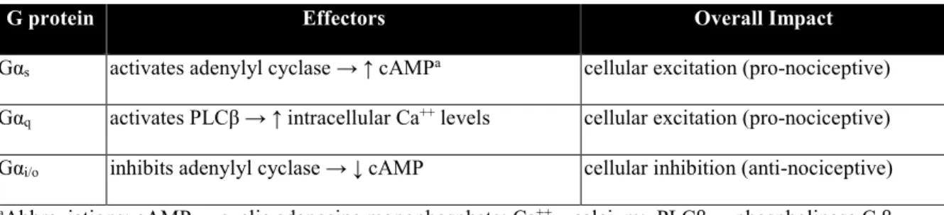
Table 1.1. Common G-proteins and Their Intracellular Effects
G protein Effectors Overall Impact
11
The resulting structure created by the transmembrane segments and loops provides interactive sites where ligands can bind. Ligands that bind to their receptor and initiate cell signaling are referred to as agonists. Upon binding, agonists produce a conformational change of the GPCR and subsequent uncoupling of the associated g-protein. Once uncoupled, the g-protein separates into two subunits (the alpha (α) and beta/gamma (β/γ) subunits), each of which initiates a chain of molecular reactions that affect cellular activity(40). Depending on the type of g-protein, the initiated downstream effects can promote cellular excitation or inhibition (Table 1.1). In general, agonists that activate pain-relevant GPCRs coupled to Gs typically produce pain, while those coupled to Gi typically inhibit pain(39).
1
2
Table 1.2. GPCRs Commonly Targeted for Clinical Pain Management
GPCR G-protein Endogenous Ligands Prescribed Analgesics Known Splice Variant
Reuptake Inhibitors Agonist Antagonist
Cannabinoid (CB) Receptors
CB1 Gαi(41) 2-AG
Anandamide
LPI
NADA
OAE
Nabilone
THC
Cannabidiol Yes
CB2 Gαi(41) Nabilone
THC
Cannabidiol Yes
Adrenergic (AR) Receptors
α1AR Gαq(42) Epinepherine
Norepinephrine
Amitriptyline (NET)
Despiramine (NET)
Desvenlafaxine (NET)
Duloxetine (NET)
Levorphanol (MAO)
Meperidine (NET)
Nortriptyline (NET)
Tapentadol (NET)
Venlafixine (NET)
Amitriptyline
Promethazine
Nortriptyline
Trazodone
Yes
α2AR Gαi(42) Clonidine Trazodone No
β1AR Gαs(43) Atenolol
Nadolol
Metoprolol
1
3
Propanolol
Timolol
β2AR Gαs,
Gαi(43)
Nadolol
Propanolol
Timolol
No
β3AR Gαs(43) Nadolol
Propanolol
Timolol
Yes
Serotonin (5-HT) Receptors
5-HT1 Gαi(44) Serotonin Amitriptyline (SERT)
Despiramine (SERT)
Desvenlafaxine (SERT)
Duloxetine (SERT)
Levorphanol (MAO)
Nortriptyline (SERT)
Trazodone (SERT)
Venlafaxine (SERT)
Almotriptan
Dihydroergotamine
Eletriptan
Frovatriptan
Naratriptan
Rizatriptan
Sumatriptan
Zolmitriptan
Trazodone No
1
4
Methylergometrine Nortriptyline
Promethazine
Trazodone
5-HT4 Gαs(44) Mosapride Yes
5-HT6 Gαs(44) Amitriptyline
Nortriptyline
Trazodone
Yes
5-HT7 Gαs(44) Amitriptyline
Trazodone
Yes
Mu-Opioid Receptor
MOR-1a Gα
i(45) α-endorphin
β-endorphin
γ-endorphin
Alfentanil
Buprenorphine
Codeine
Fentanyl
Hydrocodone
Hydromorphone
Levorphanol
Naloxone
Naltrexone
1
5
Meperidine
Methadone
Morphine
Oxycodone
Oxymorphone
Remifentanil
Sufentanil
Tapentadol
Tramadol
16
Cannabinoid receptors share similar signaling properties with MOR-1, making them attractive targets for clinical pain management. There are two cannabinoid (CB) receptor subtypes, CB1 and CB2, both of which couple to Gαi. CB receptors play a significant role in promoting analgesia
in response to endocannabinoids such as 2-Arachidonoylglycerol (2-AG), and anandamide. Commercially available CB agonists such as nabilone and tetrahydrocannabidol, which bind to both CB subtypes, are used to treat fibromyalgia and neuropathic pain(46).
Adrenergic receptors, which mediate the physiological responses to epinephrine (Epi) and norepinephrine (NE), represent another frequently targeted class of GPCRs. The adrenergic superfamily includes three subtypes respectively of α1ARs (α1AAR, α1BAR, α1DAR), α2ARs (α2AAR,
α2BAR, α2CAR), and βARs (β1ARs, β2ARs, β3ARs). The α2AR couples to Gαi and promotes analgesia via cellular inhibition. Hence α2AR agonists such as trazodone are used to promote analgesia. In
contrast, α1AR, which is coupled to Gαq, facilitates cellular excitation of pronociceptive neurons,
resulting in increased pain signaling. The βARs also facilitate pain signaling via Gαs signaling. To
attenuate their excitatory contributions, α1AR and βARs are commonly used to treat a range of
chronic pain disorders such as migraine, neuropathic pain, and fibromyalgia.
Serotonin receptors, which mediate physiological responses to the monoamine serotonin (5-HT) play an important role in pain management(44). The serotonin superfamily is quite large, including seven general members: 5-HT1 (5-HT1A, 5-HT1B, 5-HT1D, 5-HT1E, 5-HT1F), 5-HT2 (5-HT2A,
5-HT2B, 5-HT2C), 5-HT3, 5-HT4, 5-HT5, 5-HT6, and 5-HT7. With the exception of the 5-HT3 receptor, a
ligand-gated ion channel, all 5-HT receptors are GPCRs. The effects of the 5-HT receptor family on pain are heavily dependent upon the receptor subtype. Triptans target Gαi-coupled 5-HT1 receptors,
which promote analgesia via cellular inhibition, and normalize vascular changes associated with migraine headache(47). Antidepressants promote chronic synaptic serotonin release that causes the downregulation of Gαq coupled 5-HT2 receptors, thus attenuating their excitatory contributions to pain
17
gastrointestinal (GI) tract are used in the treatment of migraine(48) and IBS(49). Meanwhile, the net effect of 5-HT7 activation on pain is highly dependent on the location of the receptor. Activation of
5-HT7 receptors on peripheral nerve terminals produces pain(50,51), while activation in midbrain
structures such as the periaqueductal gray alleviates pain associated with nerve injury(52).
Finally, opioid receptors are among the most well known GPCRs that regulate the transmission and perception of pain. There are four opioid receptor subtypes, including: the mu opioid receptor (MOR-1), the delta opioid receptor, the kappa opioid receptor, and the nociceptin receptor (ORL-1). Of these subtypes, MOR-1 is the classic receptor responsible for analgesic responses to endogenous endorphins as well as exogenous drugs. Upon agonist binding to MOR-1, its associated Gαi protein is activated and produces cellular inhibition of pronociceptive neurons(7). For
this reason, opioids are used in the management of acute pain (such as that associated with surgery) as well as chronic pain disorders such as low back pain, extremity pain, and osteoarthritis(53). Opioid antagonists, usually co-administered with opioid agonists to reduce the development of unwanted opioid side effects, are also capable of producing analgesia independently of MOR-1(54).
18
1.2 The Impact of Alternative Splicing on GPCR Signaling
1.2.1 Alternative splicing adds to the diversity of GPCR signaling
19
Figure 1.2.Different types of alternative splicing. The most common type of alternative splicing in
21
Figure 1.3. Structural variations in GPCRs as a result of alternative splicing. Exons within the
23
Accumulating evidence suggests that alternative splicing significantly adds to the functional diversity of the human genome and that variations in these processes produce pathological states(58). The presence of multiple GPCR splice variants allows for essential, precisely regulated differences in expression (e.g., tissue-specific expression)(59), as well as in agonist binding(60), agonist-induced internalization21, and intracellular signaling dynamics(61,62). Some alternative splice variants even
display functional characteristics opposite to the canonical form(63-65) (Table 1.3). Polymorphisms that alter the ratio of functionally distinct protein isoforms through alternative splicing may produce changes in the direction of pain-relevant GPCR pharmacodynamics (e.g. coupling to stimulatory vs. inhibitory G protein effector systems), yet remain understudied. A PubMed search of “alternative splicing pain” yields only 87 relevant original research articles. Most are focused on ion channels such as voltage-gated calcium channels(66) and transient receptor potential channels(67,68), with only 12 articles focusing on GPCRs. This is an important area of study as identification of GPCR splice variants differentially expressed in individuals with altered pain perception and/or analgesic responses will help elucidate novel targets for the development of individualized treatment strategies.
1.2.2 Alternative splicing creates functional GPCR variants
2
4
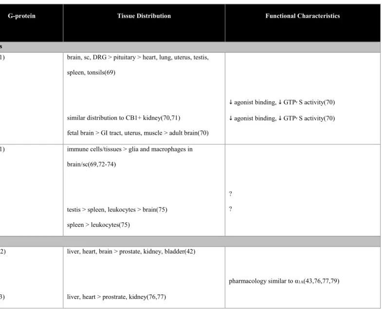
Table 1.3. Signaling, Tissue Distribution, and Function of Known GPCR Splice Variants.
Receptor
Variants
G-protein Tissue Distribution Functional Characteristics
Cannabinoid Receptors
CB1
N-term
variants
CB1a
CB1b
Gαi(41) brain, sc, DRG > pituitary > heart, lung, uterus, testis,
spleen, tonsils(69)
similar distribution to CB1+ kidney(70,71)
fetal brain > GI tract, uterus, muscle > adult brain(70)
↓ agonist binding, ↓ GTPᵞS activity(70)
↓ agonist binding, ↓ GTPᵞS activity(70)
CB2
N-term
variants
CB2A
CB2B
Gαi(41) immune cells/tissues > glia and macrophages in
brain/sc(69,72-74)
testis > spleen, leukocytes > brain(75)
spleen > leukocytes(75)
?
?
Adrenergic Receptors
α1A
C-term
variants
Gαq(42)
Gαi(43)
liver, heart, brain > prostate, kidney, bladder(42)
liver, heart > prostrate, kidney(76,77)
2
5
α1A-2
α1A-3
α1A-4
α1A-5
6TM variants
(-TM7)
α1A-6
α1A-7
α1A-8
α1A-9
α1A-10
α1A-11
α1A-12
α1A-13
α1A-14
α1A-15
α1A-16
Gαi(43)
Gαi(43)
liver > heart, prostrate (absent in kidney)(76,77)
liver, heart > prostrate, (absent in kidney)(76,77)
liver, heart, hippocampus, and prostate; expressed
intracellularly(78)
impair α1A binding & cell surface expression(78)
α1B
6TM variant
2
6
TM7)
α1B-2
expressed in hippocampus, but absent in cortex(80) ?
β3
C-term
variants
β3a (mouse)
β3b (mouse)
Gαs, Gαi(81,82)
Gαs(43,83)
Gαs, Gαi(43,83)
fat, immune cells/tissues > GI tract, DRG(81,84)
fat > ileum > brain(85)
brain > fat, ileum(85)
?
?
Serotonin Receptors
5-HT2A
6TM variant
(-TM4)
5-HT2A-tr
Gαq(44) cortex, hippocampus, brainstem, olfactory > basal ganglia,
limbic(44)
hippocampus, caudate, corpus collosum, amygdala,
substania nigra(86)
impaired 5-HT-induced Ca++ signaling(86)
5-HT2C
6TM variant
(-TM4)
5-HT2CT
Gαq(44) choroid plexus, striatum, hippocampus, hypothalamus,
olfactory, sc(44,87)
choroid plexus, striatum, hippocampus, hypothalamus,
olfactory, sc(87)
2
7
C-term variant
5-HT
2AC-R-COOH∆ sc, cortex, cerebellum, medulla, caudate, amygdala,
corpus collosum(88)
impaired 5-HT ligand binding(88)
5-HT4
C-term
variants
5-HT4a
5-HT4b
5-HT4c
5-HT4d
5-HT4e
5-HT4f
5-HT4g
5-HT4i
5-HT4n
2nd EL loop
variant
5-HT4h
Gαs(44)
Gαs(89)
Gαs, Gαi(89,90)
Gαs(89)
Gαs(89)
Gαs(91)
Gαs(92)
Gαs(93)
Gαs(94)
Gαs(95)
Gαs(92)
intestine > brain > pit > uterus, testis > spleen > heart,
kidney, lung, sc(96)
intestine, brain > pit > uterus, testis > heart > spleen, lung,
sc(96)
intestine, brain > pit > uterus > heart, spleen, lung, sc(96)
intestine > pit > brain > uterus, testis, heart, spleen, sc(96)
ileum, colon, but absent in brain(95,97)
brain > testis > sc > intestine, pit, heart, prostate ileum,
colon(97)
brain, ileum, colon(97)
brain, heart, ileum, colon(97)
brain, ileum, colon, heart(97)
brain, heart, esophagus(97)
↑ constitutive AC activity, ↑ isomerization, ↓ agonist
internalization(98,99)
↑ constitutive AC activity(89)
↑ constitutive AC activity(89)
20-fold ↑ in agonist-induced cAMP activity(100)
↑ constitutive AC activity(91)
?
?
↑ constitutive AC activity(101)
2
8
GI tract(92)
5-HT6
6TM variant
(-TM4)
5-HT6-tr
Gαs (44) cortex, hippocampus, olfactory, striatum, amygdala,
accumbens(44)
cortex, hippocampus, cerebellum, thalamus, substantia
nigra, caudate(102)
impaired binding to 5-HT and LSD(102)
5-HT7
C-term
variants
5-HT7a
5-HT7b
5-HT7d
Gαs(44)
Gαs(103)
Gαs(104)
Gαs(104)
brain, heart, GI tract, muscle, kidney, astrocytoma,
glia(105,106)
brain, heart, GI tract, spleen, lung, astrocytoma,
glia(103,105,106)
brain, heart, GI tract, spleen, lung, astrocytoma, glia(104)
-(105,106)
heart, GI tract, ovary, testis, spleen, lung,
astrocytoma(105)
?
↑ constitutive AC activity(104)
exhibit agonist-independent internalization(107)
2 9 MOR-1 C-term variants MOR-1A MOR-1B MOR-1C MOR-1D MOR-1E MOR-1F MOR-1O MOR-1P MOR-1U MOR-1V MOR-1W MOR-1X MOR-1Y N-term variants MOR-1G MOR-1H
Gαi(45)
Gαi(108)
brain, spinal cord > adrenal gland > small intestine(109)
brain(110)
brain(110)
brain(110); agonist-induced reduction(111)
brain(110) brain(110) brain(110) brain(110) brain(110) brain(110) brain(110) brain(110) brain(110) brain(110) brain(110) brain(110) brain(110) brain(110)
OP binding analgesia(113)
OP binding analgesia(113)
OP binding analgesia(113)
OP induced itch(108)
OP binding analgesia(113)
OP binding analgesia(113)
? ? ? ? ? ?
OP binding analgesia(114)
Novel opioid binding(115)
OP binding analgesia(116)
OP binding analgesia(116)
3
0
MOR-1I
MOR-1J
MOR-1K
MOR-1L
MOR-1M
MOR-1N
Single TM
variants
MOR-1Q
MOR-1R
MOR-1S
MOR-1T
MOR-1Z
MOR-1SV1
MOR-1SV2
Gαs(65)
brain(110)
brain(110)
brain(110)
brain(110)
brain(110)
brain(110)
brain(110)
brain(110)
brain(110)
brain (human neuroblastoma cell line)(112)
brain (human neuroblastoma cell line)(112)
contributes to OIH
OP binding analgesia(116)
?
?
?
Stabilization of MOR-1(117)
Stabilization of MOR-1(117)
?
?
?
?
Abbreviations: 5-HT = serotonin; AC = adenylyl cyclase; N-term = amino terminus; Ca++ = calcium; C-term = carboxyl terminus; cAMP = cyclic adenosine
31
Cannabinoid receptors
Both the CB1 and CB2 receptors undergo alternative splicing to yield variants differing at
their N-terminal region. The CB1a variant is truncated by 61 amino acids, with the first 28 amino acids
completely different from the canonical CB1(71). While its tissue distribution largely overlaps with
that of CB1, CB1a exhibits decreased agonist binding and activity, which might be due to a lack of two
glycosylation sites typically important for signal transduction(118). The CB1b variant lacks the first 33
N-terminus amino acids and although it overlaps with CB1 in a number of tissues, its abundant
expression in fetal brain suggests it may play an important role in development(70). Similar to CB1a,
CB1b exhibits decreased agonist binding and activity.
The CB2 variants are generated through the use of alternate promoters located upstream of the
major coding exon 3(75). The gene CB2A is initiated from the more distal promoter and includes
exons 1a and 1b spliced to exon 3, while CB2B is initiated from the more proximal promoter and
includes exon 2 spliced to exon 3. The CB2A variant is predominantly expressed in testes and at lower
levels in spleen and brain. In contrast, the CB2B variant is predominantly expressed in spleen with
very low expression in brain and no expression in testes. These tissue-specific distribution patterns may indicate specialized roles for the different splice variants with respect to pain modulation, immune response, and spermatogenesis.
Adrenergic receptors
Adrenergic receptors play a key role in pain processing as well as cognition and cardiovascular function. While α2ARs, β1ARs, and β2ARs are highly relevant to the modulation of
pain by endogenous and exogenous agonists, the genes encoding these receptors are intronless and not subject to alternative splicing. Among the remaining adrenergic receptors, the α1AAR subtype has
best most extensively studied with respect to alternative splicing.
32
variants(119). The canonical receptor is generated through splicing exon 1 (coding for the N-terminus and transmembranes [TM] 1 to 6) together with exon 2 (coding for TM7 and the terminus). Four C-terminus splice variants (α1A-2, α1A-3, α1A-4, α1A-5) have been identified that are generated through the
use of additional acceptor sites at varying locations within, and distal to, exon 2. The α1A-2, α1A-3, and
α1A-4 variants exhibit ligand binding properties and tissue distribution profiles similar to α1AAR,
although α1A-3 and α1A-4 are absent in kidney(76-79). In contrast to α1AAR that couples to Gαq, these
variants couple to Gαi so as to inhibit AC activity(43). This diversity in α1AAR signaling may
contribute to differential responses to α1AR antagonists used in the treatment of pain.
In addition, eleven 6TM variants (α1A-6, α1A-7, α1A-8… α1A-16) have been identified that are
generated through exon skipping. These variants lack TM7 and their C-terminal tails are located extracellularly(78). The truncated 6TM variants are expressed in similar tissues as α1AAR, but are
localized exclusively within the cell and unable to bind α1AR agonists or directly mediate signal
transduction. The 6TM variants do, however, impair α1AAR ligand binding and trafficking to the cell
surface. Thus, α1AAR 6TM variants likely play a significant physiological role by modifying the
function and expression of their parent 7TM receptors.
One α1BAR splice variant has also been identified in human brain(80). The α1BAR protein is
generated through splicing of exons 1 and 2. In contrast to the canonical receptor, the α1B-2AR
includes an immediately adjacent sequence following exon 1 in its coding sequence and excludes exon 2 that codes for TM7. Tseng-Crank and colleagues also identified low levels of a truncated
α1DAR transcript, however the result was inconclusive and naturally occurring α1DAR variants were
not observed(80). More work is required to determine the potential functional role of α1BAR and
α1DAR variants.
The β3AR is primarily known for its ability to regulate energy metabolism and
33
emerging (83,85,120). The gene encoding β3AR undergoes alternative splicing within the coding
region to yield two C-terminal splice variants differing with respect to tissue expression, g-protein signaling profiles, and regulatory properties(79,86,121). The β3AAR and β3BAR splice variants
contain completely unique terminal chains that are 13 and 17 amino acids long, respectively. The β3AAR is primarily enriched in fat tissue and couples exclusively to Gαs, while the β3BAR is primarily
enriched in brain and couples to both Gαs and Gαi. In addition, the β3AAR exhibits increased
agonist-induced extracellular acidification, a measure of cAMP-independent cellular activity. Their unique tissue distribution and signaling profiles, together with the known functional role of β3ARs, could
indicate that β3AARs play a greater role in lipolysis/thermogenesis and that β3BAR in brain mediate
pain. While these studies were conducted in mouse, it is important to note that the human β3AR
contains a significant number of genetic variants that are predicted to regulate alternative splicing(87,88).
Serotonin receptors
Serotonin receptors play a key role in pain processing as well as mood and GI function(44). Of the 5-HT1 (A, B, D-F), 5-HT2 (A-C), 5-HT4, 5-HT5, 5-HT6, and 5-HT7 GPCR family members, the
5-HT2A, 5-HT2C, 5-HT4, 5-HT6, and 5-HT7 receptors are known to undergo alternative splicing.
The human 5-HT2 receptor subtypes (5-HT2A, 5-HT2B, and 5-HT2C) couple to Gαq proteins to
promote the transient release of intracellular calcium. One truncated splice variant of 5-HT2A(5-HT 2A-tr) has been identified that utilizes alternate splice donor and acceptor sites to yield a 3TM receptor
with 57 unique amino acids in the C-terminal region(86). The 5-HT2A-tr is co-expressed with 5-HT2A
in most brain tissues, however is unable to couple to the calcium pathway. Two truncated splice variants of 5-HT2C (5-HT2CT and 5-HT2C-R-COOH∆) have also been identified. Similar to 5-HT2A-tr, the 5-HT2CT variant utilizes alternate splice donor and acceptor sites to yield a 3TM receptor with 19 unique
amino acids in the C-terminal region(87). The 5-HT2C-R-COOH∆variant retains an extra 90 nucleotides
34
Compared to the canonical 5-HT2C receptor, the truncated variants exhibit similar expression patterns
but have impaired 5-HT ligand binding and g-protein coupling(87,88). While the relative importance of these truncated 5-HT2 splice variants in humans remains unknown, they are conserved in rat and
mouse(88) where their expression levels increase following nerve injury(122).
The 5-HT4 receptor couples preferentially to Gαs and, while widely expressed, the highest
levels are found in intestine(96). Agonists targeting 5-HT4 are beneficial in alleviating abdominal pain
associated with irritable bowel syndrome. Of all the 5-HT receptors, 5-HT4 possesses the greatest
diversity in alternative splicing. At least ten splice variants have been identified that vary with respect to their tissue distribution and function. Nine C-terminus variants (5-HT4a, 5-HT4b, 5-HT4c, 5-HT4d,
5-HT4e, 5-HT4f, 5-HT4g, 5-HT4i, 5-HT4n) have been identified that are identical up to amino acid Leu358,
after which they vary in sequence and length(97). Additionally, one variant (5-HT4h) has been
identified that includes exon h coding for 14 additional amino acids in the second extracellular loop(92). The 5-HT4a, 5-HT4b, 5-HT4c, and 5-HT4e variants are expressed in most tissues, with
distribution patterns similar to the canonical form(96,97). In contrast, the 5-HT4f variant is found in
the brain and GI tract, but absent in the heart and other tissues22. Meanwhile, the 5-HT
4d and 5-HT4h
variants are expressed exclusively in the GI tract(92,94,97). While all of the 5-HT4 splice variants
display typical ligand binding properties, some show notable functional differences. Both of the GI-specific 5-HT4d and 5-HT4h variants have a tendency to recognize 5-HT antagonists as partial
agonists(92,100). Furthermore, the 5-HT4d variant exhibits a remarkable 20-fold increase in cAMP
formation following application of the 5-HT4 agonist renzapride(100). The 5-HT4b variant is unique in
its able to couple to Gαi as well as Gαs proteins, suggesting its diverse signaling capabilities in the GI
35
Collectively, these studies illustrate the high degree of tissue and signaling specificity for a number of 5-HT4 splice variants that may be represent attractive targets for the development of new more
selective drugs for the treatment of irritable bowel syndrome among other conditions.
The 5-HT6 receptor is unique in that it is expressed almost exclusively in the central nervous
system(44). A 3TM splice variant of 5-HT6 (5-HT6-tr) has been identified in brain that is generated
through different splice donor and acceptor sites(102). The corresponding receptor includes the TM1-3 and 10 unique amino acids in its C-terminus. In contrast to 5-HT6, the expression of 5-HT6-tr is
limited to substantia nigra and caudate. The 5-HT6-tr receptor is able to translocate to the membrane,
yet unable to bind serotonin. This splice variant may have a yet-to-be-determined function or be indicative of abnormalities due to pathologic state.
The 5-HT7 receptor is expressed on primary afferent nociceptors, as well as in pain-relevant
brain regions where it couples to Gαs to mediate the transmission and modulation of pain. Three splice
variants of 5-HT7 (5-HT7a, 5-HT7b, 5-HT7d) have been identified that are all generated through
alternative splicing of the second intron located near the C-terminal coding region. The 5-HT7a and
5-HT7b variants have tissue expression profiles and functional characteristics similar to the canonical
receptor, though 5-HT7b has been shown to exhibit significantly higher constitutive AC activity when
expressed in stable cell lines{Krobert:2002du}. The 5-HT7d variant is predominantly expressed in
smooth muscle tissues such as the heart and GI tract(104) and displays unique functional characteristics. Compared to the canonical 5-HT7 receptor and the 5-HT7a and5-HT7b variants, the
5-HT7d variant displays agonist-independent internalization (even in the presence of antagonist) and
associated reductions in agonist-induced AC activity(107). It has been suggested that differences in the functional characteristics of 5-HT7 variants is due to specific features of their carboxyl tails,
36
Opioid receptors
The pharmacologic manipulation of the mu opioid receptor is an essential component of clinical pain treatment. Although the signaling characteristics of MOR-1 are well established, we are just beginning to understand the complex nature of genetic variants that contribute to alternative splicing. At least 20 MOR-1 splice variants have been identified in mouse and human genomes(61), suggesting an array of potentially functional consequences that may occur with opioid administration. Pre-clinical studies within the past 15 years have begun to reveal the functional properties of specific MOR-1 splice variants. Pasternak and coworkers provide evidence that the expression of MOR-1 splice variants represent compensatory responses to chronic opioid administration that stabilize or diminish the development of tolerance(125). Additional studies investigating the functional characteristics of MOR-1 splice variants provide evidence that a set of these receptors promote opioid analgesia by providing exclusive binding sites for different opioids. Transgenic mice lacking exon 11, an exon that provides an alternative promoter region for the MOR transcript, demonstrated substantial reductions in the analgesic efficacies of heroin, fentanyl, and the morphine metabolite morphine-6β-glucuronide(60), suggesting that exon-11 containing variants play a critical role in opioid analgesia. Exon 11-containing splice variants also mediate the analgesic effects of iodobenzoylnaltrexamide (IBNtxA), a novel synthetic opioid that produces ten times the analgesic efficacy of morphine without producing respiratory distress, dependence, tolerance, or GI distress in rodents(111,115,126). MOR-1 splice variants also promote analgesia by enhancing canonical receptor function. Single-transmembrane splice variants MOR-1R and MOR-1S structurally enhance MOR-1 function by stabilizing the canonical 7TM receptor at the cellular membrane(117).
37
as MOR-1K, a truncated receptor lacking the N-terminus and first transmembrane, has been implicated in the paradoxical increase in pain sensitivity known as opioid-induced hyperalgesia (OIH). In contrast to MOR-1 which typically couples to Gαi, MOR-1K couples to Gαs to activate adenylyl
cyclase (AC) and increase intracellular calcium, thus engaging pro-nociceptive signaling events that likely drive OIH(65). A subsequent preclinical study in mice revealed that genetic knockdown of MOR-1K hindered the development of OIH and unmasked opioid analgesia(127). The relationship between MOR-1K and OIH is further discussed throughout the remainder of the manuscript.
Conclusion
G-protein coupled receptors play a major role in modulating the activity of a chorus of cells involved in the transmission, modulation and perception of pain. For this reason, GPCRs are the primary target of many pharmacologic interventions used in the management of acute and chronic pain. Nonetheless, the use of these medications is limited due to variability in analgesic efficacy and side effect profiles. These limitations are partly attributed to genetic differences that influence alternative splicing of pain-relevant GPCRs. The functional importance and implications of the diversity of GPCRs in contributing to the pathophysiology of clinical pain is just beginning to emerge. More research, especially in the clinical arena, is necessary to further investigate the functions of specific GPCR splice variants, as well as the dynamic interactions between multiple variants of the same canonical receptor, within the context of pain. This line of inquiry will evolve our understanding of pain mechanisms and inform the design of new and clinically useful drugs that target specific alternative splice variants altered in a subset of patients.
38
CHAPTER 2: MOR-1K Contributes to OIH in Genetically Susceptible Mice
22.1. Introduction
Accumulating evidence indicates that MOR-1K, a functional splice variant of the canonical mu opioid receptor (MOR-1), may contribute to the emergence of OIH. MOR-1K is a truncated six transmembrane G-protein coupled receptor (GPCR) lacking a N-terminus transmembrane due to the absence of exon 1 within its mRNA transcript (61). Replacing exon 1 are exon 11, which provides an alternative translation start site in several MOR-1 splice variants, and exon 13, which is unique to the
MOR-1K transcript. The MOR-1 transcript, which encompasses exons that encode for MOR-1K, is highly conserved across species, with a 91% nucleotide sequence homology between human and mouse. Results from a human genetic association study demonstrated that a single nucleotide polymorphism within exon 13 of the human MOR-1K transcript is associated with increased pain sensitivity and blunted morphine efficacy (128). Subsequent in vitro studies demonstrated that MOR-1K exhibits signaling properties distinct from its parent receptor MOR-1. MOR-1 utilizes Gi/o protein
to inhibit cyclic adenosine monophosphate (cAMP) levels and intracellular calcium levels to produce cellular inhibition of pronociceptive cells. In contrast, MOR-1K couples to Gs protein, leading to
increased cAMP production and intracellular calcium levels, thus promoting cellular excitation (65). Previous studies have shown that Gs-dependent increases in intracellular calcium via cAMP
production and protein kinase A activation play a critical role in central sensitization (129) and the development of inflammatory, neuropathic, and functional pain (130). The utilization of Gs signaling
2This chapter previously appeared as an article in PLoS ONE. The original citation is as follows: Oladosu F.A., Conrad M.S., O’Buckley
S.C., Rashid N.U., Slade G.D., and Nackley A.G., “Mu Opioid Splice Variant MOR-1K Contributes to the Development of Opioid-Induced
39
by MOR-1K suggests that the receptor may also contribute to central sensitization associated with OIH.
Given the receptor’s genetic association with increased pain sensitivity and its excitatory signaling profile, we hypothesize MOR-1K may contribute to OIH in genetically susceptible individuals. Here, we evaluate MOR-1K in the development of OIH using three genetically diverse mouse strains alongside small interfering RNA (siRNA) knockdown of MOR-1K. Our results demonstrate that OIH is associated with increased MOR-1K gene expression levels in a strain-specific manner. Disrupting the increase in MOR-1K gene expression levels via chronic intrathecal (i.t.) siRNA administration not only hinders the development of OIH, but also increases morphine analgesic efficacy. Collectively, these findings demonstrate that MOR-1K is likely a key contributor to OIH.
2.2. Materials and Methods
2.2.1. Ethical Statement
All procedures within this study were approved by the University of North Carolina Animal Care and Use Committee (permit number: 12-319) and adhered to the guidelines of the Committee for Research and Ethical Issues of the International Association of the Study of Pain (
http://www.iasp-pain.org/Education/Content.aspx?ItemNumber=1217). All surgeries were performed under
isofluorane anesthesia, and all efforts were made to minimize suffering.
2.2.2. Animals
Male and female C57BL/6J (http://jaxmice.jax.org/strain/000664.html) and CXB7/ByJ
(http://jaxmice.jax.org/strain/000357.html) mice were obtained from Jackson Labs (Bar Harbor, ME)
40 2.2.3. Drugs and Chemicals
Morphine sulfate (Sigma, MO) was dissolved in 0.9% sterile saline (Hospira, IL). Doses of 10 mg/kg, 20 mg/kg, or 40 mg/kg were administered via subcutaneous (s.c.) injection in a volume determined by animal weight (1µl/g). Fluorescein-tagged exon 13-antisense siRNA [5’-UCA GUC UUU AUC AGC UCA CCG CCA-3’] or fluorescein-tagged exon 13-sense siRNA (Midland Certified Reagent Co., OH) [5’-AGU CAG AAA UAG UCG AGU GGC GGU-3’] in artificial cerebrospinal fluid was administered at a rate of 0.5μl/hr; 0.291μg/hr/day for a duration of 7 days via Alzet osmotic mini-pump (Durect, CA) connected to an i.t. catheter (Durect, CA). Previous studies have successfully administered siRNA in this fashion as well (131,132). Sense siRNA was chosen as a negative control as it is related to the target mRNA sequence of interest but does not affect target mRNA expression (133).
2.2.4. Experimental Design
Experiment 1: The effects of chronic morphine administration on pain behavior and gene
expression.
41
Experiment 2: The effects of MOR-1K exon 13 siRNA knockdown on OIH and MOR-1K gene
expression.
Prior to chronic morphine administration, CXB7/ByJ mice (N=48; 8 males and 8 females per experimental condition) were assessed for baseline responses to mechanical stimuli. Following baseline assessments, mice underwent surgery for chronic i.t. administration of antisense exon 13 siRNA or sense exon 13 siRNA. Mice were anesthetized with 5% isofluorane and maintained at 2-3% isofluorane during i.t. catheter implantation, modified from Yaksh and Rudy protocol (134). A separate group of mice also underwent surgery for a sham procedure. The sham procedure, involving skin incision and muscle dissection without breakage of the arachnoid membrane to cause leakage of cerebral spinal fluid, was deemed appropriate to control for any postoperative pain. One day following surgery, mice from antisense, sense, and sham conditions began to receive either vehicle (sterile saline) or escalating doses of morphine as described above. MOR-1K gene expression levels were measured in tissues collected from separate groups of CXB7/ByJ mice (N=84; 4 males and 3-4 females per experimental condition) sacrificed on day 0, on days 1 or 3-4 following the 8am injection, or on day 5 at 8am. The experimental design is illustrated in Figure 3A.
2.2.5. Behavior
Assessment of Paw Withdrawal Threshold, Mechanical Allodynia, and Mechanical
Hyperalgesia
42
filament was presented. After the initial response threshold was crossed, this procedure was repeated in order to obtain a total of six responses in the immediate vicinity of the threshold. The pattern of withdrawals and absence of withdrawals were noted together with the terminal filament used in the series of six responses. The 50% of the paw withdrawal threshold is calculated as (10[X
f+kδ])/10,000,
where Xf = value (in log units) of the final von Frey hair used; k = tabular value of pattern of positive
(X) and negative (O) responses, and δ = mean difference (in log units) between stimuli. Mechanical allodynia was assessed by presenting a filament with bending force of 0.40 g to the hind paw 10 times for a duration of 1 s with an inter-stimulus interval of 1 s. A significant increase in the percentage frequency of paw withdrawal ([# of paw withdrawals/10] x 100) was defined as mechanical allodynia. Mechanical hyperalgesia was assessed in the same manner, using a filament with a bending force of 1.50 g.
Assessment of Thermal Heat Hyperalgesia
Thermal heat hyperalgesia was evaluated using the hot plate method (136). Mice were placed on a hot plate (Columbus Instruments, OH) maintained at a temperature of 51.5°C for one minute. Each session was videotaped and the total number of aversive responses (paw licks, paw flicks, and jumps) was measured.
2.2.6. Assessment of Gene Expression Levels
43
manufacturer protocol and concentrations were determined using a Nanodrop-1000 (Thermo Scientific, DE) and reverse transcribed using Transcriptor First Strand cDNA Synthesis kit (Roche, Switzerland) where necessary. Fast Start Universal SYBR Green Master with Rox (Roche, Switzerland) or Power SYBR Green RNA-to-CT 1-Step (Life Technologies, NY) were respectively used per manufacturer protocols to amplify cDNA and RNA, per manufacturer protocols. A 7900HT Fast Real-Time PCR system (Life Technologies, NY) and a StepOnePlus Real-Time PCR system (Life Technologies, NY) were respectively used for measuring cDNA or RNA transcripts amplification. The following primers were used for the detection of the following exon-11 containing MOR-1 splice variants: MOR-1K forward (TCCCCTCTTGAGTGTGACTAATGTC) and reverse
(GCCAGAGCAAGGTTGAAAATG); MOR-1L forward
(CAGAGCAAGGTTGAAAATGTAGATG) and reverse
(AAATCAAAATAGAAAATGGGCTAAGG); MOR-1T forward
(GAGCCACATGGAATTGCCTCTGTA) and reverse (GCATCTGCCAGAGCAAGGTTGAAA);
forward (GGGCCGATGATGGAAGCTTTCTCTAA) and reverse
(GCATCTGCCAGAGCAAGGTTGAAA) primers for splice variants that contain exons 11 and 2. Expression of the target genes was normalized to housekeeping genes RPL7 [forward (TCAATGGAGTAAGCCCAAAG) and reverse (CAAGAGACCGAGCAATCAA)] or GAPDH
[forward (TGAAGGTCGGAGTCAACGGATTTGGT) and reverse
(CATGTGGGCCATGAGGTCCACCAC)] using the 2(-ΔΔCT) method. All primers were purchased
from Integrated DNA Technologies (CA).
2.2.7. Statistical Analysis
44
45
2.3. Results
2.3.1. Strains demonstrate divergent baseline pain profiles.
46
Figure 2.1. Sex-dependent responses to mechanical and thermal heat stimuli across the three
strains. Overall, males and females displayed similar behavioral responses to mechanical and thermal
heat stimuli within strains. Female C57BL/6J mice demonstrated (A) increased paw withdrawal threshold (F(9,140) = 12.20,p<0.0001) and (B) increased responses following repeated exposure to an
innocuous mechanical stimulus (F(9,140) = 14.50, p<0.0001). Panels A-D: N=7-8/group. Males are
47 1 Po
st 2 Pre
2 Po st
3 Pre 3 Po
st 4 Pre
4 Po st 5 6 7
-100 0 100 200 300 400 5 0 % P a w W it h d ra w a l T h re s h o ld (% C h a n g e F ro m B a s e lin e ) analgesia allodynia **** **** **** ****
1 Post 2 Pr e
2 Post 3 Pr e
3 Post 4 Pr e
4 Post 5 6 7
-50 0 50 100 150 200 5 0 % P a w W it h d ra w a l T h re s h o ld (% C h a n g e F ro m B a s e lin e ) analgesia allodynia
1 P ost
2 Pre 2 P
ost 3 Pre
3 P ost
4 Pre 4 P
ost 5 6 7
-100 -50 0 50 5 0 % P a w W it h d ra w a l T h re s h o ld (% C h a n g e F ro m B a s e lin e ) *** analgesia allodynia
1 P ost
2 Pre 2 P
ost 3 Pre
3 P ost
4 Pre 4 P
ost 5 6 7
0 200 400 600 800 N u m b e r o f R e s p o n s e s (% C h a n g e f ro m B a s e lin e ) allodynia **** **** ****
1 Post 2 P
re 2 Post
3 P re
3 Post 4 P
re
4 Post 5 6 7
0.0 0.2 0.4 0.6 0.8 1.0 N u m b e r o f R e s p o n s e s (% C h a n g e f ro m B a s e lin e ) allodynia
1 Post 2 Pre
2 Post 3 Pre
3 Post 4 Pre
4 Post 5 6 7
0 200 400 600 800 1000 N u m b e r o f R e s p o n s e s (% C h a n g e f ro m B a s e lin e ) # allodynia *** * *
1 P ost
2 Pre 2 P
ost 3 Pre
3 P ost
4 Pre 4 P
ost 5 6 7
-100 0 100 200 N u m b e r o f R e s p o n s e s (% C h a n g e f ro m B a s e lin e ) analgesia hyperalgesia
1 Post 2 P
re 2 Post
3 P re
3 Post 4 P
re
4 Post 5 6 7
-100 -50 0 50 N u m b e r o f R e s p o n s e s (% C h a n g e f ro m B a s e lin e ) analgesia hyperalgesia
1 Post 2 Pre
2 Post 3 Pre
3 Post 4 Pre
4 Post 5 6 7
-50 0 50 100 N u m b e r o f R e s p o n s e s (% C h a n g e f ro m B a s e lin e ) analgesia hyperalgesia
1 P ost
2 Pre 2 P
ost 3 Pre
3 P ost
4 Pre 4 P
ost 5 6 7
-100 0 100 200 300 400 N u m b e r o f J u m p s & F lic k s (% C h a n g e f ro m B a s e lin e ) analgesia hyperalgesia
1 Post 2 P
re 2 Post
3 P re
3 Post 4 P
re
4 Post 5 6 7
-100 -50 0 50 100 N u m b e r o f J u m p s & F lic k s (% C h a n g e f ro m B a s e lin e ) analgesia hyperalgesia
1 Post 2 Pre
2 Post 3 Pre
3 Post 4 Pre
4 Post 5 6 7
-100 0 100 200 300 400 N u m b e r o f J u m p s & F lic k s (% C h a n g e f ro m B a s e lin e ) analgesia hyperalgesia
Paw Withdrawal Threshold Mechanical Allodynia Mechanical Hyperalgesia Thermal Heat Hyperalgesia
C 5 7 B L /6 J C X B 7 /B y J 1 2 9 S 6 A E
I J L
48
Our results showed that the strains exhibited baseline differences in mechanical (Figure 2.2A, F(2,43)=20.56, p<0.0001; Figure 2.2B, F(2,43)=43.77, p<0.0001), and thermal heat pain sensitivity
(Figure 2.2C; F(2,43=10.64, p=0.0002). Compared to the classic C57BL/6J inbred strain, 129S6 mice
49
Figure 2.2. Strains exhibit divergent behavioral responses to mechanical stimuli at baseline
Compared to C57BL/6J mice, 129S6, and CXB7/ByJ mice exhibit differences in (A) paw withdrawal threshold, (B) the number of responses to repeated presentation of a noxious mechanical stimulus sensitivity, and (C) the number of responses to continuous thermal heat. N=15-16/group. Data expressed as mean ± SEM. ***p<0.001, **p<0.01, *p<0.05 different from C57BL/6J. ###p<0.001