DENTAL ANKYLOSIS: CLINICAL AND MOLECULAR CHARACTERIZATION
Gabriel Senties-Ramirez
A thesis submitted to the faculty at the University of North Carolina at Chapel Hill in partial fulfillment of the requirements for the degree of Master of Science in the School of Dentistry
(Orthodontics).
Chapel Hill 2016
Approved by:
Sylvia A. Frazier-Bowers Ching Chang Ko
© 2016
ABSTRACT
Gabriel Senties-Ramirez: Dental Ankylosis: Clinical and Molecular Characterization (Under the direction of Sylvia A. Frazier-Bowers.)
Introduction: Dental ankylosis is a histological definition for a condition that is often clinically indistinguishable from other eruption disorders. C onfusion between ankylosis and PFE can result in an inaccurate diagnosis, and inappropriate orthodontic
management. In this study, we tested the central hypothesis that ankylosis is a distinct pathology compared to PFE due to PTH1R mutations. Methods: 1) We completed mutational analysis of PTH1R on patients with PFE (based on familial segregation) or
ACKNOWLEDGMENTS
TABLE OF CONTENTS
LIST OF TABLES ... VII LIST OF FIGURES ... VIII LIST OF ABBREVIATIONS ... XI LIST OF SYMBOLS ... XIII
A REVIEW OF THE LITERATURE ... 1
TOOTH ERUPTION, AND ITS DISORDERS ... 1
DENTAL ANKYLOSIS:ADIAGNOSTIC CHALLENGE ... 3
INCIDENCE AND CLINICAL PRESENTATION ... 4
ETIOLOGY OF DENTAL ANKYLOSIS:WHAT IS CURRENTLY KNOWN ... 6
Animal Studies on the Etiology of Dental Ankylosis. ... 8
THE POSSIBLE ROLE OF RANKL AND BONE MORPHOGENETIC PROTEINS (BMPS) IN DENTAL ANKYLOSIS ... 10
PRIMARY FAILURE OF ERUPTION AND ITS POSSIBLE RELATION TO DENTAL ANKYLOSIS ... 12
CONCLUSION ... 14
REFERENCES ... 16
BACKGROUND AND INTRODUCTION ... 19
MATERIALS AND METHODS ... 21
Patient Recruitment ... 21
Sample Procurement ... 24
Primary Cell Cultures ... 25
RNA Extraction ... 30
RT-PCR ... 31
Real Time PCR ... 32
Mutational Analysis ... 32
RESULTS ... 33
DISCUSSION. ... 52
LIST OF TABLES
TABLE 1:PRIMER SEQUENCES USED IN QPCR ... 32
TABLE 2:PTH1R PRIMER SEQUENCES USED DURING MUTATIONAL ANALYSIS ... 33
TABLE 3: QPCR RESULTS BY CT VALUE ... 51
LIST OF FIGURES
Figure 1 a: Patient A Intraocclusal Photographs ... 22
Figure 1 b: Patient A Panoramic Radiograph ... 22
Figure 2 a: Patient B Intraocclusal Photographs……….………23
Figure 2 b: Patient B’s sibling, Intraocclusal Photographs ... 23
Figure 2 c: Patient B’s mother, Intraocclusal Photographs ... 24
Figure 2 d: Patient B panoramic radiograph ... 24
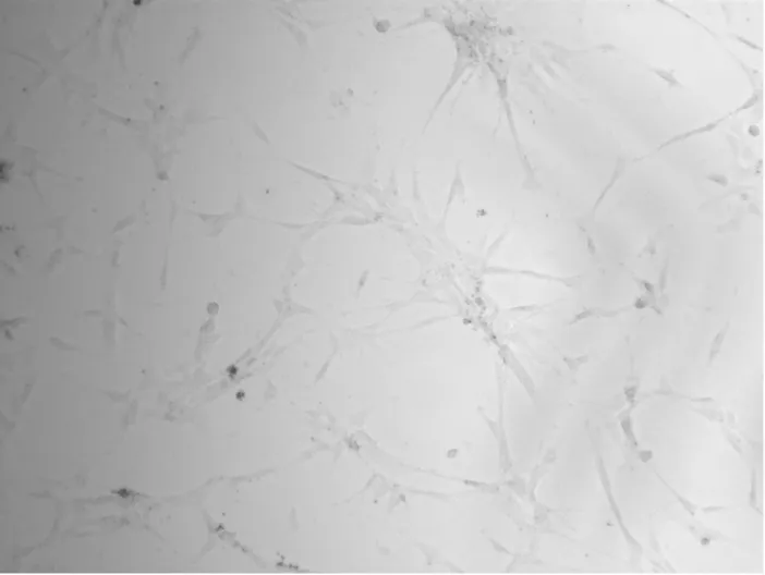
Figure 3 a: Patient A, tooth #29 cell culture……….……….26
Figure 3 b: Patient A, tooth #19 cell culture ... 27
Figure 3 c: Patient B, tooth #1 cell culture ... 28
Figure 3 d: Patient B, tooth #16 cell culture ... 29
Figure 3 e: Patient B, tooth #17 cell culture ... 30
Figure 4 a: Patient A, PTH1R Exon 8F chromatogram………..….34
Figure 4 b: Patient B, PTH1R Exon 3 chromatogram ... 34
Figure 4 c: Patient A, #29 (control), GAPDH qPCR analysis.Ct=15.68 ... 35
Figure 4 d: Patient A, #19 (ankylosed), GAPDH qPCR analysis.Ct=15.8 ... 36
Figure 4 e: Patient B, #1 (control), GAPDH qPCR analysis.Ct=15.54 ... 36
Figure 4 f: Patient B, #16 (PFE), GAPDH qPCR analysis. Ct=13.74 ... 37
Figure 4 g: Patient B, #17 (PFE), GAPDH qPCR analysis. Ct=15.39 ... 37
Figure 5 b: Patient A, #19 (ankylosed), BMP-2 qPCR analysis. ... 38
Figure 5 c: Patient B, #1 (control), BMP-2 qPCR analysis. ... 39
Figure 5 d: Patient B, #16 (PFE), BMP-2 qPCR analysis. ... 39
Figure 5 e: Patient B, #17 (PFE), BMP-2 qPCR analysis. ... 40
Figure 6 a: Patient A, #29 (control), BMP-4 qPCR analysis. Ct=15.66…………..………40
Figure 6 b: Patient A, #19 (ankylosed), BMP-4 qPCR analysis. Ct=18.78 ... 41
Figure 6 c: Patient B, #1 (control), BMP-4 qPCR analysis. Ct=15.14 ... 41
Figure 6 d: Patient B, #16 (PFE), BMP-4 qPCR analysis. ... 42
Figure 6 e: Patient B, #17 (PFE), BMP-4 qPCR analysis. ... 42
Figure 7 a: Patient A, #29 (control), BMP-6 qPCR analysis………..…………43
Figure 7 b: Patient A, #19 (ankylosed), BMP-6 qPCR analysis. ... 43
Figure 7 c: Patient B, #1 (control), BMP-6 qPCR analysis. ... 44
Figure 7 d: Patient B, #16 (PFE), BMP-6 qPCR analysis. Ct=14.17 ... 44
Figure 7 e: Patient B, #17 (PFE), BMP-6 qPCR analysis. ... 45
Figure 8 a: Patient A, #29 (control), RANKL qPCR analysis……….…….45
Figure 8 b: Patient A, #19 (ankylosed), RANKL qPCR analysis. ... 46
Figure 8 c: Patient B, #1 (control), RANKL qPCR analysis. ... 46
Figure 8 d: Patient B, #16 (PFE), RANKL qPCR analysis. ... 47
Figure 8 e: Patient B, #17 (PFE), RANKL qPCR analysis. ... 47
Figure 9 b: Patient A, #19 (ankylosed), PTH1R qPCR analysis. Ct= 14.54 ... 48
Figure 9 c: Patient B, #1 (control), PTH1R qPCR analysis. ... 49
Figure 9 d: Patient B, #16 (PFE), PTH1R qPCR analysis. ... 49
LIST OF ABBREVIATIONS ALP---Alkaline Phosphatase
BMPs---Bone Morphogenetic Proteins C---Celsius
cDNA---Complementary Deoxyribonucleic acid. DNA--- Deoxyribonucleic acid
dNTP---deoxynucleoside triphosphate DTE---Delayed Tooth Eruption
FBS---Fetal Bovine Serum
GAPDH----Glycderaldehyde-3-phosphate dehydrogenase H2O---Water
IAN---Inferior Alveolar Nerve ME---Malassez Epithelium MEM---Minimum Essential Media ml---Milliliters
PCR---Polymerase Chain Reaction PDL---Periodontal Ligament PFE---Primary Failure of Eruption
RANK--- Receptor activator of nuclear factor kappa-B
RANKL--- Receptor activator of nuclear factor kappa-B Ligand RFA---Resonance Frequency Analysis
RNA---Ribonucleic Acid
RT-PCR----Reverse transcriptase polymerase chain reaction Sec---seconds
LIST OF SYMBOLS
° Degrees
© Copyright Symbol
® Registered Trademark
A REVIEW OF THE LITERATURE
Tooth Eruption, and its disorders
Tooth eruption is the developmental process behind the movement of a dental unit from the crypt where it forms, across the bone of the jaws, and into the oral cavity, towards its place across a functional occluding counterpart 31. Some clinicians use the term eruption to refer to the appearance of a tooth in the oral cavity, but a more accurate term for this
milestone is emergence or moment of eruption. Authors in the literature advocate against using the terms “eruption” and “emergence” interchangeably in order to preserve their more specific and separate meanings31. Tooth eruption in the true sense of the word is without a doubt a complex process, involving many tissues and cell types that are coordinated with precision and specificity in time and space31. The downside of this very complexity, however, is that it
presents many elements and mechanisms that have the potential to go awry. A functional occlusion may not be maintained without proper eruption of the teeth, which is why this process is an area of study of upmost importance, not only to orthodontics, but to all of dentistry. Based on population studies, there are expected timelines for teeth to both erupt and emerge during human growth and development, either from a perspective of chronological age, or from the viewpoint of cell and tissue development. Many different conditions and phenomena have the potential to cause a deviation from the expected timelines in tooth
still a controversial topic31. The variety of conditions that may result in delayed tooth eruption is indeed vast, and it ranges from systemic conditions and affecting all of the dentition and even other organ systems as well, to localized elements that only affect one dental unit at a time31. Conditions that affect the eruption of all teeth can also affect their development, size, shape structure and color when they are syndromic; such is the case with Dentinogenesis Imperfecta, Amelogenesis Imperfecta, and Dentinal Dysplasia31. Malnutrition, Downs Syndrome and
Hypopituitarism can also result in teeth having shorter roots than expected when they erupt compared to the general population31. Localized elements that can result in physical
obstruction of dental eruption include supernumerary teeth, tumors or either odontogenic or non-odontogenic origin, cysts, scar tissue secondary trauma or surgical procedures, and gingival fibromatosis31. The current understanding of tooth eruption is surprisingly poor, as is our knowledge of many of the conditions in which this process is impaired31. Dental ankylosis is a very illustrative example of the gaps in our current knowledge with regard to tooth eruption and its related pathology.
tooth that is infra-occluded, but it can also be detrimental to the development of the alveolar process30. Although this condition was observed in many patients in the early
twentieth century, limitations in the diagnostic aides and laboratory techniques of those days narrowed the scope and depth of studies that could be carried out in an attempt to elucidate its causes and pathological process. Early research on dental ankylosis provided better
information on its incidence and clinical presentation, while theories on its etiology were more speculative in nature.
Dental Ankylosis: A Diagnostic Challenge
One of the complications in gathering data during clinical studies of dental ankylosis is that the condition can be challenging to detect and diagnose correctly compared to other causes of delayed tooth eruption31. The methods most relied upon by dental professionals to diagnose this condition in a clinical setting are percussion and mobility tests as well as
radiographic examination5. Ankylosed teeth are thought to lack any mobility due to the fusion of their root surface with the surrounding bone, and to produce a clear and high-pitched noise, which in healthy teeth would be dampened by the PDL6. The percussion, mobility, and radiographic methods however provide no histological assessment of the relationship
surface that is between ten and twenty percent of the total, and teeth with an affected surface of less than ten percent of the total show no clinical difference with unaffected teeth4. Diagnosis of dental ankylosis based on radiological evaluations can be even more problematic. When ankylosis is present at the buccal or lingual surfaces of the dental root, it is not radiologically evident; it can only be detected by that method if it is present on the proximal surfaces of the tooth, giving rise to many false negatives4. Furthermore, it has been observed that a thin layer of bone on the root surface can be linked to the surrounding alveolar bone by thin trabeculae and give the appearance of a periodontal ligament in a radiograph, which contributes to false negatives even when the condition affects proximal root surfaces4.
In 2012, Resonance Frequency Analysis was proposed as a new diagnostic tool for dental ankylosis6. This technique is most often used as a measure of stability in dental implants by measuring the implant to bone interface based on resonance frequencies during oscillations exerted into the implant and bone contact point6,21,32. Although initial studies suggest that RFA might provide greater sensitivity than more traditional methods in the diagnosis of dental ankylosis, more testing will have to take place before it is implemented widely. The practicality and economic feasibility of using this technique and the equipment it requires in a private orthodontic office is not yet clear throughout the literature. For a conclusive diagnosis of dental ankylosis, histological examination of a specimen removed from the oral cavity remains the diagnostic gold standard.
Incidence and Clinical Presentation
reports in the literature described the growing knowledge regarding the epidemiological factors of this condition in its different clinical presentations. The primary second molar was identified early on as the dental unit that was affected most often even though the condition could be observed in both permanent and deciduous dentition8. Work by
Biederman in 1956 illustrated that dental ankylosis was a selective condition with regard to its onset during dental development and the location of the dental units it affected8. By comparing 119 patients who presented with two ankylosed deciduous second molars each, he statistically determined that the clinical presentation of dental ankylosis followed a discreet pattern that was not random8. Among his findings, he showed that the condition occurred in the teeth of the mandible more than twice as frequently as the teeth of the maxillary arch and that more than ninety percent of the affected teeth were either first or second primary molars8. Biederman’s work also laid an early foundation towards
elucidating the pathophysiology of dental ankylosis through his observation that the condition being present in one dental unit did not correlate with its occluding counterpart being affected as well, thus undermining the early theory that dental ankylosis was caused by traumatic or heavy occlusal forces8. As an alternative theory, Biederman postulated that dental ankylosis was probably caused by “some metabolic disturbance of a local character” 8. In a later study, nearly ten years afterward, Biederman went to greater length to
ankylosed teeth do not have an eruption potential any longer and will not emerge into the oral cavity or occlusal plane once mechanical obstacles are cleared from their path7. The body of literature available by that time confirmed the earlier observations that dental ankylosis was specific with respect to its location in the oral cavity, with nearly all affected teeth being primary or permanent molars and mandibular teeth being represented more than twice as often as maxillary teeth7. Additionally, this phenomenon also appeared to be specific with respect to time of onset since deciduous teeth were represented over
permanent teeth by a ratio greater than ten to one7.
Two different presentations have been observed in the roots of teeth with dental ankylosis4. In some cases, resorption of cementum and dentin has been observed, while in others there is direct deposition of bone on top of cementum in the root of the ankylosed tooth4. By the nineteen-eighties, Andersson et al. had enough sources and evidence to report that ankylosis was also observed as a common complication of avulsed permanent teeth that were replanted4, while Kurol reported that a familial tendency in the pattern of ankylosis of primary molars could be demonstrated19.
Although the epidemiology and clinical presentation of dental ankylosis has been well documented in the literature, the exact pathologic mechanism that gives rise to this condition remains unclear. Advances in elucidating its pathological process are discussed next.
Etiology of Dental Ankylosis: What is Currently Known
ligament of said tooth is intact2. Biederman was among the first to speculate that ankylosis could be caused either by an incompletely developed periodontal ligament or by its partial local ossification8. He further hypothesized that a disturbed local metabolism would cause an alteration of the periodontal membrane and lead to the fusion of the surrounding bone with the tooth’s cementum by way of lysis of the membrane8. Biederman’s work also showed that dental ankylosis was not likely to have a systemic cause, since the dental units were not all affected equally nor did the condition present itself at the same rate
throughout dental development8. During the middle of the twentieth century, it was hypothesized that traumatic occlusal forces might be a causative factor for ankylosis; Biederman, however, observed that in patients who displayed multiple ankylosed teeth, these were most of the time not in occlusion with each other8. Furthermore, although molars are subjected to greater occlusal forces, and they also suffer from ankylosis at a greater rate than other types of dental units, the occlusal forces in an adult’s permanent dentition are much greater than the ones deciduous teeth experience in a primary or mixed dentition patient. The fact that dental ankylosis occurs in primary molars at ten times the rate of permanent molars suggests that traumatic occlusal forces are not a significant etiologic factor7.
describing and analyzing dental ankylosis that was produced experimentally using a rat animal model28.
Biederman’s theory of “arrhythmic metabolism” postulated that as a tooth erupts, there are alternate phases of local resorption and deposition of bone on one side of the periodontal ligament and of cementum on the other side, which allows for adjustment of Sharpey fibers along the root surface while the periodontal ligament remains intact7. Under normal circumstances, the periodontal ligament disappears after the roots of a deciduous tooth are resorbed, but if the PDL disappears or is not continuous any longer before root resorption, then cementum and bone can come into contact, leading to dental ankylosis7.
Animal Studies on the Etiology of Dental Ankylosis.
Andersson et al. made further inroads into elucidating the pathophysiology of dental ankylosis in the 1980’s when they created the condition experimentally through dental extractions and re-plantations in a monkey animal model4. After carrying out histological examinations, they further subdivided dental ankylosis into two distinct categories: the majority in which ankylosis had been preceded by resorption of both cementum and dentine, and no cementum was found at the ankylosed site; and a smaller subset in which apposition of bone on the cemental surface had occurred without previous resorption of the
periodontal trauma); a second subject had one tooth subjected to chemical trauma (using phenol) at the PDL following surgical exposure, while a second tooth was denuded of its PDL at the distal root using a dental bur; a third subject received a stainless steel crown that would contact the opposing tooth prematurely; and the final subject had a tooth luxated with forceps26. Follow up histological analysis revealed that the three animals that underwent periodontal trauma, root trauma, and hyperocclusion showed no evidence of dental ankylosis; wound repair resulted in new bone that was still separated from the root
cementum by a thin periodontal ligament26. The subject that had its tooth luxated was the only one to display histologic evidence of true ankylosis, making the case against occlusion or damage to parts of the PDL as possible etiologic factors 26.
More recently, Fujiyama et al. (2004) were able to experimentally produce dental ankylosis in the rat by transecting the Inferior Alveolar Nerve (IAN); this procedure resulted in dento-alveolar ankylosis and a decrease in width of periodontal spaces14. Malassez
Epithelium (ME) is imbedded in the periodontal ligament predominantly along the coronal root surface; after denervation, it has been observed that its distribution decreases within one week postsurgcially14. This reduction in Malassez Epithelium precedes dental ankylosis along the coronal root surface by a margin of approximately six weeks14. No degenerative changes were reported along the apical periodontal ligament region, which is densely innervated by the IAN, suggesting that it is not the denervation that directly results in
secrete molecules to inhibit osteogenesis in the periodontal space; suggesting that the ME plays an inhibitory role in the appearance of osteoclasts and odontoclasts in this anatomical region14. The ME has also been implicated in the formation of acellular cementum through epithelium to mesenchyme interactions, and it is hypothesized that its absence results in lesser cementum formation, which leads to root resorption14. In cases in which the ME recovered by regeneration after the surgical denervation, the reduced periodontal spaces increased in width once again14. The ME, however, is located predominantly on the coronal end of the PDL, which means that other mechanisms that are less well understood must play a role in maintaining the width of the periodontal space towards the apical end of the root surface14. These reports in the literature make the case for a multitude of possible etiological processes resulting in dental ankylosis and/or replacement resorption, with a compromise of the inhibitory effect of the Malassez Epithelium playing a role in the coronal end of the root surface, while other unidentified mechanisms predominate in the apical end14.
belonging to the superfamily of transforming growth factor-β (TGF- β). They play critical parts in cardiac, neural, and cartilage development and one of their main functions is to induce pluripotent cells to commit to the osteoblastic lineage and form new bone22. Because of their ability to stimulate intramembranous bone formation, BMPs are of interest in periodontal bone regeneration16; research has shown, however, that their use could result in tooth ankylosis and root resorption15. There is evidence in the literature that BMPs induce apoptosis in
progenitor cells22. With regard to the progenitor cells of the PDL specifically, Muthukuru et al. have demonstrated that their cytotoxicity when exposed to BMP-2 is ten times greater
compared to osteoblasts22. It has been postulated that disruption of PDL homeostasis by BMP-induced apoptosis could play a role in dental ankylosis22. Together with BMP-2, other members of this cytokine family such as BMP-4 and BMP-6 have been reported to have high osteoinductive potential, influencing regeneration and healing of bone27,29, bone
formation18,33, and odontogenesis1,9. The precise roles that these cytokines may or may not play during Dental Ankylosis are an active area of research where much remains to be learned.
The Receptor Activator of Nuclear factor-Kappa B, also known as RANK, is a membrane bound receptor that has been identified in many tissues of the human body, such as muscle, liver, brain, kidney, lung, and trabecular bone3. RANK is activated by binding to RANK-ligand, also known as RANKL and osteoclast differentiation factor, resulting in osteoclastic
movement10. Additionally, mice with a compromised RANKL gene have been shown to have severe osteopetrosis and defects in tooth eruption by Kong et al.17
Although the precise etiology of dental ankylosis within the larger spectrum of
eruption disorders remains unclear, the involvement of BMPs and RANKL in the homeostasis and remodeling of bone as well as their possible link to dental root resorption make them ideal subjects to study in order to confirm or rule out their relevance in the etiology of dental ankylosis.
Primary Failure of Eruption and its Possible Relation to Dental Ankylosis
Primary Failure of Eruption (PFE) was first described in the literature by Proffit and Vig24 and its subcategories have since been characterized in the literature25. These subcategories, or types, are defined in terms of timing of onset and presentation. Type I PFE is characterized by a progressive posterior open bite. In Type 1 PFE all teeth distal to the most mesial infra- occluded tooth are affected and do not erupt into occlusion. Type II PFE, on the other hand, displays greater eruption as compared to Type 1, yet it is still inadequate for the more distal teeth, such as second molars25.
PFE and Ankylosis might be indistinguishable clinically25.
Rhoads et al. described PFE as mainly affecting posterior teeth25. Moreover, the defect in eruption caused by PFE manifests as a lateral open bite and the teeth affected by it do not respond to orthodontic treatment25. Recent studies have shown that some cases of eruption failure as caused by a genetic mutation. Specifically, genetic mutations in one gene, PTH1R, have been associated with PFE and 10%-40% of cases have a familial component25. Those genetic mutations identified include but are not limited to, missense, substitution and intron-skipping mutations11-13. Rhoads et al.identified a mutation that revealed PFE in the deciduous dentition25. Although a mutation in PTH1R confirms a diagnosis of PFE, a lack of it is not definitive in ruling out this diagnosis, as PFE is believed to be caused by more than one gene since PFE cases were identified without a causative mutation in PTH1R25. Recent genotype:phenotype studies revealed that the hallmark features of PFE that provide a definitive diagnosis include: 1) involvement of the first permanent molar, supracrestal presentation of the affected teeth, and a posterior lateral open bite25. Provided that other alternative causes for it are ruled out, such as mechanical failure of eruption or skeletal discrepancies, these diagnostic criteria serve as a definitive clinical rubric25. Other clinical hallmarks associated with a mutation in PTH1R, are
involvement of the second premolar and the second molar, multiple adjacent teeth affected, supracrestal presentation of the infraoccluded teeth, bilateral presentation, involvement of teeth in both the maxilla and the mandible, frequent Class III
has been shown to be largely consistent with the diagnosis of PFE based on clinical parameters.
Gaining a deeper understanding of the relationship between ankylosis and Primary Failure of Eruption has the potential to impact the treatment decision process in
orthodontics. Ankylosis can be successfully treated by extraction of the ankylosed tooth and subsequent orthodontic movement of all other teeth. This is in contrast to patients suffering from PFE, which have more limited treatment options at their disposal, including small segmental osteotomies and prosthetic restorations of the occlusion25. For a patient suffering from PFE, orthodontic treatment with a continuous archwire results in
exacerbation of the lateral open bite by intrusion of the adjacent teeth and, frequently, ankylosis of the affected teeth regardless of whether or not the most affected of them are extracted25. Unlike cases of ankylosis, no treatment or limited esthetic treatment is often the best option for patients suffering from PFE. Cases of PFE that are misdiagnosed and treated with a continuous archwire can actually lead to an inferior occlusal end result, leaving the patient in worse condition compared to the start of treatment. Investigating the clinical and molecular differences or similarities between Dental Ankylosis and PFE will form the basis of future genetic tests that will improve the diagnosis and clinical
management of the two conditions.
Conclusion
pathology and contrasting it to other cases of delayed tooth eruption. Such diagnostic difficulties persist to this day in spite of advances in genetics, radiology, and other imaging techniques, forcing dental professionals to rely on methods that present limited sensitivity and a high degree of subjectivity. New approaches to diagnose the condition are being explored, but they are still in their infancy conceptually and technically.
Advances in biochemistry and molecular biology have led to the elucidation of many physiological pathways with regard to metabolism, remodeling and repair in the tissues of the alveolar bone, PDL and dental root surface. Progress in these areas provides us with multiple areas to study as we examine the possible factors that play a role in the
pathological process leading to dental ankylosis, a condition whose etiology is not completely understood to this day.
REFERENCES
1. Expression of bone morphogenetic proteins and msx genes during root formation - ProQuest. http://search.proquest.com/docview/209480047?pq-origsite=summon. Accessed
3/27/2016, 2016.
2. Oral pathology, 7th edition | joseph regezi, james sciubba, richard jordan | ISBN 9780323297684. http://store.elsevier.com/Oral-Pathology/Joseph-Regezi/isbn-9780323297684/. Accessed 3/26/2016, 2016.
3. Anderson D, Maraskovsky E, Billingsley W, Dougall W, Tometsko M, Roux E. A homologue of the TNF receptor and its ligand enhance T-cell growth and dendritic-cell function : Article : Nature. http://www.nature.com/nature/journal/v390/n6656/full/390175a0.html.
Accessed 3/27/2016, 2016.
4. Andersson L, Blomlöf L, Lindskog S, Feiglin B, Hammarström L. Tooth ankylosis: Clinical, radiographic and histological assessments. Int J Oral Surg. 1984;13(5):423-431. doi: http://dx.doi.org.libproxy.lib.unc.edu/10.1016/S0300-9785(84)80069-1.
5. Andreasen JO. Periodontal healing after replantation of traumatically avulsed human teeth: Assessment by mobility testing and radiography. Acta Odontol Scand. 1975;33(6):325 <last_page> 335. doi: 10.3109/00016357509004637.
6. Bertl MH, Weinberger T, Schwarz K, Gruber R, Crismani AG. Resonance frequency analysis: A new diagnostic tool for dental ankylosis. Eur J Oral Sci. 2012;120(3):255-258. doi:
10.1111/j.1600-0722.2012.00959.x.
7. Biederman W. Etiology and treatment of tooth ankylosis. Am J Orthod. 1962;48(9):670-684. doi: http://dx.doi.org.libproxy.lib.unc.edu/10.1016/0002-9416(62)90034-9.
8. Biederman W. The incidence and etiology of tooth ankylosis. Am J Orthod. 1956;42(12):921-926. doi: http://dx.doi.org/10.1016/0002-9416(56)90193-2.
9. Botchkarev VA. Bone morphogenetic proteins and their antagonists in skin and hair follicle biology. J Invest Dermatol. 2003;120(1):36 <last_page> 47. doi:
10.1046/j.1523-1747.2003.12002.x.
10. Boyle WJ, Simonet WS, Lacey DL. Osteoclast differentiation and activation. Nature. 2003;423(6937):337-342. doi: 10.1038/nature01658 [doi].
12. Frazier-Bowers SA, Hendricks HM, Wright JT, et al. Novel mutations in PTH1R associated with primary failure of eruption and osteoarthritis. J Dent Res. 2014;93(2):134-139. doi: 10.1177/0022034513513588 [doi].
13. Frazier-Bowers SA, Puranik CP, Mahaney MC. The etiology of eruption Disorders—Further evidence of a “Genetic paradigm”. Semin Orthod. 2010;16(3):180-185. doi:
http://dx.doi.org.libproxy.lib.unc.edu/10.1053/j.sodo.2010.05.003.
14. Fujiyama K, Yamashiro T, Fukunaga T, Balam TA, et al. Denervation resulting in dento-alveolar ankylosis associated with decreased malassez epithelium. J Dent Res.
2004;83(8):625-9. http://search.proquest.com/docview/209479134?accountid=14244. 15. King G, Hughes F. Effects of occlusal loading on ankylosis, bone, and cementum formation
during bone morphogenetic protein-2-stimulated periodontal regeneration in vivo. Journal of Periodontology. ;70(10):1125-1135.
16. Kobayashi M, Takiguchi T, Suzuki R, Yamaguchi A. Recombinant human bone morphogenetic protein-2 stimulates osteoblastic differentation in cells isolated from human periodontal ligament. J Dent Res. 1999;78(10):1624--33.
17. Kong YY, Feige U, Sarosi I, et al.. Activated T cells regulate bone loss and joint destruction in adjuvant arthritis through osteoprotegerin ligand. Nature. 1999;402(6759):304-309. doi: 10.1038/46303 [doi].
18. Kugimiya F, Kawaguchi H, Kamekura S, et al. Involvement of endogenous bone morphogenetic protein (BMP) 2 and BMP6 in bone formation. J Biol Chem. 2005;280(42):35704 <last_page> 35712. doi: 10.1074/jbc.M505166200.
19. Kurol J. Infraocclusion of primary molars: An epidemiologic and familial study. Community Dent Oral Epidemiol. 1981;9(2):94-102.
20. Lacey DL, Timms E, Tan H-, et al. Osteoprotegerin ligand is a cytokine that regulates osteoclast differentiation and activation. Cell. 1998;93(2):165 <last_page> 176. doi: 10.1016/S0092-8674(00)81569-X.
21. Meredith N, Alleyne D, Cawley P. Quantitative determination of the stability of the implant-tissue interface using resonance frequency analysis. Clin Oral Implants Res. 1996;7(3):261 <last_page> 267. doi: 10.1034/j.1600-0501.1996.070308.x.
22. Muthukuru M. Bone morphogenic protein-2 induces apoptosis and cytotoxicity in periodontal ligament cells. J Periodontol. 2013;84(6):829-838. doi:
10.1902/jop.2012.120339 [doi].
24. Proffit WR, Vig KWL. Primary failure of eruption: A possible cause of posterior open-bite. Am J Orthod. 1981;80(2):173 <last_page> 190. doi: 10.1016/0002-9416(81)90217-7. 25. Rhoads SG, Hendricks HM, Frazier-Bowers SA. Establishing the diagnostic criteria for
eruption disorders based on genetic and clinical data. Am J Orthod Dentofacial Orthop. 2013;144(2):194-202. doi: 10.1016/j.ajodo.2013.03.015 [doi].
26. RUBIN PL, WEISMAN EJ, BISK F. Experimental tooth ankylosis in the monkey. Angle Orthod. 1984;54(1):67-72.
http://www.angle.org/doi/abs/3219%281984%29054%3C0067%3AETAITM%3E2.0.CO%3B2. doi: 10.1043/0003-3219(1984)054<0067:ETAITM>2.0.CO;2.
27. Sakou T. Bone morphogenetic proteins: From basic studies to clinical approaches. Bone. 1998;22(6):591 <last_page> 603. doi: 10.1016/S8756-3282(98)00053-2.
28. Sharawy AM, Mills PB, Gibbons RJ. Multiple ankylosis occurring in rat teeth. Oral Surgery, Oral Medicine, Oral Pathology. 1968;26(6):856 <last_page> 860. doi: 10.1016/0030-4220(68)90360-5.
29. Shen HC, Peng H, Usas A, Gearhart B, Fu FH, Huard J. Structural and functional healing of critical-size segmental bone defects by transduced muscle-derived cells expressing BMP4. J Gene Med. 2004;6(9):984-991. doi: 10.1002/jgm.588 [doi].
30. Silver EI. Ankylosed teeth. Am J Orthod. 1951;37(1):28-34. doi:
http://dx.doi.org.libproxy.lib.unc.edu/10.1016/0002-9416(51)90145-5.
31. Suri L, Gagari E, Vastardis H. Delayed tooth eruption: Pathogenesis, diagnosis, and treatment. A literature review. American Journal of Orthodontics and Dentofacial Orthopedics. 2004;126(4):432 <last_page> 445. doi: 10.1016/j.ajodo.2003.10.031. 32. Turkyilmaz I, Sennerby L, Tumer C, Yenigul M, Avci M. Stability and marginal bone level
measurements of unsplinted implants used for mandibular overdentures: A 1-year
randomized prospective clinical study comparing early and conventional loading protocols. Clin Oral Implants Res. 2006;17(5):501 <last_page> 505. doi:
10.1111/j.1600-0501.2006.01261.x.
DENTAL ANKYLOSIS: CLINICAL AND MOLECULAR CHARACTERIZATION
Background and Introduction
Tooth eruption is the developmental process behind the movement of a dental unit from the crypt where it forms, across the bone of the jaws, and into the oral cavity, towards its place across a functional occluding counterpart 15. It is without a doubt a complex process, involving many tissues and cell types that are coordinated with precision and specificity in time and space 15. The downside of this very complexity, however, is that it presents many elements and mechanisms that have the potential to go awry. Based on population studies, there are expected timelines for teeth to both erupt and emerge during human growth and development, either from a perspective of chronological age, or from the viewpoint of cell and tissue development. Many different conditions and phenomena have the potential to cause a deviation from the expected timelines in tooth eruption, but in spite of their extensive study, the nature of the forces behind dental eruption is still a controversial topic 15. The current understanding of tooth eruption is surprisingly poor, as is our knowledge of
many of the conditions in which this process is impaired 15. Dental ankylosis is a very
illustrative example of the gaps in our current knowledge with regard to tooth eruption and its related pathology. Biederman compared 119 patients who presented with two ankylosed deciduous second molars each, and he statistically determined that the clinical
his findings, he showed that the condition occurred in the teeth of the mandible more than twice as frequently as the teeth of the maxillary arch and that more than ninety percent of the affected teeth were either first or second primary molars 4. In the 1980s, Andersson et al. had enough sources and evidence to report that ankylosis was also observed as a common complication of avulsed permanent teeth that were replanted 3, while Kurol reported that a familial tendency in the pattern of ankylosis of primary molars could be demonstrated 9.
Dental ankylosis has been reported in the scientific literature at least since the early twentieth century. The clinical presentation and epidemiology of this phenomenon were documented and characterized early on in spite of the difficulties in diagnosing this pathology and contrasting it to other cases of delayed tooth eruption. Such diagnostic difficulties persist to this day in spite of advances in genetics, radiology, and other imaging techniques, forcing dental professionals to rely on methods that present limited sensitivity and a high degree of subjectivity. New approaches to diagnose the condition are being explored, but they are still in their infancy conceptually and technically.
Advances in biochemistry and molecular biology have led to the elucidation of many physiological pathways with regard to metabolism, remodeling and repair in the tissues of the alveolar bone, PDL and dental root surface. Progress in these areas provides us with multiple areas to study as we examine the possible factors that play a role in the
pathological process leading to dental ankylosis, a condition whose etiology is not completely understood to this day.
is that the condition can be challenging to detect and diagnose correctly compared to other causes of delayed tooth eruption 15. For a conclusive diagnosis of dental ankylosis,
histological examination of a specimen removed from the oral cavity remains the diagnostic gold standard.
By comparing dental ankylosis with other eruption disorders such as Primary Failure of Eruption at a molecular and genetic level, it will be possible to establish whether these conditions are completely distinct and unrelated or part of a wider spectrum of aberrant physiology. A greater understanding of their differences and similitudes as well as the exact processes that give rise to them will lead to improved diagnosis in terms of sensitivity and accuracy, resulting in better-informed clinical decisions and improved outcomes for all dental patients.
Materials and Methods
Patient Recruitment
presentation, and the patient’s family history was not indicative of any recurring eruption disorders across generations, putatively ruling out the possibility of a familial case of Primary Failure of Eruption (PFE) and suggesting Patient A suffered from dental ankylosis.
Figure 1 a: Patient A Intraocclusal Photographs
Patient B on the other hand, did present with a family history of dental eruption issues. The patient’s dental presentation showed a supracrestal but infraoccluded first molar, as well as posterior teeth being affected distal to that tooth. Both Patient B’s dental and family history suggested the patient suffers from a familial case of PFE. Not only did Patient B’s mother and sibling were affected by a lateral open bite like Patient B was, but they all shared a presentation in which the same side of the dental arches was consistently affected across generations. This inherited specificity in the location of PFE affected teeth has not been reported in a familial case in the literature before. The presence of a contralateral unaffected tooth allowed for comparison to a control during the study as well. Based on this dental presentation and family history, Patient B was recruited to serve as a putative PFE patient in this study.
Figure 2 a: Patient B Intraocclusal Photographs
Figure 2 c: Patient B’s mother, Intraocclusal Photographs
Figure 2 d: Patient B panoramic radiograph
Sample Procurement
mandibular left first molar (#19) were collected to serve as the control and affected tooth for Patient A, respectively. Patient B had all third molars extracted in preparation for corticotomies to address a lateral open bite. The maxillary left, mandibular left, and maxillary right third molars were collected from Patient B for use in the study; teeth #16 and #17 served as affected or experimental, while #1 served as the unaffected control (tooth #32 was too damaged during extraction to be useful in the study). Collected specimens were placed in Minimal Essential Medium (MEM) and immediately used to start a primary cell culture following the protocol of Scanlon et al. 14. After plating of the primary cell culture, the extracted teeth were transferred to RNAlater in order to preserve the RNA in the PDL tissue.
Primary Cell Cultures
Tissues from the Periodontal Ligament (PDL) of the extracted teeth specimens were used to start primary cell culture lines following the protocol of Scanlon et al. 14. Sections of PDL tissue were dissected from the root surfaces using a sterile scalpel blade and pinned down against the bottom of a 100 mm petri dish using a glass microscope cover slip that was slightly smeared with sterile petroleum jelly in one of its sides for retention purposes. 12 mL of
Minimum Essential Media containing 10% Fetal Bovine Serum, 1% antimycotic and 1%
Figure 3 e: Patient B, tooth #17 cell culture
RNA Extraction
temperature for 3 min. Next, the samples were centrifuged at 12,000g for 15 min at 4°C. The aqueous phase of the sample was removed by pipetting and placed in a new tube. 4mL of 100% isopropanol was added to the aqueous phase of each sample, incubated for 10 min at room temperature and centrifuged at 12,000g for 10 min at 4°C.
After the supernatant was discarded, the RNA pellet was washed with 8 mL of 75% ethanol, vortexed, and centrifuged at 7500g for 5 min at 4°C. The wash was discarded and the pellets were air dried for 10 minutes before being reuspended in 50 μL of RNase-free water. The RNA samples were incubated at 60°C for 10 min and stored at -70°C afterwards.
RT-PCR
Real Time PCR
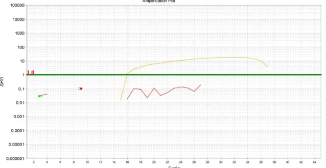
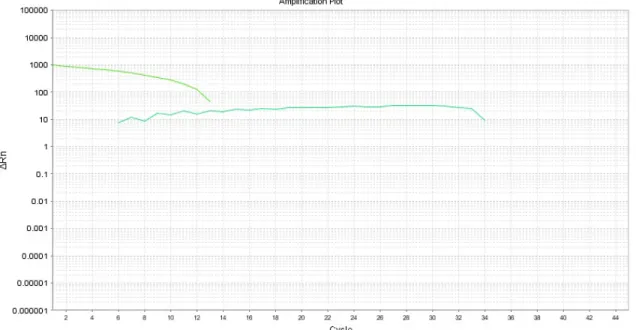
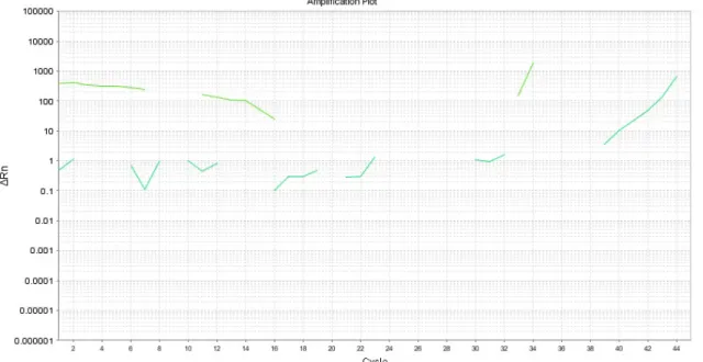
The resulting cDNA samples were amplified by quantitative real time polymerase chain reaction (qPCR) using primers specific for the housekeeping gene Glycderaldehyde-3-phosphate dehydrogenase (GAPDH), as well as the candidate genes PTH1R, RANKL, BMP2, BMP4, and BMP6. A StepOnePlus™ Real-Time PCR System (Applied Biosystems, Foster City, CA) was
employed for the qPCR under the following conditions: 10 min at 95°C activation/premelt, then 45 cycles of 10 sec at 95°C, 10 sec at 60°C and 20 sec at 72°C. The Melt Curve stage consisted of 15 sec at 95°C, then 1 min at 60°C, and 15 sec at 95°C. Primer sequences are displayed in Table 1:
Table 1: Primer sequences used in qPCR
Gene Forward Primer (5’-3’) Reverse Primer (5’-3’) Annealing (°C) Reference BMP-2 atggattcgtggtggaagtg gtggagttcagatgatcagc 58 7
BMP-4 agcagccaaactatgggcta tggttgagttgaggtggtca 60 20
BMP-6 cgtgaaggcaatgctcacct cctgtggcgtggtatgctgt 64 6 RANKL ctatttcagagcgcagatggat tatgagaacttgggattttgatgc 61 16 PTH1R ggggcttcacagtcttcg tggccagggtagctctga 60 Probefinder Roche
NM_000316.2 GAPDH acacccactcctccaccttt tgacaaagtggtcgttgagg 60
Mutational Analysis
activation/premelt, followed by 35 cycles of 30 sec at 95°C melt, 1 min at 60°C anneal, and 3 min of 72°C extension. PCR Products were then purified using ExoSaplt (USB, Cleveland OH), and sequenced at the University of North Carolina at Chapel Hill Genome Analysis Core facility. Primer sequences are listed in Table 2.
Table 2: PTH1R primer sequences used during mutational analysis PTH1R Primer Sequence (5' to 3')
PTHR1050MutF gaatgaccttgtggacagca PTHR1050MutR gaagcctcctaggtccctgt PTHR1543AND463Mut
F gccctgtccggactacatt
PTHR1543AND463Mut
R cagggagagcatcagggtaa
PTHR1ex3F ctctgcaccccctacca PTHR1ex3R cccaaacgaggcttcag PTHR1ex4F aaatcccaccttccctct PTHR1ex4R ccttcacctggctctgtat PTHR1ex5F cacctggcaagcacttta PTHR1ex5R aagagccaagaagcatgag PTHR1ex6F ttcatccttctgggtcacta PTHR1ex6R aggttgctggaggagtca PTHR1ex8F tgactcctccagcaacct PTHR1ex8R cgccgtaggtagggaca PTHR1ex10F ccactctacctcttcactttg PTHR1ex10R ggaatagggtcaggatcac PTHR1ex11F aagctgttagggcaccac PTHR1ex11R cttgaggctggactgagaa PTHR1ex12F ttctcagtccagcctcaag PTHR1ex12R gctctgtcactgcatctctg PTHR1ex14F tattagcacttagccaggaca PTHR1ex14R agggtggaagaatggagaa Results
revealed a possible frameshift mutation due to insertion at gene PTH1R, and further deep sequencing will be carried out to corroborate this finding (Figure 4b).
Figure 4 a: Patient A, PTH1R Exon 8F chromatogram
Figure 4 b: Patient B, PTH1R Exon 3 chromatogram
Candidate gene BMP-4, however, was found to be expressed in the control samples for both Patient A and Patient B as well as in the ankylosed tooth of Patient A; this candidate gene was not found to be expressed in the teeth affected by PFE of Patient B (Figures 6a-e). Additionally, candidate gene BMP-6 was found to be expressed in one of the teeth affected by PFE on
Patient B, #16, while it was not found to be expressed in any of the other samples (Figure 7a-e). Candidate gene PTH1R, was only found to be expressed in tooth #19 of Patient A (Figures 9a-e).
GAPDH
Figure 4 d: Patient A, #19 (ankylosed), GAPDH qPCR analysis.Ct=15.8
Figure 4 f: Patient B, #16 (PFE), GAPDH qPCR analysis. Ct=13.74
BMP-2
Figure 5 a: Patient A, #29 (control), BMP-2 qPCR analysis.
Figure 5 c: Patient B, #1 (control), BMP-2 qPCR analysis.
Figure 5 e: Patient B, #17 (PFE), BMP-2 qPCR analysis.
BMP-4
Figure 6 b: Patient A, #19 (ankylosed), BMP-4 qPCR analysis. Ct=18.78
Figure 6 d: Patient B, #16 (PFE), BMP-4 qPCR analysis.
BMP-6
Figure 7 a: Patient A, #29 (control), BMP-6 qPCR analysis.
Figure 7 c: Patient B, #1 (control), BMP-6 qPCR analysis.
Figure 7 e: Patient B, #17 (PFE), BMP-6 qPCR analysis.
RANKL
Figure 8 b: Patient A, #19 (ankylosed), RANKL qPCR analysis.
Figure 8 d: Patient B, #16 (PFE), RANKL qPCR analysis.
PTH1R
Figure 9 a: Patient A, #29 (control), PTH1R qPCR analysis.
Figure 9 c: Patient B, #1 (control), PTH1R qPCR analysis.
Table 3: qPCR results by Ct value GAPDH
(Ct) BMP-2 (Ct) BMP-4 (Ct) BMP-6 (Ct) RANKL (Ct) PTH1R (Ct)
Patient A,
#29
15.68 Negative 15.66 Negative Negative Negative
Patient A,
#19
15.8 Negative 18.78 Negative Negative 14.54
Patient B,
#1
15.54 Negative 15.14 Negative Negative Negative
Patient B,
#16
13.74 Negative Negative 14.17 Negative Negative
Patient B,
#17
15.39 Negative Negative Negative Negative Negative
Table 4: ΔCt values of positive qPCR results (ΔCt = Cttest – CtGAPDH)
Sample and gene
tested Positive Ct Value GAPDH value ΔCt value
Patient A, #29,
BMP-4 15.66 15.68 -0.02
Patient A, #19,
BMP-4 18.78 15.8 2.98
Patient A, #19,
PTH1R 14.54 15.8 1.26
Patient B, #1,
BMP-4 15.14 15.54 -0.4
Patient B, #16,
BMP-6 14.17 13.74 0.43
Candidate gene BMP-4 was found to be expressed at a higher rate with respect to GAPDH across all positive samples with exception of Patient A’s ankylosed tooth (#19). Candidate gene BMP-6 was expressed at a higher rate compared to GAPDH for Patient B’s affected upper PFE tooth, #16. Candidate gene PTH1R was expressed at a higher rate than the housekeeping gene in the ankylosed tooth of Patient A only at a higher rate than the
housekeeping gene.
Discussion.
Bone Morphogenetic Proteins are key players in many processes of bone metabolism such as formation, repair, regeneration, healing and callus formation. Although the
BMP-2 was found not to be expressed across any of the samples tested. We speculate that this result is due to the fact that no active bone formation was being induced. BMP-2 is osteoinductive and it has been shown to induce osteoblast differentiation in a variety of cell types 11 , whereas RANKL stimulates progenitor cells to differentiate into osteoclasts, leading to bone resorption 10. BMP-2 has been shown to down-regulate RANKL in vitro and it has been proposed to promote growth of alveolar bone around a dental unit 10. Just like RANKL,
expression of BMP-2 has been shown to go through a temporal peak in a rat animal model, in this case around day 9 post-natally in the dental follicle 17,19. The developmental stage of the teeth in this study and level of differentiation of the dental follicle concur with the finding that BMP-2 was not expressed in any of the samples tested.
PTH1R was another candidate gene with observed differences in expression across the samples tested. In bone, PTH1R is a receptor expressed on the cell surface. Activation of the receptor by binding to its substrate, PTH, leads to expression of RANKL, which leads to an increase in the differentiation of osteoclasts and bone resorption. Bone resorption and
remodeling have been observed to accompany root replacement resorption in teeth that suffer from dental ankylosis 3. In a patient that suffers from a tooth with dental ankylosis, such as Patient A, PTH1R would be expected to be expressed in order to see the changes in vertical height of the alveolar bone and replacement resorption that often coincides with dental ankylosis. PTH1R was found to be expressed in the putatively ankylosed tooth, we therefore speculate that this tooth does not suffer from PFE.
REFERENCES
1. BMP4 bone morphogenetic protein 4 [homo sapiens (human)] - gene - NCBI. http://www.ncbi.nlm.nih.gov/gene/652. Accessed 4/3/2016, 2016.
2. The role of RANKL (TRANCE/TNFSF11), a tumor necrosis factor family member, in skeletal development: Effects of gene knockout and transgenic rescue. - PubMed - NCBI.
http://www.ncbi.nlm.nih.gov/pubmed/12952207. Accessed 4/4/2016, 2016.
3. Andersson L, Blomlöf L, Lindskog S, Feiglin B, Hammarström L. Tooth ankylosis: Clinical, radiographic and histological assessments. Int J Oral Surg. 1984;13(5):423-431. doi: http://dx.doi.org.libproxy.lib.unc.edu/10.1016/S0300-9785(84)80069-1.
4. Biederman W. The incidence and etiology of tooth ankylosis. Am J Orthod. 1956;42(12):921-926. doi: http://dx.doi.org/10.1016/0002-9416(56)90193-2.
5. Botchkarev VA. Bone morphogenetic proteins and their antagonists in skin and hair follicle biology. J Invest Dermatol. 2003;120(1):36 <last_page> 47. doi:
10.1046/j.1523-1747.2003.12002.x.
6. Crews L, Adame A, Patrick C, et al. Increased BMP6 levels in the brains of alzheimer's disease patients and APP transgenic mice are accompanied by impaired neurogenesis. J Neurosci. 2010;30(37):12252-12262. doi: 10.1523/JNEUROSCI.1305-10.2010 [doi].
7. Kochanowska I, Chaberek S, Wojtowicz A, et al. Expression of genes for bone morphogenetic proteins BMP-2, BMP-4 and BMP-6 in various parts of the human skeleton. BMC
Musculoskelet Disord. 2007;8:128. doi: 1471-2474-8-128 [pii].
8. Kriangkrai R, Iseki S, Eto K, Chareonvit S. Dual odontogenic origins develop at the early stage of rat maxillary incisor development. Anat Embryol. 2006;211(2):101 <last_page> 108. doi: 10.1007/s00429-005-0068-7.
9. Kurol J. Infraocclusion of primary molars: An epidemiologic and familial study. Community Dent Oral Epidemiol. 1981;9(2):94-102.
10. Liu D, Yao S, Pan F, Wise GE. Chronology and regulation of gene expression of RANKL in the rat dental follicle. Eur J Oral Sci. 2005;113(5):404 <last_page> 409. doi: 10.1111/j.1600-0722.2005.00245.x.
12. Reddi AH. Interplay between bone morphogenetic proteins and cognate binding proteins in bone and cartilage development: Noggin, chordin and DAN. Arthritis Res. 2001;3(1):1-5. doi: 10.1186/ar133 [doi].
13. Rhoads SG, Hendricks HM, Frazier-Bowers SA. Establishing the diagnostic criteria for eruption disorders based on genetic and clinical data. Am J Orthod Dentofacial Orthop. 2013;144(2):194-202. doi: 10.1016/j.ajodo.2013.03.015 [doi].
14. Scanlon C, Marchesan J, Soehren S, Matsuo M, Kapila Y. Capturing the regenerative potential of periodontal ligament fibroblasts. J Stem Cells Regen Med. 2011;7(1):54-56. 15. Suri L, Gagari E, Vastardis H. Delayed tooth eruption: Pathogenesis, diagnosis, and
treatment. A literature review. American Journal of Orthodontics and Dentofacial Orthopedics. 2004;126(4):432 <last_page> 445. doi: 10.1016/j.ajodo.2003.10.031. 16. Wara-aswapati N, Surarit R, Chayasadom A, Boch JA, Pitiphat W. RANKL upregulation
associated with periodontitis and porphyromonas gingivalis. J Periodontol. 2007;78(6):1062-1069. doi: 10.1902/jop.2007.060398 [doi].
17. Wise GE, Ding D, Yao S. Regulation of secretion of osteoprotegerin in rat dental follicle cells. Eur J Oral Sci. 2004;112(5):439-444. doi: 10.1111/j.1600-0722.2004.00156.x [doi].
18. Wise GE, Lumpkin SJ, Huang H, Zhang Q. Osteoprotegerin and osteoclast differentiation factor in tooth eruption. J Dent Res. 2000;79(12):1937 <last_page> 1942. doi:
10.1177/00220345000790120301.
19. Wise GE, Yao S. Expression of tumour necrosis factor-alpha in the rat dental follicle. Arch Oral Biol. 2003;48(1):47 <last_page> 54. doi: 10.1016/S0003-9969(02)00153-X.
20. Wu Q, Yao J. BMP4, a new prognostic factor for glioma. World J Surg Oncol. 2013;11:264-7819-11-264. doi: 10.1186/1477-7819-11-264 [doi].