STUDIES TOWARD UNDERSTANDING THE BIOSYNTHESIS OF SACTIPEPTIDES AND THE CREATION OF PEPTIDE NATURAL PRODUCT LIBRARIES THROUGH MRNA
DISPLAY
Paul Michael Himes
A dissertation submitted to the faculty at the University of North Carolina at Chapel Hill in partial fulfillment of the requirements for the degree of Doctor of Philosophy in the Pharmaceutical Sciences in the Doctoral Program of the UNC Eshelman School of Pharmacy
(Division of Chemical Biology and Medicinal Chemistry).
Chapel Hill 2017
iii
ABSTRACT
Paul Michael Himes: Studies toward Understanding the Biosynthesis of Sactipeptides and the Creation of Peptide Natural Product Libraries through mRNA Display
(Under the direction of Albert A. Bowers)
Ribosomally-synthesized and post-translationally modified peptides (RiPPs) are a class of natural products that are an attractive starting point for new antibiotics due to their wide range of structural diversity and biological activities. The post-translational modifications imparted upon the peptide substrate are carried out by promiscuous RiPP enzymes. Sactipeptides are members of the RiPPs family that are made through radical-mediated cysteine sulfur to α-carbon coupling reactions. The resulting thioether linkages give rise to sactipeptides defined structures and concomitant biological activities. The research presented here focuses on the biochemical and structural characterization of CteB, a radical SAM enzyme that imparts a single sactionine bridge, the development of an E. coli heterologous expression system for sactipeptides and the combination of RiPPs and mRNA display for the production of modified peptide libraries.
We have biochemically and structurally characterized CteB, a radical SAM enzyme that imparts a sactionine bridge on its corresponding peptide substrate. A crystal structure was obtained at 2.04 Å and showed a RiPPs recognition element connected to a (β/α)6-TIM barrel fold, followed by an SPASM domain that houses two auxiliary [4Fe-4S] clusters, one of which contains a free coordination site for potential peptide ligation.
iv
substrate and radical SAM enzyme (AlbA) are expressed together and the modified sactipeptide is produced and isolated. This system was used to probe the substrate promiscuity of AlbA, and determine what changes it can tolerate. Additionally, an unnatural amino acid, O-Me-tyrosine, was able to be incorporated into the peptide substrate while also forming a thioether bridge at that position.
v
vi
ACKNOWLEDGMENTS
Graduate school has been a long and arduous task, and I would not have been able to complete it without the many people who helped me along the way. First, I would like to thank my wife, Avery, who has stood by my side through the good and bad times and tolerated the late days and nights in the lab, all I do is for ours and our son’s future. Second, I would like to thank my parents and my brother who were always supportive, encouraging, and believed in me even when my own belief waned. I love you all.
I would like to thank my mentor and advisor, Dr. Albert Bowers, for his tutelage and guidance throughout my thesis work and graduate school studies. You have taught me the many things that go into being a successful and productive scientist, including diligence and the passion for the work we do. I am very honored to have learned as much as I could have from you and I hope to take the lessons you have conveyed to me onward into the next chapter of my life.
vii
Lastly, thank you, Dr. Kevin Weeks, for bringing insightful discussion to each and every committee meeting and helping me grow as a critical thinker in those moments.
Thanks to all who have been members of the Bowers Laboratory while I was there: Nicoleta Economou, Scott Allen, Walter Wever, Rachel Bleich, Jonathan Bogart, Swapnil Ghodge, Sungwon Hwang, Kelly Bird, and Steven Fleming. Each one of you helped me become the scientist that I am today and I hope that I will leave having a positive impact on your lives and your work as scientists. Thank you to Dr. Bo Li and Dr. Eric Brustad, and their respective laboratories whose help with the LC-MS was always appreciated. Thank you to Dr. Rita Tamayo and her laboratory for their help with anaerobic procedures and continued use of the anaerobic chamber. A special thanks to Dr. Steve Almo and Dr. Tyler Grove at Albert Einstein School of Medicine, whose crystallography expertise was invaluable on the CteB paper and whose helpful discussions made working with them a true pleasure.
viii
PREFACE
Parts of this dissertation work were done in collaboration with other, talented scientists. Chapter 2 represents a submitted journal article for which I was one or two co-first authors. My contributions to the work focused on the cloning, expression, and reconstitution of activity of the sactionine synthase, CteB. I also characterized all activity of the enzyme by mass spectrometry while performing bioinformatic analysis of the enzyme and related proteins. These results are shown in Tables 2.1-2.2, Figures 2.2, and 2.5-2.6 and Appendix Figures A.1-A.3 and A.11-A.16. Dr. Tyler Grove performed all the crystallography on both the apo and peptide bound forms of CteB, structural comparisons to other known enzymes in the same class, as well as size exclusion chromatography. These experiments are shown in Figures 2.3, 2.4, 2.8 and Appendix Table A.1, Figures A.4-A.10. With the help of Dr. Bowers and Dr. Almo, the co-first authors designed experiments, communicated and divided the work, and then wrote the paper. This work has been submitted as a full article to JACS and has been through two rounds of review and resubmission:
Grove, T.L., Himes, P.M., Hwang, S., Yumerefendi, H., Bonanno, J.B., Kuhlman, B., Almo, S.C., Bowers, A.A. Structural Insights into Thioether Bond Formation in the Biosynthesis of Sactipeptides. JACS, 2017, resubmission
Dr. Tyler Grove and Dr. Steve Almo, co-first author on the paper and his PI respectively, have given permission for me to include this work in my dissertation.
ix
helpful discussion, experimental design and mass spectrometry analysis. Sungwon Hwang helped me clone the library of peptides into the system I had developed for the production of sactipeptides in E. coli. The paper was published previous to the writing of this thesis with the following citation:
Himes, P.M., Allen, S.E., Hwang, S., Bowers, A.A. Production of Sactipeptides in Escherichia coli: Probing the Substrate Promiscuity of Subtilosin A Biosynthesis. ACS Chem Biol. 2016, 11, 1737-1744
Permission to include the article in its entirety in this dissertation was retained from ACS Publications. Copyright (2016) American Chemical Society
Chapter 4 represents unpublished research that was designed and preformed primarily by myself with help from Steven Fleming also of the Bowers lab.
x
TABLE OF CONTENTS
LIST OF TABLES ... xiv
LIST OF FIGURES ...xv
LIST OF ABBREVIATIONS ... xvii
CHAPTER 1: INTRODUCTION ...1
1.1The Promiscuity of RiPPs Biosynthesis and the Potential for Natural Product Libraries ...1
1.2References ...8
CHAPTER 2: STRUCTURAL INSIGHTS INTO THIOETHER BOND FORMATION IN THE BIOSYNTHESIS OF SACTIPEPTIDES ...11
2.1 Introduction ...11
2.2 In vitro reconstitution of CteB: a sactionine synthase ...16
2.3 Crystal structure of CteB ...21
2.4 Homology and Comparison to other SPASM and Twitch Domains ...30
2.5 Contributions to Binding Affinity of CteA ...32
2.6 Summary and Discussion ...33
2.7 Experimental ...42
2.7.1 General Cloning and Molecular Biology Techniques ...42
xi
2.7.3 In vitro Reconstitution. Assays and Characterization
of Products ...48
2.7.4 Fluorescence Polarization Assays ...52
2.7.5 Oxidation of CteB with Glutathione ...54
2.7.6 Size Exclusion Chromatography...55
2.7.7 Crystallography Methods ...55
2.7.8 Structural Modeling of the CteB Catalytic Site ...59
2.8 References ...71
CHAPTER 3: PRODUCTION OF SACTIPEPTIDES IN ESCHERICHIA COLI: PROBING THE SUBSTRATE PROMISCUITY OF SUBTILOSIN A BIOSYNTHESIS ...76
3.1 Introduction ...76
3.2 Heterologous Production of Pre-subtilosin A ...79
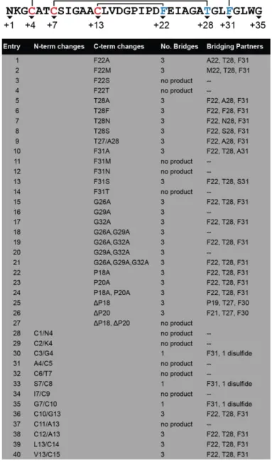
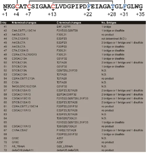
3.3 Design and Evaluation of SboA Mutants...81
3.4 Substrate Tolerance at Bridging Partners ...86
3.5 Substrate Tolerance at Unmodified Positions ...88
3.6 Substrate Tolerance for Cysteine Placement ...89
3.7 Unpublished Mutants Tested ...90
3.8 Incorporation of Unnatural Amino Acids into Sactipeptides ...91
3.9 Design, production, and analysis of potential sactipeptide MDM2-p53 inhibitor ...94
3.10 Other Sactipeptide Systems: Thurincin H and 4BD1 ...97
3.11 Summary and Discussion ...101
3.12 Experimental ...103
xii
3.12.2 Expression and purification of sactipeptides
from Duet System ...108
3.12.3 Production and Extraction of 4BD1-Natural Product (NP) ...110
3.12.4 NEM Modification of free cysteines ...111
3.12.5 Incorporation of unnatural amino acids into Duet System ...111
3.12.6 Characterization of sactipeptides by mass spectrometry ...112
3.12.7 Synthesis of p5315-29 and derivatives by Solid Phase Peptide Synthesis ...113
3.12 References ...115
CHAPTER 4: PROBING THE PROMISCUITY OF RIPPS ENZYMES USING MRNA DISPLAY TECHNOLOGIES AND NEXT-GENERATION SEQUENCING ...119
4.1 Introduction ...119
4.2 Design of Targeted RiPP libraries for mRNA Display ...123
4.3 Creation of Displayed-Peptide Libraries ...125
4.4 Selection Procedures ...127
4.4.1 PaaA Activity ...127
4.4.2 PaaA and TbtF Binding ...129
4.5 Summary and Discussion ...131
4.6 Experimental ...133
4.6.1 Design of DNA templates and mRNA transcription ...133
4.6.2 Synthesis of DNA-Puromycin Linker (P-link) ...136
4.6.3 Preparation of puromycin-fused mRNA libraries ...139
4.6.4 PURE System Translation of Displayed Peptide Libraries ...140
xiii
4.6.6 Protein Expression and purification of PaaA and TbtF ...144 4.7 References ...147 CHAPTER 5: CONCLUSION ...151 APPENDIX A: SUPPLEMENTARY FIGURES AND
TABLES FOR CHAPTER 2 ...154 APPENDIX B: SUPPLEMENTRAY FIGURES AND
xiv
LIST OF TABLES
Table 2.1 – Mass Spec. table for peptide modification assays treated with NEM ...19 Table 2.2 – Tandem MS/MS table for peptide modification assays
treated with NEM ...19 Table 2.3 – List of CteB(RRE) and CteA(M1-G9) hydrogen bond interactions ...29 Table 2.4 – Gene-blocks ordered from Integrated DNA Technologies (IDT)
codon optimized used in CteB study ...45 Table 2.5 – Plasmids, sites, and primers (ordered from Eton Bioscience, Inc.)
used in CteB study ...45 Table 3.1a – SboA mutants analyzed and sites of linkages
identified by MS-MS ...83 Table 3.1b – Unplublished SboA mutants analysis ...92 Table 3.3 – Plasmids, sites, and primers used in pETDuet-SboA-AlbA
heterologous system ...106 Table 4.1 – DNA sequences used to create template DNA
xv
LIST OF FIGURES
Figure 1.1 – Overview of ribosomally synthesized and post-translationally
modified peptides ...4
Figure 1.2 – Sactipeptide secondary structure ...4
Figure 1.3 – Overview of natural product peptide libraries ...6
Figure 2.1 – Introduction to sactipeptides ...13
Figure 2.2 – MS analysis of CteA modified by CteB ...20
Figure 2.3 – Structure of CteB ...27
Figure 2.4 – Leader peptide and binding to RRE of CteB ...29
Figure 2.5 – Comparison of Aux I and Aux II clusters ...31
Figure 2.6 – Fluorescence Polarization binding of CteA to CteB. ...33
Figure 2.7 – Proposed mechanisms of sactionine bridge formation. ...35
Figure 2.8 – Conservation of CteB homologs. ...41
Figure 3.1 – Sactipeptide biosynthesis...77
Figure 3.2 – Expression of sactipeptides in Escherichia coli ...80
Figure 3.3 – Confirming bridge formation in mutant sactipeptides...87
Figure 3.4 – Incorporation of unnatural amino acids (UAAs) into sactipeptides in E. coli. ...93
Figure 3.5 – Overview of proposed inhibitor SboA-2xMut ...95
Figure 3.6 – Fluorescence Polarization assay of MDM2-p53 interaction ...96
Figure 3.8 – Surface Plasmon Resonance binding assays of MDM2-p53 interaction ...98
Figure 3.9 – Isolation of 4BD1 Natrual Product (4BD1-NP) ...99
xvi
Figure 4.1 – Overview of mRNA display combined with RiPPs ...123
Figure 4.2 – Covalent attachment of DNA-Puromycin linker to mRNA ...126
Figure 4.3 – Translation and purification of “display” peptide ...127
Figure 4.4 – Selection for PaaA activity ...129
Figure 4.5 – Selection for binding to RiPPs enzymes ...130
xvii
LIST OF ABBREVIATIONS
5’-dA• 5’-deoxyadensoyl radical
Å angstrom (1Å = 1x10⁻¹⁰ meters)
amu atomic mass unit
anSME anaerobic sulfatase maturating enzyme
ATP adenosine 5’-triphosphate
Aux auxiliary cluster
bp(s) base pair(s)
BME 2-mercaptoethanol
Cα alpha-carbon atoms
cte thermocellin
DIPEA N,N-diisopropylethyl amine
DCM dichloromethane
DMF dimethylformamide
DMSO dimethyl sulfoxide
DT dithionite
DTT dithiothreitol
EIC extracted ion chromatogram
ESI electrospray ionization
FA formic acid
FP fluorescence polarization assay
GSH reduced glutathione
xviii
HATU 2-(7-Aza-1H-benzotriazole-1-yl)-1,1,3,3-tetramethyluronium hexafluorophosphate
HCl hydrochloric acid
HEPES N-(2-Hydroxyethyl)piperazine-N′-(2-ethanesulfonic acid) IPTG isopropyl β-D-1-thiogalactopyranoside
Kd dissociation constant
kDa kilodalton
LC-MS liquid chromatography–mass spectrometry LIC ligation independent cloning
MCS1 multiple cloning site 1 MCS2 multiple cloning site 2
MBP maltose binding protein
min minute(s)
MS/MS yandem mass spectrometry
NEM N-ethylmalemide
NGS next-generation sequencing
O-Me-Tyr O-methyl tyrosine
ppm parts per million
P-linker DNA-Puromycin linker
RBS ribosomal binding site
RiPPs ribosomally synthesized and post-translationally modified peptides RMSD root mean square deviation
RRE RiPPs recognition element
xix
rSAMs radical SAM enzymes
sactionine cysteine-sulfur to alpha-carbon thioether
sactipeptides sulfur-to-alpha carbon thioether cross-linked peptides
SAM S-adenosyl-L-methionine
SCIFF six cysteines in forty-five residues
SEC size exclusion chromatography
Skf sporulation killing factor
SPASM subtilosin A/PQQ/anaerobic sulfates maturing SPPS solid phase peptide synthesis
SPR surface plasmon resonance
SSN sequence similarity network
TAMRA 5-(and 6)-carboxytetramethylrhodamine TCEP tris(2-carboxyethyl)phosphine
TIM triose phosphate isomerase
TEV tobacco etch virus
TFA trifluoroacetic acid
Tris tris(hydroxymethyl)aminomethane UAA(s) unnatural amino acid(s)
UV ultra-violet
wHTH winged helix-turn-helix
WT wild-type
1
CHAPTER 1
INTRODUCTION
1.1 The Promiscuity of RiPPs Biosynthesis and the Potential for Natural Product Libraries
Natural products, for more than a century, have advanced the understanding of biology and have been at the forefront of the development of novel medicines for the world’s most
2
Ribosomally-synthesized and post-translationally modified peptides (RiPPs) are a class of natural products defined by their unique biosynthetic pathways as well as their modifying enzymes (Figure 1.1).1 Unlike polyketide synthases (PKS) or non-ribosomal peptide synthases (NRPS), both of which use separate active module-like assembly to create their natural products, RiPPs use the ribosome to create the precursor peptide.10,11 This precursor peptide can house up to three domains, termed the leader, core, and follower peptides respectively. The leader peptide is used by the RiPPs modifying enzymes to recognize the peptide substrate, while the core peptide is where the modifications are imparted by the enzyme. The follower, if the peptide has one, can play the same role as the leader peptide in terms of recognition. After the modifications are imparted, the leader and/or follower sequence is removed by a peptidase and the modified core is released to give the biologically active product.1,12 RiPPs modifying enzymes impart extensive post-translational/co-translational modifications that give these peptides structures that are not directly accessible by natural ribosomal synthesis or by the modular synthesis related to PKS or NRPS. These modifications, which are typically conformationally constraining, allow a) better target recognition and higher binding affinity, b) metabolic and chemical stability, and c) a change in chemical functionality by altering the side chains of the canonical amino acids.1Due to their structural diversity, wide range of biological activities, and conformational constraining structures, RiPPs are an attractive starting point for novel therapeutics for anti-cancer and antibiotic therapies.
A member of the RiPPs family of natural products is a class of diverse modified peptides known as sactipeptides. Sactipeptides are characterized by their unique thioether bridges that form intramolecular bridges between the sulfurs of cysteine residues and the unreactive α-carbon
3
partner’s amino acid side chain. Therefore, sactipeptides tend to have highly defined regions of
secondary structure due to the distribution and number of thioether or sactionine bridges as well as the stereochemistry at the α,α-disubstituted bridging partner residues.13-20 Subtilosin A, a founding member of sactipeptides, adopts a 310 helix within its structure while other sactipeptide can adopt α-helical structures. The amphipathic helicity of these regions within sactipeptides is
thought to grant subtilosin A and others narrow spectrum activity through the ability to interact and disrupt bacterial cell walls resulting in cell death through membrane disruption 16,21,22 This activity, as well as their added stability to heat and proteases due to their thioether bridges, make sactipeptide an attractive biological scaffold for the development of novel therapeutics and chemical probes. Previously in our lab, a system was developed to predict and estimate these highly defined secondary structures using analyses generated from replica exchange molecular dynamics (REMD) trajectories using AMBER 14 and various constraints and implicit solvent conditions.23-25 In REMD, multiple molecular dynamic simulations are run simultaneously at varying temperatures, and these temperatures are exchanged between replicas at set intervals over the course of the entire simulation. This exchange of temperatures can allow the simulation to overcome energy wells and barriers that cannot be overcome at lower temperatures. After the simulation, the likelihood of each residue adopting a particular secondary structure over the course of the simulation will be identified by hydrogen bonding patterns and angles of that residue. We did this simulation for subtilosin A and found it agreed well with the NMR structure reported by Vederas and co-workers in 2004 (Figure 1.2).19 Using these simulations, we found that continuous stereochemistry (all D or all L) is required to propagate helicity (either α or 310)
4
can be used to help design and predict highly defined secondary structures that could be used as biologically active scaffolds for the grafting of known epitopes for desired function and biologically activity (i.e. inhibition, binding, cell-death, etc.) if it could be paired with a robust expression system.
Figure 1.1. Overview of ribosomally synthesized and post-translationally modified peptides (RiPPs).
5
RiPPs machinery, including the radical SAM enzymes in sactipeptide biosynthesis, that impart the modifications necessary for biological activity have only recently been isolated and studied in a way that sheds light on how the RiPPs modifying enzyme recognizes and imparts the aforementioned modifications. Through crystallography of these enzymes as well as mutational analysis (in vitro and in vivo), it has been determined there is a specialized recognition domain termed the RiPPs recognition element (RRE) that allows the RiPPs enzymes to recognize and coordinate to their intended peptide substrate.26 This RRE recognizes sequences at either the leader or follower sequence of the precursor peptide. This gives RiPPs one of their most impressive abilities, their promiscuity within their own biosynthesis. It has been shown that as long as the recognition element within the leader peptide is intact, the core peptide can be mutated and the RiPPs modifying enzymes can still impart their modification on this “new” core peptide.27-32 While not every change is allowed, this system houses much more flexibility in the identity of its substrates than most enzymes could tolerate. This gives RiPPs the advantage of creating a wide range of distinct, yet similar peptides that can be tested and altered for specific activities or properties.
6
differing activities (Figure 1.3) than its natural counterpart giving promising new leads for potential therapeutics against cancer and bacterial infections.
Figure 1.3. Overview of natural product peptide libraries. a) By using the same leader peptide and the innate promiscuity of RiPPs biosynthesis, a natural compound library can be created. b) Proposed workflow for the modification and selection of RiPPs natural products by mRNA display.
New antibiotics are desperately needed due to the rise of antibiotic resistance and the severe lack of new antibiotics. A recent study by the PEW Charitable Trust reported that there has not been a new class of antibiotics registered since 1984.42 A potential work-around would be to use RiPPs as a starting point and using the power of mRNA display, test on the order of trillions molecules for activity against bacterial species. This can give rise to novel therapeutics in a high-throughput manner.
7
8
1.2 REFERENCES
1. Arnison, P. G.; Bibb, M. J.; Bierbaum, G.; Bowers, A. A.; Bugni, T. S.; Bulaj, G.; Camarero, J. A.; Campopiano, D. J.; Challis, G. L.; Clardy, J.; Cotter, P. D.; Craik, D. J.; Dawson, M.; Dittmann, E.; Donadio, S.; Dorrestein, P. C.; Entian, K.-D.; Fischbach, M. A.; Garavelli, J. S.; Göransson, U.; Gruber, C. W.; Haft, D. H.; Hemscheidt, T. K.; Hertweck, C.; Hill, C.; Horswill, A. R.; Jaspars, M.; Kelly, W. L.; Klinman, J. P.; Kuipers, O. P.; Link, A. J.; Liu, W.; Marahiel, M. A.; Mitchell, D. A.; Moll, G. N.; Moore, B. S.; Muller, R.; Nair, S. K.; Nes, I. F.; Norris, G. E.; Olivera, B. M.; Onaka, H.; Patchett, M. L.; Piel, J.; Reaney, M. J. T.; Rebuffat, S.; Ross, R. P.; Sahl, H.-G.; Schmidt, E. W.; Selsted, M. E.; Severinov, K.; Shen, B.; Sivonen, K.; Smith, L.; Stein, T.; Süssmuth, R. D.; Tagg, J. R.; Tang, G.-L.; Truman, A. W.; Vederas, J. C.; Walsh, C. T.; Walton, J. D.; Wenzel, S. C.; Willey, J. M.; van der Donk, W. A. Nat. Prod. Rep. 2012, 30, 108
2. Sengupta, S.; Chattopadhyay, M.; Grossart, H. Front. Microbiol. 2013, 4, 1 3. Fleming, A. Br. J. Exp. Pathol. 1929, 10, 226
4. Brown D.; Lister, T.; May-Dracka, T. Bioorg. Med. Chem. Lett. 2014, 24, 413 5. Silver, L. Future Microbiol. 2015, 10, 1711
6. Newman, D.J.; Cragg, G.M. J. Nat. Prod. 2016, 79, 629 7. Maier, M.E. Org. Biomol. Chem. 2015, 13, 5302
8. Nicolaou, K.C.; Sorensen, E.J.; Winssinger, N. J. Chem. Educ. 1998, 75, 1225
9. Forsberg, K.; Patel, S.; Gibson, M.; Lauber, C.; Knight, R.; Fierer, N.; Dantas, G. Nature. 2014, 509, 612
10.Dutta, S.; Whicher, J.; Hansen, D.; Hale, W.; Chemler, J.; Congdon, G.; Narayan, A.; Håkansson, K.; Sherman, D.; Smith, J.; Skiniotis, G. Nature. 2014, 510, 512
11.Reimer, J.; Aloise, M.; Harrison, P.; Schmeing, T. Nature. 2016, 529, 239 12.Dunbar, K. L.; Mitchell, D. A. ACS Chem. Biol. 2013, 8, 473
13.Jarrett, J. T. J. Biol. Chem. 2015, 290, 3972
14.Sit, C. S.; van Belkum, M. J.; McKay, R. T.; Worobo, R. W.; Vederas, J. C. Angew. Chem. Int. Ed. 2011, 50, 8718
9
16.Rea, M. C.; Sit, C. S.; Clayton, E.; O’Connor, P. M.; Whittal, R. M.; Zheng, J.; Vederas, J. C.; Ross, R. P.; Hill, C. Proc. Natl. Acad. Sci. U. S. A. 2010, 107, 9352
17.Sit, C. S.; McKay, R. T.; Hill, C.; Ross, R. P.;Vederas, J. C. J. Am. Chem. Soc. 2011, 133, 7680
18.Babasaki, K.; Takao, T.; Shimonishi, Y.; Kurahashi, K. J. Biochem. 1985, 98, 585
19.Kawulka, K. E.; Sprules, T.; Diaper, C. M.; Whittal, R. M.; McKay, R. T.; Mercier, P.; Zuber, P.;Vederas, J. C. Biochem. 2004, 43, 3385
20.Liu, W.T.; Yang, Y.L.; Xu, Y.; Lamsa, A.; Haste, N. M.; Yang, J.Y.; Ng, J.; Gonzalez, D.; Ellermeier, C. D.; Straight, P. D.; Pevzner, P. A.; Pogliano, J.; Nizet, V.; Pogliano, K.;Dorrestein, P. C. Proc. Natl. Acad. Sci. U. S. A. 2010, 107, 16286
21.Thennarasu, S.; Lee, D.K.; Poon, A.; Kawulka, K. E.; Vederas, J. C.;Ramamoorthy, A. Chem. Phys. Lipids. 2005, 137, 38
22.Wang, G.; Feng, G.; Snyder, A. B.; Manns, D. C.; Churey, J. J.;Worobo, R. W. FEMS Microbiol. Lett. 2014, 357, 69
23.Case, D. A.; Berryman, J. T.; Betz, R. M.; Cerutti, D. S.; Cheatham, T. E., III; Darden, T. A.; Duke, R. E.; Giese, T. J.; Gohlke, H.; Goetz, A. W.; Homeyer, N.; Izadi, S.; Janowski, P.; Kaus, J.; Kovalenko, A.; Lee, T. S.; LeGrand, S.; Li, P.; Luchko, T.; Luo, R.; Madej, B.; Merz, K. M.; Monard, G.; Needham, P.; Nguyen, H.; Nguyen, H. T.; Omelyan, I.; Onufriev, A.; Roe, D. R.; Roitberg, A.; Salomon-Ferrer, R.; Simmerling, C. L.; Smith, W.; Swails, J.; Walker, R. C.; Wang, J.; Wolf, R. M.; Wu, X.; York, D. M.; Kollman, P. A. AMBER 2015, 2015.
24.(a) Hornak, V.; Abel, R.; Okur, A.; Strockbine, B.; Roitberg, A.; Simmerling, C. Proteins. 2006, 65, 712 (b) Wickstrom, L.; Okur, A.; Simmerling, C. Biophys. J. 2009, 97, 853
25.Ryckaert, J.-P.; Ciccotti, G.; Berendsen, H. J. C. J. Comput. Phys. 1977, 23, 327 26.Burkhart, B.J.; Hudson, G.A.; Dunbar, K.L.; Mitchell, D.A. Nat. Chem. Biol. 2015, 11,
564
27.Donia, M. S.; Hathaway, B. J.; Sudek, S.; Haygood, M. G.; Rosovitz, M. J.; Ravel, J.; Schmidt, E. W. Nat. Chem. Biol. 2006, 2, 729
28.Himes, P. M.; Allen, S. E.; Hwang, S.; Bowers, A. A. ACS Chem. Biol. 2016, 11, 1737 29.Melby, J. O.; Dunbar, K. L.; Trinh, N. Q.; Mitchell, D. A. J. Am. Chem. Soc. 2012, 134,
10
30.Melby, J. O.; Nard, N. J.; Mitchell, D. A. Curr. Opin. Chem. Biol. 2011, 15, 369
31.Tianero, M. D.; Donia, M. S.; Young, T. S.; Schultz, P. G.; Schmidt, E. W. J. Am. Chem. Soc. 2012, 134, 418
32.Velasquez, J. E.; van der Donk, W. A. Curr. Opin. Chem. Biol. 2011, 15,11 33.Bashiruddin, N.K.; Suga, H.; Curr. Opin. Chem. Biol. 2015, 24, 131
34.Takahashi, T.T.; Austin, R.J.; Roberts, R.W. Trends Biochem. Sci. 2003, 28, 159
35.Barendt, P.A.; Ng, D.T.W.; McQuade, C.N.; Sarkar, C.A.; ACS Comb. Sci. 2013, 15, 77 36.Guillen Schlippe, Y.V.; Hartman, M.C.T.; Josephson, K.; Szostak, J.W.; J. Am. Chem.
Soc. 2012, 134, 10469
37.Horiya, S.; Bailey, J.K.; Temme, J.S.; Guillen Schlippe, Y.V.; Krauss, I.J. J. Am. Chem. Soc. 2014, 136, 5407
38.Millward, S.W.; Takahasi, T.T.; Roberts, R.W. J. Am. Chem. Soc. 2005, 127, 14142 39.Hayashi, Y.; Morimoto, J.; Suga, H. ACS Chem. Biol. 2012, 7, 607
40.Jalai-Yazdi, F.; Lai, L.H.; Takahasi, T.T.; Roberts, R.W. Angew. Chem. Int. Ed. 2016, 55, 4007
41.Maini, R.; Umemoto, S.; Suga, H. Curr. Opin. Chem. Biol. 2016, 34, 44 42.The PEW charitable trust.
http://www.pewtrusts.org/~/media/assets/2016/05/ascientificroadmapforantibioticdiscove ry.pdf (Accessed June 6, 2017)
43.Ghodge, S.V.; Biernat, K.A.; Bassett, S.J.; Redinbo, M.R.; Bowers, A.A. J. Am. Chem. Soc. 2016, 138, 5487
44.Hudson, G.A; Zhang, Z.; Tietz, J.I.; Mitchell, D.A.; van der Donk, W.A. J. Am. Chem. Soc. 2015, 137, 16012
45.Wever, W.J.; Bogart, J.W.; Bowers, A.A. J. Am. Chem. Soc. 2016, 138, 13461
11
CHAPTER 2
STRUCTURAL INSIGHTS INTO THIOETHER BOND FORMATION IN THE
BIOSYNTHESIS OF SACTIPEPTIDES
2.1 Introduction
Enzymes that belong to the S-adenosylmethionine (SAM) radical superfamily are capable of catalyzing a wide array of radical mediated reactions utilizing the 5’-deoxyadenosyl radical (5’-dAdo•) as a radical intermediate. These reactions include modification to not only DNA and
RNA, but also complex peptide modifications such as the formation of carbon-carbon bonds and quaternary carbon-sulfur bonds.1,2 These radical SAM (RS) enzymes (rSAMs) contain conserved domains and motifs that unite the family. rSAMs bind several [4Fe-4S] clusters that carry out the chemistry of the enzyme. One [4Fe-4S] cluster is bound by a CX3CXφC motif where φ is an
aromatic residue. This motif is present in a conserved partial ()6 triose-phosphate isomerase
12
regenerated by a chemical reductant such as dithionite, or, in some cases, by the enzymatic NADPH/flavodoxin-flavodoxin reductase system.1 Sequence homology suggests that many rSAMs contain a unique C-terminal extension, termed a SPASM domain in addition to a conserved RS domain.2,3,6-9 The SPASM domain (named for the biochemically characterized members, AlbA, PqqE, anSME, and MftC which are involved in subtilosin A, pyrroquinoline quinone, anaerobic sulfatase, and mycofactocin maturation respectively) is involved in the coordination of auxiliary [4Fe-4S] clusters by cysteine residues, which are thought to expand and enhance the range of chemistries accessible by the RS domain.2,7,8 All known SPASM domain-containing enzymes catalyze overall oxidation of their respective substrates by two electrons, yet there appears to be significant sequence and structural variation among SPASM domains, namely in the state and arrangement of cysteine residues that coordinate to the auxiliary iron-sulfur clusters.7,8 The crystal structure of anSME was solved in 2013 and showed that the SPASM domain housed two additional, fully ligated auxiliary [4Fe-4S] clusters that were important for the enzymatic reaction of anSME. anSME co-translationally catalyzes the formal 2-electron oxidation of a cysteine residue found in the active site of its sulfatase substrate to yield a formyl glycine residue (Figure 2.1b).9-11 To date this is the only modifying enzyme belonging to the rSAM superfamily, with a full SPASM domain, whose structure has been solved.
13
promiscuous enzymes that are readily exploited for combinatorial biosynthesis as well as other applications.16-20 The leader peptide is responsible for binding to the post-translational modifying enzymes while the chemical modification is done on the core peptide. Recently it has been shown that leader peptide interacts with a conserved motif present in RiPPs enzymes known as a RiPP precursor peptide recognition element (RRE). These RREs have been found in a wide variety of RiPPs enzymes such as LynD, a cyclodehyratase involved in cyanobactin biosynthesis, and NisB, a dehydratase involved in the biosynthesis of the lantibiotic nisin.21 These domains are based on the structure of PqqD which associates with the rSAM PqqE to allow the formation of the carbon-carbon bond between a glutamic acid and tyrosine residues.
14
Not only can rSAMs catalyze the formation of carbon-carbon bonds such as those found in PQQ biosynthesis by PqqE and the recently characterized StrB from streptide biosynthesis 22-25
, they can also form sulfur to α-carbon bonds to form thioether (sactionine) bridges, a
characteristic of the subclass of RiPPs known as sactipeptides. Sulfur-to-α-carbon-antibiotics
(sactibiotics), also known as sactipeptides, are in the family of sulfur bridged bacteriocins, but unlike their lantipeptide cousins, sactipeptides are formed by making sulfur to α-carbon bonds
(sactionine bridges) through the use of rSAMs, termed sactionine synthases.26-28 These sactionine bridges impart conformational strain on the peptide, giving rise to unique secondary structures forming natural “stapled” helices. Sactipeptide may contain one or more of these sactionine
15
modification to sactionine linkages.43 These efforts are limited to substrates that fortuitously undergo modification by the native enzyme, but engineering and rational design of sactipeptide libraries will require better understanding of both sactisynthase structure and mechanism of thioether bond formation. Not only are these rSAMs predicted to hold SPASM domains (only a few of which has been structurally characterized)2,3,6-9, but they are also RiPPs enzymes and predicated to hold RRE domains attached to, not separate, of the active site of the enzyme. That makes any structural information on these thioether bond forming sactionine synthases extremely important. A crystal structure of a sactionine synthase can shed light on a) how the SPASM domain of a peptide modifying enzyme compares to known SPASM domains, b) how the RRE domain interacts with the peptide substrate, c) insights into the mechanism of sactionine bond formation, and d) what this can tell us about other known sactionine synthases.
Recent bioinformatic efforts predicted a number of sactipeptide clusters in a wide array of bacterial genomes, including several from thermophiles.7,8,44 We anticipated that sactionine synthases from thermophilic bacteria might have the desired stability for efficient heterologous expression and crystallization. In particular, the sactionine synthase from Clostridium thermocellum ATCC 27405, Cthe_0906, here referred to as CteB, looked to be a member of the newly defined family of sactipeptides being called SCIFF (or six cysteines in forty-five residues) peptides.7 CteB is co-located with the short peptide Cthe_0907, here referred to as CteA, in the C. thermocellum ATCC 27405 genome and is therefore predicted to chemically modify CteA to form its SCIFF peptide known as thermocellin (cte). Although no native natural products belonging to this family have been isolated to date, Bandarian and co-workers reconstituted
16
enzymatic reconstitution and potential crystallographic investigation aimed at understanding the mechanism of sactionine synthases. In this study, we reconstitute the activity of the sactisynthase, CteB and through a combination of chemical modification and tandem mass spectrometry, we demonstrate that CteB installs a single sactionine thioether linkage between Cys32 and Thr37 of its precursor peptide, CteA, and that the remaining five cysteines in CteA go unmodified. We also report two structures of CteB: a 2.70-Å-resolution structure of CteB with SAM bound and a 2.04-Å-resolution structure of CteB with both SAM and the leader peptide of CteA bound. These structures define all three [4Fe-4S] clusters predicted by bioinformatics, one of which, auxiliary cluster I (Aux I), displays a novel open coordination site on one of its constituent iron ions. These structures, together with substrate binding assays and a computational model based on the crystal structure, provide insights into the mechanism of thioether bond formation for CteA and other members of the sactisynthase family.
2.2 In vitro reconstitution of CteB: a sactionine synthase
17
Using Fluhe et al. as a basis37, we ran a series of anaerobic assays to determine a) whether CteB could reductively cleave SAM to generate 5’-deoxyadenosine or 5’-dA and b) if/how it modified CteA. When there is no peptide substrate for a sactisynthase, the 5’-dA• abstracts a hydrogen from nearby solvent, forming 5’-dA. The production of 5’-dA is a clear
indication of proper folding, redox state, and activity of the enzyme. When CteB was incubated in the presence of SAM and the strong non-physiological reductant sodium dithionite, we observed a distinctive mass (252.1108) corresponding to 5’-dA (within <10 ppm error) in LC-MS traces of the assay supernatants (see Appendix Figure A.1). This product mass was not observed in control reactions without CteB or SAM, suggesting that reconstituted CteB carries out this characteristic reductive cleavage of radical SAM enzymes.
18
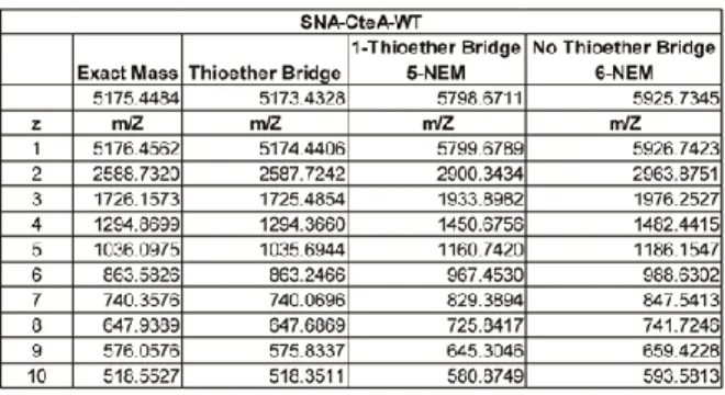
m/z values for the various charge states corresponded to the mass of the peptide plus six molecules of NEM (Figure 2.2c, black trace and Table 2.1). In contrast, CteA that was modified by CteB before being quenched with NEM exhibited masses corresponding to m/z for peptide with five alkylated cysteine residues and a loss of two hydrogens (Figure 2.2c, red trace and Table 2.1), confirming that a single thioether had been installed by CteB under these conditions.
In order to identify the location of the single sactionine thioether linkage, we used tandem mass spectrometry. CteA that was modified with CteB and treated with NEM (CteA-mod-5NEM) was fragmented by collision induced dissociation (CID). Based on the pattern of b- and y- ions, the newly formed thioether bridge was found to reside between residues Cys32 and Thr37 of CteA (Figure 2.2d). We found that the b- and y- ions for fragments containing Cys32 lack one NEM group and two hydrogens corresponding to the formation of the sactionine thioether linkage at this position. A full table of observed masses and the residues to which they correspond is provided in Table 2.2.
19
the assignment of the sactionine linkage, the Cys32Ala mutant of CteA was prepared via Gibson Assembly mutagenesis, and purified similar to wild type. Assays with CteA-C32A in presence of CteB and SAM yielded only the unmodified precursor peptide, consistent with thioether formation at this position (see Appendix Figure A.3). Peptide products with only one sactionine bridge were observed regardless of whether CteB was limited or used in large excess. It cannot be completely ruled out that multiple thioether bridges may be formed in the cellular environment of the native producer with the native reductant. Whether this is the active form of CteA in vivo remains to be determined.
Table 2.1. Mass Spec. table for peptide modification assays treated with NEM
20
21
2.3 Crystal structure of CteB
We solved the crystal structures of CteB, from anomalous iron edge datasets, in two different states: CteB bound to SAM and no substrate was solved at 2.7 Å resolution while CteB bound to both SAM and a 21-residue N-terminal fragment of CteA (M1-C21) at 2.04 Å resolution. We attempted co-crystallization with the full-length CteA precursor peptide, but were unable to obtain diffraction quality crystals. As of the time of writing, there have been no structures reported for any RiPP enzyme and its full-length precursor peptide substrate bound, presumably due to the dynamic nature of the interactions between the core peptide and the RiPP enzyme.
22
maturases (Appendix A.15), which may form a disulfide bond with the adjacent Cys336 from a symmetry mate (see Appendix Figure A.8). In addition, this entire region (CteB residues 330-341) is disordered in the CteB+SAM structure. Interestingly, the crystallization solution contained about 500 μM dithiothretol (DTT) that carried over with the CteB added to the
23
the non-reducing SDS-PAGE gel. With these results, we concluded that this disulfide is most likely an artifact of crystallization. Due to the weak electron density and the evidence that in solution, the enzyme behaves as a monomer, we have decided not to model this disulfide bond forming a dimer (see Appendix Figure A.8).
24
the substrate and enzyme, replacing a disulfide loss upon mutation. Multiple, subsequent mutations within the CteA substrate would be required to answer this question. Further experiments are underway to elucidate the role Cys336 plays in complex formation and activity of CteB.
The structure of CteB exhibits three discernable domains (Figure 2.3a and b): (1) a partial (β/α)6 triose phosphate isomerase (TIM) barrel (residues 95-319) containing one [4Fe-4S]
cluster (canonical radical SAM domain) in green, which is flanked by (2) an N-terminal winged helix-turn-helix (wHTH) motif (residues 1-71) in purple and (3) a C-terminal extension (residues 338-450), which chelates two additional [4Fe-4S] clusters in orange. These are discussed individually below.
The central portion of the CteB structure exhibits the characteristic )6-TIM barrel
(residues 95-319), common to nearly all other members of the radical SAM superfamily. This barrel is also known as the AdoMet or radical SAM (RS) domain for the fact is holds the [4Fe-4S] cluster that binds and reductively cleaves SAM. The [4Fe-[4Fe-4S] cluster motif (CX3CXφC) is found within the RS domain, in the loop between α1 helix and β1 loop (residues 100-125). This cluster is ligated by three cysteines (residues 104, 108, and 111), leaving one site open to chelate the α-aminonitrogen and α-carboxyl oxygen of the SAM co-factor.3,46
25
Interestingly there are two new SAM binding pocket interaction found in the CteB structure: Arg253 and Thr255 in the β5 strand form hydrogen bonds to N3 of the adenine base within
SAM. These residues reside in a highly conserved RGT motif found in thermophilic sactisynthases. In summary, a total of eight residues make side chain or backbone polar contacts with SAM (see Appendix Figure A.9). Presumably these numerous interactions and motifs correctly position and orient SAM for radical-based hydrogen abstraction from its substrate in a very specific manner.
Through a partially ordered loop, the RS domain is connected to the C-terminal SPASM domain which spans the residues 338-450. It holds the conserved seven-cysteine motif found in SPASM domains, CX9-15GX4CXnCX2CX5CX3CXnC and coordinates two additional [4Fe-4S] clusters known as auxiliary clusters. The CteB SPASM domain exhibits structural homology (see Appendix Figure A.9, R.M.S.D. of 2.3 Å over 113 Cα) to the SPASM domain from anSME
26
ansME, where both Aux I and Aux II are fully ligated, Aux I of CteB is left with an open
coordination site. The fourth coordinating ligand, present in anSME but absent in CteB, to Aux I, besides leaving an open coordination site, results in the positioning of the [4Fe-4S] cluster AuxI closer to the RS cluster in CteB. The RS cluster resides 14.4 Å from the open coordination site of Aux I (Figure 2.3c), which is ~ 2.5 Å closer than seen in the structure of anSME (16.9 Å). The distance between Aux I and Aux II in CteB is 11.6 Å, which is slightly compressed compared to that seen in the SPASM domain of anSME (12.9 Å). These differences indicate that the overall arrangement and separation of all [4Fe-4S] clusters within these SPASM family proteins is likely to support the different chemistries that are catalyzed by these different proteins.
enzyme-27
substrate interactions that would occur during the catalytic cycle, involving one of the six cysteines from CteA and Aux I.
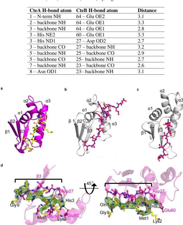
Figure 2.3. Structure of CteB. a) Overall structure of CteB. The β6/α6 core of the RS domain (green) contains one [4Fe-4S] cluster that coordinates one molecule of SAM. The C-terminal SPASM domain (orange) contains the [4Fe-4S] clusters Aux I and Aux II and comprises of residues 344 - 432. The N-terminal RRE domain (purple) of CteB provides the binding specificity for the peptide substrate leader sequence of CteA (yellow, stick representation). b) Topology figure of CteB with matching color scheme as a). c) Zoom of [4Fe-4S] clusters present in CteB along with their distances from one another. The distance from RS to Aux I is 14.4 Å while the distance from Aux I to Aux II is 11.6Å. RS, radical SAM cluster, Aux I, and, Aux II d) Omit map (2Fo-Fc) contoured to 1.5 σ of Gly20 and Cys21 from CteA-M1-C21 substrate bound to Aux I. The distance between the Fe and Sϒ of Cys21 is 2.7 Å.
28
represents just the fourth reported structure of a leader-bound RRE structure (Figure 2.4a and d).48,49 These reported structures all exhibit a common pattern in the conserved wHTH domain architecture and β-strand conformation when bound to peptide substrate (Figure 2.4a-c). While
the RREs vary greatly in sequence, they are predicted based upon secondary structure. Despite sharing only 13% sequence identity to the RRE domains of LynD and NisB, the RRE domain of CteB exhibits a relatively small overall R.M.S.D at 2.16 Å and 3.05 Å, respectively over 71 Cα. The RRE domain provides one of the primary structural motifs for leader peptide recognition. The three-stranded β-sheet, or wing, of the RRE interacts with the backbone of the N-terminus of the CteA fragment in the co-crystal structure in a manner similar to LynD and NisB (Figure 2.4a-c). A very extensive hydrogen bond network is formed by backbone carbonyl and amide interaction of the RRE with CteA (Figure 2.4d and Table 2.3). Hydrogen bonds can be seen between side-chain and main-chain atoms of CteA. His3 from CteA forms a series of salt bridges with the CteB residues Asp27, Glu60 and Glu64. CteA also makes favorable van der Waals interactions with the RRE domain via Ile4 and Ile6, both of which fit into hydrophobic pockets found in the cleft between α3 and β3 strands. The RRE is connected to the N-terminus of β1 of
the partial (β/α)6 TIM barrel by a long, flexible linker, which passes across the face of the SPASM domain to position the RRE next to the α6’ helix (Figure 2.3a and b). β1 and β2 of the
RRE also make hydrophobic contacts with the α6’ helix coming from the C-terminus of the
SPASM domain, which weakly stabilizes its position relative to the active site. In addition, the RRE domain makes limited crystallographic contacts with symmetry molecules and, as a result, shows higher than average β-factors than the core of CteB. This explains why the density for the
29
Table 2.3. List of CteB(RRE) and CteA(M1-G9) hydrogen bond interactions
CteA H-bond atom CteB H-bond atom Distance
1 – N-term NH 64 – Glu OE2 3.1
2 – backbone NH 64 – Glu OE1 3.3
3 – backbone NH 64 – Glu OE1 2.8
3 – His NE2 60 – Glu OE1 3.3
3 – His ND1 27 – Asp OD2 2.7
3 – backbone CO 27 – backbone NH 3.2 5 – backbone NH 25 – backbone CO 2.9 5 – backbone CO 25– backbone NH 2.7 7 – backbone NH 23 – backbone CO 2.6
8 – Asn OD1 23– backbone NH 3.1
30
2.4 Homology and Comparison to other SPASM and Twitch Domains
There are not many proteins to which CteB can be compared to as it is the first of its kind to be structurally characterized. CteB is only the second example of a SPASM domain to be structurally characterized, the first being anSME. In addition, BtrN55 and MoaA56 exhibit smaller, single [4Fe-4S] cluster domains dubbed “Twitch” domains.2 Taken together, the four structures of the SPASM and Twitch domains provide four different coordination architectures for Aux I (Figure 2.5). All four of these enzymes use the two conserved cysteines present on either side of the β1’/β2’ hairpin loop (Cys344 and Cys362 in the case of CteB) but differ in the
positioning of the remaining coordinating cysteines. MoaA has an open coordination site on Aux I, similar to that of CteB. However, the open iron sites in these two structures differ as they are on alternate sides of Aux I. While Cys413 from the CX2CX5CX3C SPASM motif loops back to provide the third coordination in CteB, this cysteine motif is not present in MoaA’s twitch domain, and MoaA’s corresponding Aux I is instead ligated by an additional cysteine, Cys264, upstream of the β1’/β2’ hairpin loop. Cys264 in MoaA is analogous to the cysteine, Cys261,
present in anSME, but absent in CteB. The difference in coordination pattern results in the open coordination site of CteB’s Aux I being oriented towards the active site entrance, favorably
31
could facilitate the pseudo-intermolecular bond formation reaction between Cys32 and Thr37 of the CteA peptide substrate.
Figure 2.5. Comparison of Aux I and Aux II clusters. a) Topology diagrams of known crystallized enzymes that hold either one or both Aux I and Aux II clusters. Yellow-BtrN, gray-MoaA, red anSME, and orange-CteB. b) Sequence alignments of those domains.
32
2.5 Contributions to Binding Affinity of CteA
A series of CteA derivatives were prepared in order to assess the separate contributions of leader peptide, core, and cysteine residues to affinity of CteA for CteB. A fluorophore-labeled probe was prepared by SPPS; specifically CteA-M1-C21 was synthesized with a TAMRA label on the N-terminus for use in fluorescence polarization (FP) binding assays. The leader peptide alone exhibits a 0.7 ± 0.2 μM binding affinity, in good agreement with affinities for similar
33
Figure 2.6. Fluorescence Polarization binding of CteA to CteB. a) Binding curve of 2 nM of TAMRA-CteA-M1-C21 to CteB. To produce the curve, two replicates done in triplicate and analyzed by GraphPad Prism 5 (One site- Specific Binding with Hill Slope). b) Competition Assay with full length competitors (CteA (WT or C32A) and leader peptide truncates (GGSSG-CteA (M1-G20) or (GGSSG-CteA (M1-G20)-H3A). The fluorophore concentration was set at 5 nM while the protein concentration was set at 5 μM. To produce individual curves, one set of data was done in triplicate and analyzed by GraphPad Prism 5 (log (inhibitor) vs. response-Variable slope (four parameters)). Kd values were calculated using the following equation: Kd=IC50/(1+[L]/Kd,labled) where Kd is the dissociation constant of for the unlabeled peptide, [L] is the concentration of labeled peptide (5 nM), and Kd,labeled is the dissociation constant for the labeled peptide (0.7 μM from Figure 2.6a).
2.6 Summary and Discussion
34
respectively, whereas Drennan and co-workers hypothesized that an open site on the [4Fe-4S] cluster might be involved in substrate binding as in MoaA.2 The structures of CteB are consistent with a mechanism in which the open coordination of Aux I in CteB is involved in substrate binding, namely through one of the six cysteines present in CteA. Substrate coordination at this open site also appears to be consistent with spectrophotometric data reported by Marahiel et al. for AlbA, where substrate titration into a solution of enzyme was accompanied by a shift in the UV-spectrum where the [4Fe-4S] clusters absorb (300-500 nm), which is absent in enzyme mutants that disrupt the predicted Aux I present in AlbA.37 Although the current structure shows a terminal cysteine, Cys21 from the peptide fragment, coordinating to Aux I, we hypothesize that in the full-length, native substrate, coordination of the reacting cysteine (Cys32) would serve to orient and activate the cysteine for thioether bridge formation.
Two mechanisms have been proposed for enzymatic formation of sactionine bridges
(Figure 2.7a). The first mechanism, involves separate activation of the bridging partner α
-carbon and the cysteine sulfur by distinct [4Fe-4S] clusters, followed by attack of the -carbon centered radical on the coordinated/activated sulfur atom (Figure 2.7a, Mechanism A). An alternative mechanism, in which the intermediate α-carbon radical undergoes a one-electron
35
low barrier to inversion of a carbon centered radical, especially when it may proceed via the enol radical tautomer cannot realistically rule out Mechanism A. The open coordination site on Aux I neither refutes nor supports either of the proposed mechanisms. Both mechanisms can reasonably be drawn, as in Figure 2.7a, with a substrate Cys-ligated Aux I.
36
with Cys32 ligated to Aux I. This computational model shows possible interactions between CteA (yellow) and CteB. In the model Cys32 from CteA ligates the free coordination site on Aux I and Thr37 is placed in close proximity to where the 5’-dA radical is formed from SAM (gray)
This newly discovered free ligation site on Aux I has other potential ramifications for the mechanism of thioether bond formation. Based on coordination of Cys21 in the peptide bound structure of CteB, a site that does not make a thioether bridge in CteA in vitro, it seems possible that the observed coordinating cysteine could be artificial. An alternative is that the bound Cys21 in the current structure mimics the physiologically relevant cysteine in CteA, Cys32, which would be activated for crosslinking by coordinating to this [4Fe-4S] cluster. By analogy to anSME, CteA would bind the RRE with its N-terminus (residues 1-9) and likely project down into the bowl-like active site, where the reactive Cys32 sulfur may coordinate to Aux I (Figure 2.7b). Substrates then make an abrupt turn and climb out of the active site, aided by a number of
conserved H-bonding residues on the β5 and β6 strands of the TIM barrel. The peptide
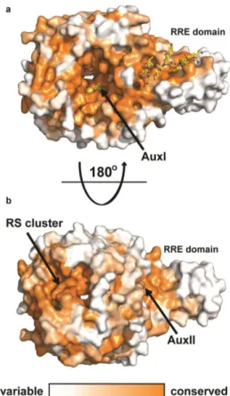
trajectory presumably places the bridging partner residue in front of the SAM binding pocket for activation, but there are no obvious pockets capable of dictating stereochemistry. The open coordination site on Aux I present in CteB could also provide either an electron sink for the radical mechanism (Mechanism A) or an oxidant and intermediate Lewis acid for the polar/ketoimine intermediate mechanism (Mechanism B). The CteB structure demonstrates that Aux I and Aux II of CteB are in sufficient proximity to act as electron transfer partners.60 Patches of highly conserved surface residues border the RS and Aux II clusters of CteB (Figure 2.8), showing possible recognition surfaces for single electron donors and acceptors, such as ferrodoxins or the flavodoxin-flavodoxin reductase system.1
With the help of the Kuhlman Lab at UNC, Rosetta3 macromolecular modeling suite was
37
38
side chains of CteA and CteB. First there is a polar interaction predicted between the carboxylic acid in Asp150 from CteB and the terminal amine in Lys33 from CteA. Second there is another polar interaction predicted between the guanidinium group from Arg182 of CteB and the carbonyl in the terminal amide group of Gln41. The model can be tested by canceling out and/or flipping these interactions by exchanging the interacting residues between CteA and CteB. If activity or binding is altered, it is a strong possibility that the interactions predicted by the model are real and the model is a good representation of how the substrate sits in the active site of the enzyme. Experiments are ongoing to determine if the aforementioned interactions are a true representation of substrate-enzyme interaction.
39
formation of multiple nested thioether linkages. For example, AlbA catalyzes formation of three sactionine linkages in subtilosin A and ThnB makes four in thurincin H biosynthesis. Dynamics in the active site and a degree of substrate control could both play roles in the formation of additional thioethers linkages. Initial substrate coordination may act to “set the register” for
thioether positioning in these multiply bridged systems. Along with this, the long RRE linker would presumably allow greater flexibility of the N-terminus, and enable a more diverse ensemble of approaches to the catalytic site.
40
41
Figure 2.8. Conservation of CteB homologs. Surface map (ConSURF server) of sequence conservation based on 150 sequences with homology ranging from 35% to 90% identity. Conservation scores are based on Bayesian method. a) The highest sequence conservation can be found around the active site and peptide binding surface of the RRE domain. b) 180 ° rotation showing the bottom of CteB. A patch of highly conserved residues are found around the RS and Aux II clusters. These sites may have a role in the recognition of redox partners.
42
for the role of SPASM auxiliary clusters in direct substrate ligation and potential residue activation required to facilitate product formation. We anticipate that this structure will have utility for the continued mechanistic understanding and engineering of sactionine synthases and other PqqE-like RiPP enzymes.
2.7Experimental
2.7.1 General Cloning and Molecular Biology Techniques
Cloning of cteA and cteB into pMCSG7
43 Generation of CteA and CteB variants
The cteA or cteB gene-blocks were used as templates to produce mutations with primers from Table 2.5. PCR was performed with Q5® High-Fidelity DNA Polymerase following the manufacturer’s manual. Primer 1 and corresponding reverse mutant primer (Table 2.5) were
used to create Piece I for CteA. Primer 2 and corresponding forward mutant primer (Table 2.5)
were used to create Piece II for CteA. Primer 3 and corresponding reverse mutant primer (Table 2.5) were used to create Piece I for CteB. Primer 4 and corresponding forward mutant primer
(Table 2.5) were used to create Piece II for CteB. The two PCR pieces were purified and kept in water. In parallel, pMCSG7 was prepared as above. Piece I and II were mixed with linearized pMCSG7, then ligated using Gibson Assembly® Master Mix according to the manufacturer’s protocol (NEB). Then, 3 μL of reaction mixture was then transformed into 50 μL of One-Shot ®
Top 10 cells. A single colony we used to inoculate 5 mL of LB culture. The plasmid was purified as above.
Cloning cteA and cteB into Duet plasmid
44
The resulting plasmids were purified as above. cteB was cloned into MCS2 first and once the resulting plasmid was sequencing confirmed, His-cteA was cloned into MCS1.
Expression of His-CteA peptides
His-CteA and its variant plasmids were transformed into BL-21 (DE3) or BL-21 (DE) cells harboring the pPH151 corrector plasmid by electroporation. The cells stocks were made electrocompetent according to standard molecular biology protocols found in Green et al.61 The electroporation was carried out in a 0.1cm cuvette, at 1.8 kV, 200 Ω, and 20 μFD. His-CteA
precursor peptide (in pETDuet-His-CteA-1, CteB-2) was heterologously expressed in E. coli (pPH-151/BL21 DE3) while His-CteA (in pMCSG7) variants were heterologously expressed in E. coli (BL21 DE3) cells. LB media was supplemented with ampicillin (100 μg/mL) with or without chloramphenicol (34 μg/mL). A 5 mL LB overnight culture was used to inoculate a 1 L
LB culture. Cultures are grown at 37 oC and 200 rpm to an OD600~0.6-0.7, at which point IPTG was added to a final concentration of 0.5 mM and the culture was grown at 18 oC, 200 rpm for 22-24 hours.
Expression of His-CteB proteins
His-CteB and its mutant plasmids were transformed into BL-21 (DE) cells harboring the pPH151 corrector plasmid by electroporation. His-CteB enzyme (in pMCSG7-CteB) was heterologously expressed in E. coli (pPH-151/BL21 DE3) cells using 1 L of auto-induction media, adapted from Studier.62 Auto-induction media was supplemented with ampicillin (100 μg/mL) and chloramphenicol (34 μg/mL). A 5 mL overnight culture of ZYP-0.8G was used to
inoculate 1 L of ZYP-5052. Cultures were grown at 37 oC and 200 rpm to an OD600 ~ 0.6-0.8, at which point the culture was cooled to 30 oC for 30 minutes. After cooling, cysteine was added to
45 hours before harvest.
Table 2.4. Gene-blocks ordered from Integrated DNA Technologies (IDT), codon optimized used in CteB study
CteA 5’-ATG AAG CAC ATT AAA ATT TTG AAC GGG TCA ACA CTG AAA GAC AGC CTG AAA AAA GGT GGG TGT GGG GAA TGT CAA ACC TCT TGC CAG TCA GCT TGC AAG ACC TCA TGT ACC GTT GCT AAT CAG TCA TGC GAA AAG CGT TAA -3’
CteB 5’-ATG GCG ATG ATC CAC AAA TTC TCG ATG ATG GGC ACA AAC ATT GTG GTG GAC GTA AAT TCA GGT GCT GTA CAC GTG GTT GAT GAT ATC AGT TTT GAT ATC CTT GAT TAT TAC AAG AAT TTT ACC GCG GGT GAG ATC AAG AAC AAG TTG GCG CAT AAG TAC AAT GCC GAC GAA ATC GAC GAA GCG TTA CGC GAA ATC GAG TCA TTA GAA GCT GAG GGC CTG TTA TTT TCA GAG GAC CCG TAT AAA GAA TAC GTA TCA TCT ATG GAC CGC AAG TCC GTC GTA AAA GCG TTG TGT CTT CAT ATC TCA CAC GAC TGT AAT CTG CGC TGC AAA TAT TGT TTT GCT TCG ACA GGA AAT TTC GGG GGC CAG CGT AAT ATG ATG TCC CTG GAG GTT GGA AAG AAG GCT ATT GAC TTC CTT ATT TCG GAA TCA GGT AAC CGC AAG AAT CTT GAG ATC GAT TTC TTT GGG GGC GAG CCC ATG ATG AAC TTC GAC GTC GTA AAG GGT ATT ATT GAG TAT GCC CGT CAG AAA GAG AAG GAG CAT AAT AAA AAC TTT CGC TTT ACA TTG ACT ACT AAT GGT CTG CTT CTG AAT GAT GAA AAT ATT AAG TAC ATT AAC GAA AAC ATG CAG AAT ATC GTT TTA TCG ATC GAC GGT CGC AAG GAA GTC AAT GAC CGT ATG CGC ATT CGC ATT GAC GGA TCC GGT TGT TAT GAT GAC ATT CTG CCC AAA TTC AAA TAT GTA GCC GAA AGC CGC AAT CAA GAC AAT TAC TAT GTT CGT GGC ACG TTC ACA CGC GAG AAT ATG GAC TTT TCA AAT GAC GTG TTA CAC TTG GCC GAC GAA GGG TTC CGT CAA ATT AGC GTT GAA CCG GTG GTT GCT GCT AAA GAC TCT GGT TAC GAC CTT CGT GAA GAA GAT CTG CCT CGT CTT TTT GAG GAA TAT GAA AAG CTG GCG TAC GAG TAC GTG AAA CGT CGT AAG GAG GGA AAT TGG TTT AAT TTC TTC CAC TTC ATG ATT GAC TTA ACA CAA GGT CCA TGT ATT GTA AAG CGC CTT ACC GGA TGT GGT AGC GGA CAC GAA TAT TTG GCC GTC ACG CCT GAA GGG GAT ATT TAC CCA TGC CAC CAA TTC GTA GGG AAT GAG AAG TTC AAG ATG GGC AAT GTA AAG GAG GGC GTC CTT AAC CGC GAT ATC CAA AAC TAC TTC AAA AAC AGC AAT GTA TAC ACT AAG AAG GAA TGT GAT TCC TGT TGG GCT AAA TTC TAT TGC AGT GGA GGC TGT GCA GCG AAC TCC TAC AAT TTC CAC AAA GAC ATT AAT ACG GTG TAC AAA GTT GGT TGT GAA TTG GAA AAG AAA CGT GTG GAG TGC GCT TTA TGG ATC AAG GCG CAA GAG ATG TAA -3’
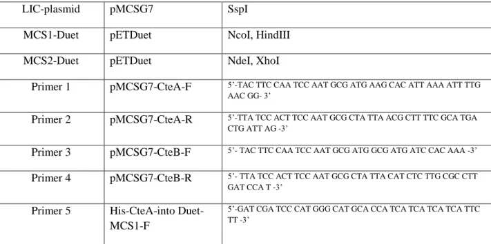
Table 2.5. Plasmids, sites, and primers (ordered from Eton Bioscience, Inc.) used in CteB study
LIC-plasmid pMCSG7 SspI
MCS1-Duet pETDuet NcoI, HindIII
MCS2-Duet pETDuet NdeI, XhoI
Primer 1 pMCSG7-CteA-F 5’-TAC TTC CAA TCC AAT GCG ATG AAG CAC ATT AAA ATT TTG AAC GG- 3’
Primer 2 pMCSG7-CteA-R 5’-TTA TCC ACT TCC AAT GCG CTA TTA ACG CTT TTC GCA TGA CTG ATT AG -3’
Primer 3 pMCSG7-CteB-F 5’- TAC TTC CAA TCC AAT GCG ATG GCG ATG ATC CAC AAA -3’ Primer 4 pMCSG7-CteB-R 5’- TTA TCC ACT TCC AAT GCG CTA TTA CAT CTC TTG CGC CTT
GAT CCA T -3’ Primer 5 His-CteA-into
Duet-MCS1-F
46 Primer 6 His-CteA-into
Duet-MCS1-R
5’-GAT CGA TCA AGC TTT TAA CGC TTT TCG CAT GAC -3’
Primer 7 CteB-into Duet-MCS2-F
5’-GAT CGA TCC ATA TGG CGA TGA TCC ACA AA -3’
Primer 8 CteB-into Duet-MCS2-R
5’-GAC TGA TCC TCG AGT TAC ATC TCT TGC GC -3’
Primer 9 CteB-Y350A-F 5’-GTG GTA GCG GAC ACG AAG CGT TGG CCG TCA CGC CTG-3’ Primer 10 CteA-Y350A-R 5’-CAG GCG TGA CGG CCA ACG CTT CGT GTC CGC TAC CAC -3’ Primer 11 CteB-H363A-F 5’-GGA TAT TTA CCC ATG CGC GCA ATT CGT AGG GAA TG-3’ Primer 12 CteB-H363A-R 5’-CAT TCC CTA CGA ATT GCG CGC ATG GGT AAA TAT CC -3’ Primer 13 CteA-C32A-F 5’-CCT CTT GCC AGT CAG CTG CTA AGA CCT CAT GTA CCG -3’ Primer 14 CteA-C32A-R 5’-CGG TAC ATG AGG TCT TAG CAG CTG ACT GGC AAG AGG -3’
2.7.2 Purification of Substrates and Enzymes
Purification of His-CteA (WT and variants) peptide and cleavage to SNA-CteA (WT and variants)
47
to an FPLC (NGC-Quest-10 Bio-Rad). The Ni2+ IMAC column was washed with 6 column volumes (CV) of IB Buffer. The peptide was eluted with a gradient of 0-100% of elution buffer (25 mM Tris-HCl, pH 8.0, 150 mM NaCl, 500 mM imidazole) over 10 CV. The peptide eluted between 15-25% elution buffer. Fractions containing the peptide were combined and dialyzed against 25 mM Tris-HCl, pH 7.0 and 150 mM NaCl in a 2000 MWCO cassette from Thermo Fischer. The buffer exchange was repeated three times, while being maintained at 4 oC. The peptide solution was then collected and tobacco etch virus (TEV) protease was added at approximately a 1:15 ratio of TEV to peptide. The protease reaction was incubated overnight at 4 oC. The cleaved CteA peptide was further purified by preparative HPLC. Preparative HPLC was performed on a Shimadzu UFLC CBM-20A with a dual channel wavelength detector at 220 nm and 280 nm with a Luna® 10 µm, 100 Å, 250 x 30 mm) AXIA™ (Phenomenex®)
semipreparatory column. Purification was carried out with a two solvent system (solvent A = 0.1% trifluoroacetic acid (TFA) in water; solvent B = 0.1% TFA in acetonitrile) using gradient of 30-60% B over 20 min at a flow rate of 15 mL/min. The peptide eluted from the column between 40-45% solvent B. The peptide product after these steps is CteA with three additional amino acids (SNA) at the N-terminus, and is hereby denoted as CteA. The fractions containing the CteA (or its variants) were pooled and partially concentrated with a rotary evaporator, followed by flash freezing and lyophilization to obtain the purified solid product. The yield of peptide was ~ 1mg of CteA per 1 L of culture. The peptide was then dissolved in dimethyl sulfoxide (DMSO) to a final concentration of 1.25 mM (based on mass).
Purification of His-CteB protein