RELATIONSHIP BETWEEN MUSCLE STIFFNESS OF THE SUPERFICIAL SHOULDER MUSCULATURE AND SUBACROMIAL SPACE DISTANCE
Pamela Rachelle Young
A thesis submitted for defense to the faculty of the University of North Carolina at Chapel Hill in partial fulfillment of the requirements for the degree of Master of Arts in the Department of Exercise & Sport Science in the College of Arts & Sciences.
Chapel Hill 2014
Approved by:
Joseph Myers
Troy Blackburn
Elizabeth Hibberd
Timothy Mauntel
ii ©2014
iii ABSTRACT
Pamela Rachelle Young: Muscle Stiffness of the Superficial Shoulder Musculature and its Relationship to Subacromial Space Distance
(Under the direction of Dr. Joseph Myers)
Side-to-side differences in subacromial space distance, muscle stiffness, and
pectoralis minor length (PML) and the predictive ability of these physical characteristics to
predict subacromial space distance in overhead athletes were investigated. Fifty collegiate
overhead athletes completed one testing session of bilateral measurements of the subacromial
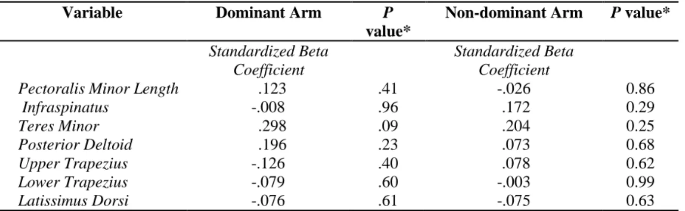
space distance, muscle stiffness, and PML. The dominant arm exhibited a shorter PML
(p=0.02) and greater stiffness of the teres minor (1.50kg: p<0.005; 1.75kg: p<0.005; 2.0kg:
p<0.005), posterior deltoid (1.50kg: p<0.005; 1.75kg: p=0.02; 2.0kg: p<0.005), and lower
trapezius (1.50kg: p=0.04; 1.75kg: p=0.03; 2.0kg: p=0.03) compared to the non-dominant
arm. Neither stiffness nor PML predicted subacromial space distance in either limb of
healthy overhead athletes. These side-to-side differences could provide clinicians with a
screening tool to identify individuals with asymmetries. Further research is needed to
determine the relationship between stiffness of the superficial shoulder musculature and
iv
ACKNOWLEDGEMENTS
A piece of work as large as this one cannot be accomplished without the diligence
and effort of many people. I would like to thank all of you who supported me throughout this
process. Specific to my research, I would like to thank my advisor Dr. Joe Myers for his
dedication to my hopes for this project and Lizzie Hibberd for her incredible patience and
commitment to seeing me succeed. My thesis committee: Dr. Troy Blackburn, Tim Mauntel,
and Terri Jo Rucinski were instrumental in developing this project into something
exceptional. Many of the department staff, faculty, and my classmates at the University of
North Carolina at Chapel Hill are to thank for their amazing support and encouragement
along the way.
I would also like to thank my athletes for whom this project was ultimately designed.
It is for them that I was motivated to see this to its completion. I have to thank my family:
Dan, Beth, Krysti, and Carli Young for their support and love. Without them, I could not
have journeyed this far and accomplished all that I have. To my friends who sent me their
love and prayers, thank you. Finally, I have to thank God for the grace that He has shown me
v
TABLE OF CONTENTS
LIST OF FIGURES ... viii
LIST OF TABLES ... ix
CHAPTER I ... 1
An Overview ... 1
Subacromial Impingement Syndrome ... 1
Subacromial Space Distance ... 3
Muscle Stiffness and Abnormal Shoulder Kinematics ... 4
Research Questions ... 7
RQ 1 ... 7
RQ 2 ... 7
RQ 3 ... 7
RQ 4 ... 7
Variables ... 7
Hypotheses ... 9
H1 ... 9
H2 ... 9
H3 ... 9
H4 ... 9
Null Hypotheses ... 9
H1 ... 9
H2 ... 10
H3 ... 10
H4 ... 10
Statistical Hypotheses ... 10
Operational Definitions ... 10
vi
Delimitations ... 11
Limitations ... 11
CHAPTER II ... 12
Introduction ... 12
Muscle Stiffness ... 13
Subacromial Impingement Syndrome ... 16
Epidemiology ... 16
Pathoanatomy and Biomechanics of the Shoulder ... 17
Etiology ... 20
Altered Glenohumeral and Scapular Kinematics ... 22
Altered Muscle Recruitment ... 26
Intervention Programs ... 26
Instrumentation... 28
Myotonometer ... 28
Diagnostic Ultrasound ... 29
Vernier Caliper ... 29
Summary ... 30
CHAPTER III ... 32
Population and Recruitment ... 32
Subject Inclusion Criterion ... 32
Subject Exclusion Criterion ... 32
Instrumentation... 33
Myotonometer ... 33
Diagnostic Ultrasound ... 34
Vernier Caliper ... 35
Procedures ... 35
Subacromial Space Distance ... 36
Muscle Stiffness ... 38
Pectoralis Minor Length ... 40
Data Reduction ... 40
vii
Summary of Research Questions ... 45
CHAPTER IV... 46
INTRODUCTION... 48
MATERIALS AND METHODS ... 51
Participants ... 51
Procedures ... 51
Acromiohumeral Distance ... 52
Muscle Stiffness ... 54
Pectoralis Minor Length ... 56
Statistical Analysis ... 57
RESULTS ... 57
DISCUSSION ... 59
CONCLUSION ... 64
viii
LIST OF FIGURES
FIGURE 1: Proposed Subacromial Impingement Cascade of Injury ... 15
FIGURE 2: Transducer Locations ... 37
FIGURE 3: Subacromial Space Distance ... 37
FIGURE 4: Myotonometer Probe Locations ... 39
ix
LIST OF TABLES
TABLE 1: Muscles and their Function ... 15
TABLE 2: Participant Demographics ... 33
1 CHAPTER I INTRODUCTION
An Overview
Shoulder pain is common among overhead athletes, particularly among competitive
baseball, volleyball, tennis, and swimming athletes (Borich et al., 2006). The prevalence of
shoulder pain among competitive overhead athletes is reported to be between 10-30%
(Diederichsen et al., 2009). It is common for overhead athletes to describe a vague sense of
discomfort, often an achy pain that developed over time, in their shoulder. This has often been
attributed to several different pathological findings with subacromial impingement syndrome (SAIS) being one of the more frequently reported causes of shoulder pain (McClure, Bialker,
Neff, Williams, & Karduna, 2004).
Subacromial Impingement Syndrome
SAIS accounts for 44-65% of all shoulder pain related doctors’ visits (de Witte et al.,
2011; McClure et al., 2004; Michener, McClure, & Karduna, 2003; Umer, Qadir, & Azam,
2012). SAIS was first described by Neer (Neer, 1983) as three progressive stages of rotator
cuff tendinopathy. Stage I involves inflammation of the subacromial bursa and the rotator
cuff, particularly the supraspinatus, with minor evidence of tendon degeneration and
2
repetitive overload and are more common in people ages 25 and older (Neer, 1983).
Collegiate overhead athletes primarily experience the early symptoms of SAIS and it is
relatively uncommon for Stage III impingement to occur in the collegiate athlete (Cowderoy,
Lisle, & O'Connell, 2009).
Currently, SAIS is classified into two main categories: structural and functional
(Page, 2011). Structural impingement, or primary impingement, stems primarily from
anatomical factors that predispose the athletic shoulder to subacromial impingement
including acromion morphology and coracoacromial ligament thickening (Bigliani & Levine,
1997; Magaji, Singh, & Pandey, 2012; Neer, 1983; Tibone et al., 1985). The current method
of treatment for primary impingement includes surgical intervention with subacromial
decompression and/or anterior acromioplasty (Bigliani & Levine, 1997; Magaji et al., 2012;
Neer, 1983). However, the overhead athlete more commonly experiences the effects of
functional rather than structural impingement due to the repetitive nature of his/her sport
(Cowderoy et al., 2009; Page, 2011). Functional impingement, or secondary impingement, is
the compression of the long head of the biceps tendon, the subacromial bursa, and/or the
supraspinatus tendon between the humeral head and the acromion process as a result of
superior migration of the humeral head during elevation of the arm (Cools, Cambier, &
Witvrouw, 2008; Desmeules, Minville, Riederer, Cote, & Fremont, 2004; Diederichsen et al.,
2009; Ludewig & Cook, 2000; Neer, 1983; Page, 2011). Secondary impingement manifests
as a result of altered glenohumeral and scapular kinematics (Burkhart, Morgan, & Kibler,
2003; Cools et al., 2008; Diederichsen et al., 2009; Ludewig & Cook, 2000; Maenhout, Van
Eessel, Van Dyck, Vanraes, & Cools, 2012; McClure et al., 2004; Page, 2011). Recent
3
rotation range of motion (ROM), has been linked with SAIS (Maenhout et al., 2012; Myers,
Laudner, Pasquale, Bradley, & Lephart, 2006; Tyler, Nicholas, Roy, & Gleim, 2000). The
overall effect of each of these etiologies is a narrowing of the subacromial space distance,
ultimately increasing the likelihood of pathological compression of the structures within
(Burkhart et al., 2003; Maenhout et al., 2012).
Subacromial Space Distance
The subacromial space is defined as the space between the humeral head and
coracoacromial arch (Cowderoy et al., 2009; Neer, 1983). The coracoacromial arch is formed
by the acromion process, the coracoid process, and the coracoacromial ligament (Bigliani &
Levine, 1997; Cowderoy et al., 2009; Michener et al., 2003; Neer, 1983). The subacromial
bursa, supraspinatus tendon, and long head of the biceps tendon lie within this space and are
susceptible to pathological compressions with subacromial space distance reductions
(Michener et al., 2003). At 0° of flexion and abduction, healthy shoulders demonstrate a
subacromial space distance of approximately 10mm which narrows to approximately 5mm
with further arm elevation to 60° and 120° of abduction (Flatow et al., 1994; Ludewig &
Cook, 2000). Shoulders with impingement demonstrate even further reductions of this space
at 90° of shoulder abduction (mean 1.4 mm ± 1.1 mm) (Graichen, Bonel, et al., 1999).
Narrowing of the subacromial space distance has been partially attributed to abnormal
glenohumeral and scapular kinematics, such as increased superior translation of the humeral
head (Deutsch, Altchek, Schwartz, Otis, & Warren, 1996), decreased internal rotation
(Borich et al., 2006; Maenhout et al., 2012), increased anterior scapular tilting (Borich et al.,
4
scapular upward rotation (Karduna, Kerner, & Lazarus, 2005), and increased protraction of
the scapula (Solem-Bertoft, Thuomas, & Westerberg, 1993). Altered scapular kinematics that
are related to subacromial impingement have also been linked with altered muscle activity
and a shortened pectoralis minor length (PML). Graichen et al. (Graichen et al., 1998;
Graichen, Stammberger, Englmeier, Reiser, & Eckstein, 1999) identified increased muscle
activity of the shoulder abductors while Borstad et al. (Borstad & Ludewig, 2005) identified
a shortened PML as contributing factors to a narrower subacromial space distance. In
addition to these known contributors, it is likely that muscle stiffness of the superficial
shoulder musculature may also play a role in reducing the subacromial space distance (Hung,
Hsieh, Yang, & Lin, 2010). A narrower subacromial space distance increases the risk of
injury because the limited available space increases the compressive contact of the
aforementioned structures, ultimately predisposing the shoulder to SAIS.
Muscle Stiffness and Abnormal Shoulder Kinematics
Muscle stiffness is the resistance of tissue to change in position or length and is
defined as the ratio of change in force to change in length (Blackburn, Norcross, & Padua,
2011; Hung et al., 2010; Huxel et al., 2008; Myers & Lephart, 2000; Oatis, 1993; Olds,
McNair, Nordez, & Cornu, 2011). Much of the research in regards to stiffness and the
shoulder concerns either the pathological “frozen,” or stiff shoulder (Hung et al., 2010), or
the benefits of muscle stiffness in relation to pathological instability of the glenohumeral
joint (Huxel et al., 2008; Olds et al., 2011). Stiff shoulder occurs as the result of muscular
5
Hung et al. (Hung et al., 2010) reported significant glenohumeral internal rotation deficits in
participants with stiff shoulder.
Several other studies have examined the influence of muscle stiffness in subjects with
glenohumeral instability. These studies found dynamic muscle stiffness at the shoulder is
essential for maintaining glenohumeral stability during functional activity (Huxel et al.,
2008; Myers & Lephart, 2000; Olds et al., 2011). Patients with recurrent glenohumeral
instability have demonstrated significantly less active muscle stiffness and a relative increase
in dislocation episodes (Olds et al., 2011). Active muscle stiffness also assists in resisting
stretching episodes, heightens muscle spindle sensitivity, and reduces the amount of delay
prior to reflexive stabilization of a joint, overall creating a more functionally stable joint
(Myers & Lephart, 2000).
Research clearly identifies the cascade of subacromial impingement as a progression
from posterior shoulder tightness to internal rotation deficits (Hung et al., 2010) to altered
glenohumeral and scapular kinematics (i.e. increased scapular upward rotation (Karduna et
al., 2005), anterior tilting (Borich et al., 2006), and internal rotation (Ludewig & Cook,
2000)) and finally to subsequent reductions in subacromial space (Graichen, Bonel, et al.,
1999; Maenhout et al., 2012). Because glenohumeral internal rotation deficits are theorized
to be a major contributing factor to alterations in kinematics and ultimately reduction in
subacromial space distance, stiffness in muscles that function to externally rotate the
shoulder (thus limiting internal rotation range of motion) may potentially be correlated to
decreased subacromial space distance and a greater risk of SAIS. Theoretically, stiffness of
the infraspinatus, teres minor, and posterior deltoid would contribute to limited internal
6
glenohumeral abduction and external rotation of the humerus, and increase scapular upward
rotation during abduction (Laudner & Williams, 2013), stiffness of the upper trapezius would
create an elevated scapular posture, stiffness of the lower trapezius would increase scapular
upward rotation, and stiffness of the pectoralis major and pectoralis minor would limit
external rotation of the humerus and scapular posterior tilting during shoulder abduction
(Terry & Chopp, 2000). Therefore, it is possible to theorize that each of these can contribute
to a functional narrowing of the subacromial space distance. Theoretically, muscle stiffness
of the superficial shoulder musculature could be the predisposing factor that instigates this
cascade of injury.
Purpose and Clinical Relevance
Research clearly identifies the cascade of subacromial impingement as a progression
from posterior shoulder tightness to internal rotation deficits (Hung et al., 2010) to altered
glenohumeral and scapular kinematics (i.e. increased scapular upward rotation (Karduna et
al., 2005), anterior tilting (Borich et al., 2006), and internal rotation (Ludewig & Cook,
2000)) and finally to subsequent reductions in subacromial space (Graichen, Bonel, et al.,
1999; Maenhout et al., 2012). Because posterior shoulder tightness and alterations in
glenohumeral and scapular kinematics are related to reductions in subacromial space
distance, stiffness in the muscles that can contribute to abnormal glenohumeral and scapular
kinematics may potentially be correlated to decreased subacromial space distance and an
increase in SAIS. While there is a theoretical link between muscle stiffness and subacromial
space distance, to date there are no previous studies that identify this relationship in either
7
relationship within the healthy overhead athlete’s shoulder. The purpose of this study was to
evaluate side-to-side differences in subacromial space distance, muscle stiffness, and PML,
as well as to determine the ability of these physical characteristics to predict subacromial
space distance. Understanding the contribution of each of these to subacromial space distance
may provide clinicians with valuable information regarding potential risk factors for
decreasing subacromial space distance and developing SAIS. Through a better understanding
of these possible risk factors, clinicians could develop better intervention and prevention
programs that could ultimately reduce the likelihood of instigating the subacromial
impingement cascade of injury.
Research Questions
RQ 1: What are the relative contributions of superficial shoulder musculature stiffness and
PML to subacromial space distance?
RQ 2: Is there a difference in muscle stiffness values between dominant and non-dominant
shoulders?
RQ 3: Is there a difference in subacromial space distance between dominant and
non-dominant shoulders?
RQ 4: Is there a difference in pectoralis minor length between dominant and non-dominant
shoulders?
Variables
Predictor:
8
Teres minor
Infraspinatus
Posterior deltoid
Upper trapezius
Lower trapezius
Latissimus dorsi
o Pectoralis minor length
Criterion:
o Subacromial space distance
Independent:
o Dominant Arm (DOM)
o Non-dominant Arm (NON)
Dependent:
o Muscle Stiffness
Teres minor
Infraspinatus
Posterior deltoid
Upper trapezius
Lower trapezius
Latissimus dorsi
9 o Subacromial space distance
Hypotheses
H1: There will be a set of variables that significantly predict subacromial space distance with
relative contributions from greatest to smallest as:
Pectoralis minor length
Posterior deltoid stiffness
Infraspinatus stiffness
Teres minor stiffness
Upper trapezius stiffness
Latissimus dorsi stiffness
Lower trapezius stiffness
H2: The dominant arm will demonstrate greater muscle stiffness compared to the
non-dominant arm.
H3: The dominant arm will demonstrate lesser subacromial space distance compared to the
non-dominant arm.
H4: The dominant arm will demonstrate a shorter pectoralis minor length compared to the
non-dominant arm.
Null Hypotheses
H1: Greater muscle stiffness of the infraspinatus, teres minor, upper and lower
trapezius, and latissimus dorsi and a shorter pectoralis minor length will not predict a
10
H2: There will be no significant difference in muscle stiffness values between
dominant and non-dominant arms.
H3: There will be no significant difference in subacromial space distance values
between dominant and non-dominant arms.
H4: There will be no significant difference in pectoralis minor length values between
dominant and non-dominant arms.
Statistical Hypotheses - Hypothesis 1:
o H0: r = 0 o HA: 0 > r > -1.0
- Hypothesis 2:
o Muscle Stiffness H0: µDom = µNon o Muscle Stiffness HA: µDom > µNon
- Hypothesis 3:
o Subacromial space distance H0: µDom = µNon o Subacromial space distance HA: µDom < µNon
- Hypothesis 4:
o Pectoralis Minor Length H0: µDom = µNon o Pectoralis Minor Length H0: µDom < µNon
Operational Definitions
Healthy shoulders: Participants without any history of shoulder surgery and without
current or history of shoulder injury within the previous year.
Shoulder injury: Shoulder impairments in either the dominant or non-dominant arm
which limited their normal activities for three consecutive days within the past six
months.
Dominant arm: The arm with which the participant would throw a ball for maximal
11
Subacromial space distance: The space between the proximal humerus, most lateral
portion of the acromion, and coracoacromial ligament.
Muscle stiffness: The resistance of muscle tissue to changes in length or position. The
ratio of change in force to the change in muscle length.
Pectoralis minor length: The measurement of the pectoralis minor from the
sternocostal junction of the fourth rib to the coracoid process.
Assumptions
Participants will follow directions when completing the tasks required during the
study.
A myotonometer is a valid and reliable tool used to measure muscle stiffness.
A Vernier caliper is a valid and reliable tool used to measure pectoralis minor length.
A digital inclinometer is a reliable measure of glenohumeral range of motion.
Delimitations
Only subjects between the ages of 18-25 years will be used in order to control for
possible degenerative changes that occur with age.
The shape of the acromion will not be investigated.
Limitations
The 2D US measurements of subacromial space cannot capture the effects on
subacromial space during 3D movement normal to the athletic shoulder.
12 CHAPTER II
A REVIEW OF THE LITERATURE
Introduction
Shoulder pain is frequently reported among collegiate overhead athletes, particularly
among those involved in swimming, baseball, volleyball, and tennis due to the demands of
their sport (Diederichsen et al., 2009). Lo et al. (Lo, Hsu, & Chan, 1990) reported that the
prevalence of shoulder pain in Chinese athletes involved in upper arm sports was 43.8% with
66.1% of them were under the age of 25 and 41.9% having competed at the elite or collegiate
level. Of the athletes reporting shoulder pain as their primary complaint, volleyball and
swimming ranked the highest with tennis, basketball, and badminton equally distributed with
10 athletes each. One of the more common injuries reported in conjunction with shoulder
pain is shoulder impingement (McClure et al., 2004). This pathology can be debilitating to an
athlete’s performance, activities of daily living, and overall feelings of well being. The
pathological anatomical and biomechanical contributing factors to subacromial impingement
have been addressed throughout the literature. These contributors include acromion
morphology (Bigliani & Levine, 1997), abnormal glenohumeral and scapular kinematics
(Deutsch et al., 1996; Ludewig & Cook, 2002; Yamaguchi et al., 2000), and posterior
shoulder tightness (Maenhout et al., 2012; Myers et al., 2006). The current literature has only
13
narrowing of the subacromial space distance. The purpose of this review of the literature is to
analyze and discuss each of these factors as well as others that may be considered
predisposing risk factors for developing subacromial impingement. This review of the
literature will seek to demonstrate the gaps in knowledge and understanding of how
subacromial space is directly affected by modifiable physical characteristics of the shoulder.
Muscle Stiffness
Muscle stiffness is the resistance of tissue to change in position or length and is
defined as the ratio of change in force to change in length (Blackburn et al., 2011; Oatis,
1993). This infers that stiffer muscles surrounding the shoulder girdle may limit the amount
of free movement of the scapula and humerus as compared to more compliant/less stiff
muscles, ultimately affecting normal glenohumeral and scapular kinematics. However,
research has elucidated the need for dynamic muscle stiffness as it relates to dynamic
stability of the shoulder. Dynamic muscle stiffness at the shoulder is essential for maintaining
glenohumeral stability during functional activity, protecting the joint from instability
episodes (Huxel et al., 2008; Myers & Lephart, 2000; Olds et al., 2011). Huxel et al. (Huxel
et al., 2008) noted that shoulder stiffness was 77% greater with active contraction as
compared to passive rest regardless of joint position and suggested that moderate levels of
torque production and stiffness remain relatively constant. The authors went on to suggest
that consistent levels of stiffness are more desirable and can contribute to supplementing
joint stability, particularly within the unstable joint. Olds et al. (Olds et al., 2011) observed a
lower level of stiffness in unstable shoulders at 30% and 50% maximal voluntary strength
14
comments on the influence of the sensorimotor system on the functional stability of the
shoulder and suggests that the preparatory muscle activation component of neuromuscular
control contributes to increasing active muscle stiffness and subsequently improves dynamic
glenohumeral stability. Less research has been conducted considering the effects of muscle
stiffness on the stable glenohumeral joint and scapulothoracic joint and particularly how it
relates to subacromial impingement syndrome (SAIS).
Determining the influence of stiffness of muscles acting on the glenohumeral and
scapulothoracic joints is essential to further understanding of SAIS. In particular, the stiffness
of the infraspinatus, teres minor, posterior deltoid, upper and lower trapezius, and latissimus
dorsi. Each of these muscles contributes to overhead motion and may affect subacromial
space distance (Table 1). Greater stiffness of each of these muscles, theoretically, will create
abnormal glenohumeral and scapular kinematics during overhead movements. For instance,
greater infraspinatus, teres minor, and posterior deltoid stiffness will create glenohumeral
internal rotation deficits (GIRD) (Hung et al., 2010). Greater latissimus dorsi stiffness and
lower trapezius stiffness will increase scapular upward rotation (Karduna et al., 2005;
Laudner & Williams, 2013) and greater upper trapezius stiffness will posture the scapula in a
position of elevation. A shortened PML, which may be as a result of pathological increases in
tissue stiffness, also contributes to greater anterior tilting and internal rotation of the scapula,
subsequently decreasing the subacromial space distance (Borstad & Ludewig, 2005; Ludewig
& Cook, 2000). Theoretically, these limitations induced by tissue stiffness can functionally
narrow the subacromial space. Overall, this paper proposes a cascade of injury that stems
from muscle stiffness of the superficial shoulder musculature and ultimately leads to SAIS
15 TABLE 1: Muscles and their Function
Muscle Function
Infraspinatus Externally rotates the humerus; cuffs the humeral head into the glenoid fossa
Teres Minor Externally rotates the humerus; cuffs the humeral head into the glenoid fossa
Posterior Deltoid Extends and externally rotates the humerus
Upper Trapezius Elevates and upwardly rotates the scapula
Lower Trapezius Depresses and upwardly rotates the scapula
Latissimus Dorsi Adducts, extends, and internally rotates the humerus
Pectoralis Minor Protracts and downwardly rotates the scapula
FIGURE 1: Proposed Subacromial Impingement Cascade of Injury
SAS
Distance
Posterior
Shoulder
Tightness
Muscle
Stiffness
Abnormal
GH/Scapular
Kinematics
GIRD
Shortened
Subacromial Impingement Syndrome Epidemiology
The term “shoulder impingement” encompasses three main pathologies of the
shoulder: 1) internal impingement, 2) coracoid impingement, and 3) subacromial
impingement. Internal impingement is the compression of the articular surface of the
supraspinatus and infraspinatus between the humeral head and posterior superior glenoid rim
with the shoulder in a position of 90° of abduction and external rotation (Davidson,
Elattrache, Jobe, & Jobe, 1995). Coracoid impingement is the compression of the
subscapularis tendon between the coracoid process and lesser tuberosity of the humerus
typically with the shoulder in a position of glenohumeral elevation, horizontal adduction, and
internal rotation (Okoro, Reddy, & Pimpelnarkar, 2009). Subacromial impingement is the
compression of the long head of the biceps tendon, the supraspinatus, and the subacromial
bursa between the humeral head and the acromion process. Although each of these
impingements may be present in the overhead athlete, the focus of this research project is to
evaluate the relationship between subacromial space distance and the development of SAIS.
SAIS is a common pathology of shoulder pain accounting for 44-65% of all shoulder
pain related doctor’s visits (de Witte et al., 2011; McClure et al., 2004; Michener et al., 2003;
Umer et al., 2012). SAIS commonly affects populations in which a primary function of daily
activities includes repetitive overhead activity. This is most commonly seen in competitive
overhead athletes, particularly those involved in swimming, tennis, baseball, and volleyball
(Borich et al., 2006), and in the industrial workplace, particularly among construction
workers, welders, and steelworkers (Ludewig & Cook, 2000). Tibone et al. (Tibone et al.,
17
involving 35 shoulders, the authors identified 17 pathological shoulders in baseball, 6 in
swimming, and 4 in tennis, with the remaining distributed between football, skiing, surfing,
and racquetball. Shoulder pain, often linked with SAIS, in USA competitive swimming has
been reported at rates as high as 38-75% (McMaster & Troup, 1993). Other studies have also
directly examined the incidence of shoulder impingement in competitive baseball athletes
(Mihata et al., 2012; Myers et al., 2006).
Pathoanatomy and Biomechanics of the Shoulder
The glenohumeral joint and scapulothoracic joints are the primary joints involved in
SAIS. The alteration of normal movement at these joints contributes to the development of
SAIS in the overhead athlete. The ball and socket glenohumeral joint has six degrees of
freedom allowing a variety of movement necessary for activities of daily living. This is
particularly important in facilitating the motions commonly utilized in overhead dominant
athletics. Throwing and hitting athletes often operate out of a position of abduction and
external rotation, a position often implicated in pathologic conditions such as SAIS. Normal
glenohumeral kinematics requires external rotation in order for the greater tuberosity to clear
the acromion and therefore enable optimal shoulder flexion and abduction (Flatow et al.,
1994; Neagle & Bennett, 1994). The infraspinatus, teres minor, and posterior deltoid function
as primary external rotators as well as humeral head stabilizers and experience a resultant
increase in eccentric load during the deceleration phase of throwing. The deltoid and
supraspinatus are the primary movers for humeral abduction. These muscles work in concert
with each other in order to abduct the humerus while the infraspinatus, teres minor, and
subscapularis function as opposing forces that simultaneously keep the humeral head
18
concurrent superior translation of the humeral head 1-3mm on the glenoid fossa occurs in
order to facilitate elevation of the glenohumeral joint. For the remainder of the movement,
the humeral head remains relatively centered on the glenoid fossa (Neumann, 2010; Terry &
Chopp, 2000; Umer et al., 2012). These dynamic force couples, the deltoid and supraspinatus
in conjunction with the other three rotator cuff muscles, serve to stabilize the humeral head
on the glenoid fossa effectively limiting the amount of pathological superior humeral
translation that would contribute to reducing the subacromial space distance and increasing
the risk of SAIS (Terry & Chopp, 2000).
Normal scapulothoracic (ST) joint function is a crucial component of enabling normal
movements of the shoulder in overhead activity. In order to achieve optimal shoulder
elevation the scapula must elevate, upwardly rotate, externally rotate, and posteriorly tilt. The
primary muscles responsible for these movements are the trapezius, rhomboids, levator
scapulae, serratus anterior, and pectoralis minor (Terry & Chopp, 2000). The trapezius is a
broad tri-portioned muscle that extends from the base of the skull to the scapular spine,
clavicle, acromion, and spinous processes of the lower thoracic vertebrae, functioning as a
scapular retractor and upward rotator. The rhomboids work concurrently with the middle
trapezius as scapular retractors, while the levator scapulae work in conjuction with the upper
trapezius to upwardly and internally rotate the scapula. The serratus anterior originates on the
first nine ribs and inserts from the superior to inferior angle on the scapula. Contraction of
the serratus anterior causes protraction and upward rotation of the scapula. The pectoralis
minor also originates on the ribs and inserts at the coracoid process of the scapula and
functions to protract, and downwardly rotate the scapula. These normal scapular movements
19
maintaining normal subacromial space distance (Hébert et al., 2002; Ludewig & Braman,
2011; Terry & Chopp, 2000).
The subacromial space is defined as the space between the humeral head and the
coracoacromial arch. The coracoacromial arch is formed by the acromion process, the
coracoid process, and the coracoacromial ligament (Bigliani & Levine, 1997; Cowderoy et
al., 2009; Michener et al., 2003; Neer, 1983). The subacromial space houses three primary
structures often compromised in SAIS including the supraspinatus tendon, the long head of
the biceps tendon, and the subacromial bursa (Michener et al., 2003). In a healthy shoulder, a
normal subacromial space distance is between 6-14mm, but is affected by normal overhead
movements. At 30° of abduction, the subacromial space is at its maximum width, whereas it
narrows to its minimum at 120°, with the majority of spatial reductions occurring between
60° and 120° of abduction. Rotation at 90° of abduction also has a significant effect on
subacromial space distance. The subacromial space is at its maximum width in internal
rotation and at its minimum in external rotation. However, the vector of the minimal distance
of the subacromial space in internal rotation passes directly through the supraspinatus tendon
at the location where most rotator cuff tears occur, indicative of greater risk of injury during
internal rather than external rotation (Graichen, Stammberger, et al., 1999). The width of this
space is affected by overhead movements and subsequently can affect the aforementioned
structures.
One example of functional overhead movement is exemplified in the baseball pitch.
The throwing motion involves complex coordination of movement of the humerus and
scapula. During the cocking phase the humerus is abducted, externally rotated, and
20
humerus to act upon. The acceleration phase begins when the humerus begins to internally
rotate in order to generate and transfer force to the ball upon release. Maintaining a position
of abduction, the humerus internally rotates while the scapula protracts, preserving that stable
base for the humerus, and begins the conversion of eccentric to concentric force at the
anterior shoulder and concentric to eccentric force at the posterior shoulder. The final phase
of the throwing motion is the violent and forceful deceleration phase. The humerus begins its
migration from horizontal abduction to horizontal adduction while continuing its internal
rotation moment about the shoulder. Meanwhile the scapula continues to protract and the
posterior shoulder muscles create a forceful eccentric contraction to slow down the rotational
velocity generated during the acceleration phase (Dillman, Fleisig, & Andrews, 1993;
Meister, 2000).
These dynamic and functional motions at the shoulder ultimately affect the
subacromial space. When the humerus abducts and the scapula upwardly rotates and
protracts as seen in the throwing motion, the subacromial space naturally narrows, but
maintains a width that will not predispose the internal structures to pathological compression
(Graichen, Stammberger, et al., 1999; Ludewig & Cook, 2002). During abduction, normal
translations of the humerus on the glenoid involve a superior humeral glide approximately
1-3mm within the first 30-60° of glenohumeral elevation (Ludewig & Cook, 2002; Umer et al.,
2012). For the remainder of the movement, the humeral head remains relatively centered on
the glenoid fossa. However, functional narrowing of the subacromial space can become
injurious with alterations in glenohumeral and scapular kinematics.
Etiology
21
glenohumeral and scapulothoracic joints. SAIS is often divided into two categories based on
these anatomical versus biomechanical differentiations that predispose the athletic shoulder
to pathological impingement: 1) Primary and 2) Secondary impingement. Primary
impingement is the result of variations in the coracoacromial arch that impinge on the
structures occupying the subacromial space. Secondary impingement, however, occurs as the
result of a cascade of biomechanical abnormalities at the shoulder. The most common cause
of secondary impingement is the instability of the glenohumeral joint commonly observed in
the high school and collegiate overhead throwing athlete (Cowderoy et al., 2009; Tyler et al.,
2000).
The structural changes of the coracoacromial arch associated with primary
impingement most frequently involve variations in the inherent shape of the acromion
process. Bigliani et al. (Bigliani & Levine, 1997) classified three different types of acromion
morphology: Type I (flat), Type II (curved), and Type III (hooked). Research has also
identified a pseudo-Type III acromion morphology resulting from an increase in osteoblastic
activity at the anterior acromion contributing to the formation of an exostosis. This spurring
of the anterior acromion is not typically present in the younger athletic shoulder, but rather is
seen in middle aged adults (Cowderoy et al., 2009). Subacromial impingement has been
attributed to the encroachment of the acromion process into the subacromial space (Neer,
1983). The hooked acromial morphology protrudes into the subacromial space thereby
increasing the compressive forces on the structures located within that space (Bigliani &
Levine, 1997). Subacromial decompression and anterior acromioplasty are common surgical
techniques utilized to reduce the compressive forces applied on the subacromial structures by
22
ligament thickening is another less common anatomical variation that can contribute to
impingement of the structures within the subacromial space (de Witte et al., 2011). Surgical
intervention is the only option for correcting bony abnormalities; therefore, the focus of this
study will be on the modifiable muscular characteristics commonly implicated in SAIS.
Secondary impingement, unlike primary impingement, involves biomechanical
abnormalities that lead to compression of the structures within the subacromial space.
Secondary impingement can be further subdivided into two other categories: intrinsic and
extrinsic impingement. Intrinsic impingement is the degeneration of the rotator cuff,
particularly the supraspinatus, as a result of overuse, tensile overload, and/or insufficient
stability and excessive mobility of the glenohumeral joint. This ultimately engenders
imbalances of the scapular muscles and abnormal scapulohumeral rhythm contributing to
ischemic changes in the supraspinatus tendon (de Witte et al., 2011; Michener et al., 2003).
Extrinsic impingement is the narrowing of the subacromial space thereby causing a
mechanical compression of the rotator cuff, subacromial bursa, and long head of the biceps
tendon (de Witte et al., 2011; Umer et al., 2012). These typically stem from alterations in the
biomechanics and kinematics of the glenohumeral and scapulothoracic joints.
Altered Glenohumeral and Scapular Kinematics
Alterations in glenohumeral kinematics often involve pathological superior
translations of the humeral head on the glenoid fossa; an alteration often observed within
individuals with impingement. Individuals with impingement demonstrate excessive superior
translation of 1.0-1.2mm as evidenced on radiographic images (Deutsch et al., 1996). Those
unaffected by impingement and those with stage II impingement demonstrate a centrally
23
as compared to those with stage III impingement (full rotator cuff tears) that presented with
the humerus located above the glenoid’s center (mean +0.3mm) (Deutsch et al., 1996). Other
studies have also identified excessive and abnormal superior humeral head translation during
glenohumeral elevation in subjects with impingement (Ludewig & Cook, 2002; Yamaguchi
et al., 2000).
In addition to alterations in humeral head movement, aberrations in scapular
kinematics are related to SAIS. SICK scapula, first defined by Burkhart et al. (Burkhart et al.,
2003), refers to Scapular malposition, Inferior medial border prominence, Coracoid pain and malposition, and dysKinesis of scapular movement. There are three primary patterns of scapular dyskinesis and Type III is most often related to SAIS. In Type III SICK scapula, the
malpositioned scapula sits in a protracted and anteriorly tilted position making the
inferomedial border appear more prominent and makes the affected shoulder appear lower
than the contralateral side. As a result of this protraction and anterior tilt, the pectoralis minor
and short head of the biceps become adaptively tight and short serving to maintain and
increase the malposition of the scapula. This altered scapular kinematic decreases the
available subacromial space and subsequently increases the risk of impingement (Burkhart et
al., 2003).
Abnormal muscle activation of the serratus anterior, upper and lower trapezius,
rotator cuff, and middle deltoid contributes to alterations in scapular kinematics such as
decreased posterior tipping, increased upward rotation, and elevation of the scapula during
glenohumeral abduction increasing the risk of impinging the subacromial structures
(Ludewig & Cook, 2000). Upper crossed syndrome, first described by Vladimir Janda, refers
24
and cervical spine. These imbalances of tight pectorals, suboccipitals, upper trapezius, and
levator scapulae, and weak cervical flexors, rhomboids, and lower trapezius create a forward
head and rounded shoulders posture often implicated in SAIS (Janda, 1988; Page, 2011).
Individuals with greater forward head and rounded shoulders posture demonstrate greater
anterior tilting, internal rotation, and upward rotation of the scapula as well as concurrent
reductions in serratus anterior activation (Thigpen et al., 2010). A shortened PML orients the
scapula in a more protracted position. Protraction of the scapula diminishes the subacromial
space thereby increasing the amount of contact pressure on the structures within (Borstad &
Ludewig, 2005). Internal rotation of the scapula also decreases the subacromial space and is a
patterned behavior in shoulders with symptoms of SAIS (Ludewig & Cook, 2000). Recent
research has also identified an increase in latissimus dorsi tightness, or stiffness, in swimmers
that contributes to greater upward rotation of the scapula during the humeral elevation that
occurs during the repetitive performance of the swimming strokes (Laudner & Williams,
2013). Greater scapular upward rotation decreases the amount of subacromial clearance and
subsequently increases subacromial contact forces (Karduna et al., 2005). A study by
McClure et al. (McClure, Michener, & Karduna, 2006) demonstrated slightly greater upward
rotation in subjects with SAIS. Interestingly enough, other studies have found that shoulders
with impingement typically demonstrate decreased scapular upward rotation (Ludewig &
Cook, 2000; Su, Johnson, Gracely, & Karduna, 2004) and this may be a compensatory
reaction in order to decrease the amount of subacromial contact occurring during humeral
elevation.
Research has also identified posterior shoulder tightness as a predominant factor
25
shoulder stems from a tight posterior capsule, posterior rotator cuff, and posterior deltoid
(Harryman et al., 1990; Myers et al., 2006; Tyler et al., 2000). Stiffness of the infraspinatus,
teres minor, and posterior deltoid has a high correlation with GIRD in patients with
pathological stiff shoulder (Hung et al., 2010) and GIRD is correlated with a greater number
of shoulder injuries within throwing athletes (Myers et al., 2006). In a study by Tyler et al.
(Tyler et al., 2000), participants (non-throwers) with subacromial impingement in their
dominant arm demonstrated significant internal rotation deficits (mean of -22.29°) as
compared contralaterally, as well as greater posterior capsule tightness than the control
group. It has also been suggested that anterior and superior humeral head translation on the
glenoid fossa increases as a result of posterior capsular tightness. One cadaveric study
operatively tightened the posterior capsule and demonstrated a significant increase in anterior
translation (mean of 7.27mm) and slight increase in superior translation (mean of 2.13mm) of
the humeral head on the glenoid fossa during flexion (Harryman et al., 1990). GIRD is often
present in patients involved in regular overhead activity and subsequently affects scapular
kinematics by increasing anterior scapular tilt during glenohumeral flexion and abduction,
thereby reducing subacromial space distance (Borich et al., 2006; Hébert et al., 2002). Most
importantly, GIRD also contributes to a reduction in the acromiohumeral distance (AHD), or
subacromial space distance, in overhead athletes (Maenhout et al., 2012) ultimately
predisposing the supraspinatus, long head of the biceps tendon, and subacromial bursa to
pathologic compression and injury within the subacromial space. All of these factors
considered, it is likely that posterior shoulder muscle stiffness and subsequent internal
26 Altered Muscle Recruitment
Normal glenohumeral abduction involves a complex synchronization of the forces
elicited by the supraspinatus, infraspinatus, and deltoid as they work in opposition to one
another during the first phase of abduction. As the deltoid creates a superiorly directed vector
of force on the humerus, the supraspinatus and infraspinatus apply a medially directed line of
pull on the humerus in order to center it on the glenoid and prevent excessive superior
humeral migration. This force couple enables partial stabilization of the glenohumeral joint
during the beginnings of overhead activities. However, alteration of this force couple through
the degeneration, inhibition, or fatigue of the rotator cuff muscles results in a domination of
the deltoid during abduction consequently generating a relative increase in the superior
translation of the humeral head (Deutsch et al., 1996). Theoretically, facilitation or stiffness
of the posterior deltoid, infraspinatus, and teres minor may also alter the functions of this
force couple, creating pathological movement patterns and abnormal humeral head
translations. As a result, this causes a functional narrowing of the subacromial space
contributing to the development of SAIS.
Intervention Programs
SAIS in overhead athletes establishes a need to address predisposing factors such as
GIRD, muscle imbalances, and abnormal scapular and glenohumeral kinematics. Fortunately,
these are all modifiable physical characteristics, ultimately making it possible to formulate
intervention programs to decrease the risk of developing SAIS. In order to reduce the amount
of GIRD in athletic shoulders, research has studied the effects of stretching the posterior
27
internal rotation ROM (McClure et al., 2007); additionally, the sleeper stretch also increases
glenohumeral internal rotation, acromiohumeral distance (AHD), or subacromial space
distance, in overhead athletes at 0°, 45°, and 60° of shoulder abduction (Maenhout et al.,
2012). Other stretching interventions have examined the effect of stretching the pectoralis
minor in order to correct the forward head and rounded shoulders posture observed in
shoulders with adaptive pectoralis minor shortening (Thigpen et al., 2010). A self stretch
procedure, where the patient places the affected arm in a position of 90° of abduction and 90°
of elbow flexion on a planar surface and rotates the trunk away from the targeted side thereby
increasing the amount of horizontal abduction, has been demonstrated as the most effective
stretch for lengthening the pectoralis minor (Borstad & Ludewig, 2006). Evidence indicates
that increasing the length of the pectoralis minor will assist in correcting the abnormal
scapular kinematics, such as decreased posterior tipping and external rotation, that contribute
to SAIS (Borstad & Ludewig, 2005; Ludewig & Cook, 2000).
Scapular stabilization exercises are also necessary to correct deviations in scapular
posture that contribute to reductions in subacromial space distance and development of SAIS.
Başkurt et al. (Başkurt, Başkurt, Gelecek, & Ozkan, 2011) determined the effectiveness of
scapular stabilization exercises on pain, ROM, joint position sense, muscle strength, and
quality of life in patients’ with SAIS and found that each of these factors improved as a result
of the 6 week intervention program. Wilk et al. (Wilk, Meister, & Andrews, 2002) outlined
the following 5 step program for nonoperative treatment of SAIS: 1) Rest for 7-10 days from
repetitive overhead athletic activity. 2) Restore normal glenohumeral and scapular kinematics
by stretching the posterior shoulder. 3) Increase stability of glenohumeral joint as well as
28
strengthening. 4) Emphasize scapular retraction, and 5) gradually return to throwing. These
are common therapeutic strategies used in athletic training rehabilitation programs for the
athlete with SAIS; however, other recent research attempted to validate these common
rehabilitation strategies and found little success with the interventions. Hibberd et al.
(Hibberd, Oyama, Spang, Prentice, & Myers, 2012) analyzed the effects of a 6-week
preventative intervention on scapular and shoulder girdle strengthening and scapular
kinematics in competitive collegiate swimmers and found the intervention program was
unsuccessful in correcting and/or preventing a rounded shoulder posture. Not many other
studies have been conducted on the efficacy of certain rehabilitation exercises in the
treatment of subacromial impingement and there is a lack of current evidence for anecdotal
treatment strategies. Further research is necessary to ascertain which rehabilitation strategies
are effective for both treatment and prevention of SAIS.
Instrumentation Myotonometer
A myotonometer (Neurogenic Technologies Inc., Missoula, MT) will be used to
collect measurements of active and passive muscle stiffness. The myotonometer is a patented
and computerized meter-type device that effectively and efficiently measures tissue
compliance and stiffness. The myotonometer measures the amount of resistance encountered
by the probe when it is applied to the muscle and underlying tissue and subsequently
quantifies the amount of tissue displacement which is then used to calculate stiffness
29
muscle stiffness using a myotonometer have been proven valid and reliable (Leonard,
Stephens, & Stroppel, 2001; Rydahl & Brouwer, 2004).
Diagnostic Ultrasound
A diagnostic ultrasound (US) (Model: Sonosite, Sonosite, Inc., Bothella, WA) unit
will be used to collect measurements of the subacromial space distance via measurements of
the AHD. The AHD is defined as the shortest distance between the humeral head and most
inferior and lateral portion of the acromion process (Desmeules et al., 2004). Coronal axis
views of the subacromial space with the transducer positioned according to the methods
described by Desmeules et al. (Desmeules et al., 2004) and Azzoni et al. (Azzoni, Cabitza, &
Parrini, 2004) will allow for visualization and accurate measure of the AHD. Previous studies
have measured subacromial space distance and AHD with the arm positioned at 0°, 45°, and
60° of abduction, but have been unable to collect measurements in greater degrees of
humeral abduction because of the limitations of the US unit created by beam reflection on
bone interfering with visual clarity and inhibiting accurate measurements. However, a recent
study by Timmons et al. (Timmons et al., 2013), measured AHD at 90° of abduction in
positions of clinical full can (neutral humeral rotation) and empty can tests (humeral internal
rotation). These methods for US measurement of the AHD and for quantifying the
subacromial space distance have been found both valid and reliable (Azzoni et al., 2004;
Desmeules et al., 2004; Maenhout et al., 2012).
Vernier Caliper
A vernier caliper will be used to measure PML (Westward Tools, Edmonton, AB,
30
are the sternal aspect of the fourth rib and the coracoid process respectively. The vernier
caliper will then be used to measure the distance between these and calculate the length of
the pectoralis minor. These procedures are outlined by the validation and reliability study of
PML measurement conducted by Borstad et al (Borstad, 2008) in which they used an
electromagnetic motion capture system, a vernier caliper, and a cloth tape measure to
measure the pectoralis minor and established relatively high intraclass correlation
coefficients (ICC) between the electromagnetic motion capture system and caliper as well as
between the electromagnetic motion capture system and tape measure. Therefore, the vernier
caliper has been found to be a clinically valid assessment tool for the measurement of PML
(Borstad, 2008).
Summary
Subacromial impingement syndrome is a common pathologic condition of the
shoulder, particularly within the overhead athletic population (Diederichsen et al., 2009;
McClure et al., 2004). Studies have identified modifiable physical characteristics of the
superficial shoulder musculature that contribute to subacromial impingement. These
contributors include posterior shoulder tightness and GIRD (Harryman et al., 1990; Hung et
al., 2010; Myers et al., 2006; Tyler et al., 2000), altered glenohumeral and scapular
kinematics such as greater superior humeral head translation (Deutsch et al., 1996; Ludewig
& Cook, 2002), anterior tilting and upward rotation of the scapula (Burkhart et al., 2003;
Ludewig & Cook, 2000), shortened PML (Borstad & Ludewig, 2005), and muscle
imbalances (Page, 2011). However, very little research has identified direct effects of these
modifiable characteristics on subacromial space distance. Muscle stiffness has primarily been
31
Myers & Lephart, 2000; Olds et al., 2011); however, evidence points towards the influence
of muscle stiffness on subacromial impingement particularly through its affect on
subacromial space distance (Laudner & Williams, 2013; Maenhout et al., 2012). As such, it
is important to consider the effects of greater stiffness of the infraspinatus, teres minor,
posterior deltoid, upper and lower trapezius, and latissimus dorsi on the functional narrowing
of the subacromial space and the potential for predisposition to SAIS as a result. It is also
apparent that intervention programs have little basis and the literature is lacking in
rehabilitation protocols for SAIS. Therefore, the purpose of this study was to evaluate
side-to-side differences in subacromial space distance, muscle stiffness, and PML, as well as
determine the ability of these physical characteristics to predict subacromial space distance.
Through a better understanding of these possible risk factors, clinicians could develop better
intervention and prevention programs that could ultimately reduce the likelihood of
32 CHAPTER III METHODOLOGY
Population and Recruitment
Fifty male and female participants, all of whom were overhead athletes at the division
I level, were recruited to participate (Table 2). Individuals were recruited via flyers, word of
mouth communication, and presentations by the primary investigator. Potential participants
met with the primary investigator, received explanation regarding the study, and, once
enrolled, provided Institutional Review Board (IRB) approved informed consent.
Subject Inclusion Criterion
Participants were included in this study if they met the following criteria:
- Varsity overhead athlete between the ages of 18-25 years
- Currently participating in one of the following varsity sports: baseball,
softball, tennis, swimming, volleyball.
- No history of shoulder surgery, no current shoulder pain, and were not
receiving rehabilitation for shoulder injury/pain.
Subject Exclusion Criterion
Participants were excluded from this study if they met the following criteria:
33 TABLE 2: Participant Demographics
Participant Demographics
Number of Participants (n) 50
Males/Females 19/31
Age (yrs) 19.4±1.2
Height (cm) 176.4±8.0
Weight (kg) 75.6±9.8
Arm Dominance
Right/Left 44/6
Subjects per sport
Baseball 10
Softball 10
Volleyball 10
Swimming 10
Tennis 10
Years of playing experience 11.8±2.7
Instrumentation Myotonometer
A myotonometer (Neurogenic Technologies Inc., Missoula, MT) was used to collect
measurements of active muscle stiffness. The myotonometer is a patented and computerized
meter-type device that effectively and efficiently measures tissue compliance and stiffness.
The myotonometer measures the amount of resistance encountered by the probe when it is
applied to the muscle and underlying tissue and subsequently quantifies the targeted tissue’s
stiffness (Hung et al., 2010). Measurements of muscle stiffness using a myotonometer have
been shown to be valid and reliable in lower extremity muscles (Leonard et al., 2001; Rydahl
& Brouwer, 2004). We established the reliabilityand validity of the myotonometer
measurements of muscle stiffness of the muscles we proposed to assess in the current study
34
TABLE 3: Intraclass Correlations of Myotonometric Measurements of Muscle Stiffness
Muscle Stiffness Intrasession ICC
Intrasession SEM (mm)
Mean Detectable Difference
INFRA 1.50 .984 0.65 1.79
INFRA 1.75 .981 0.67 1.85
INFRA 2.0 .978 0.68 1.89
TM 1.50 .955 1.02 2.82
TM 1.75 .957 1.03 2.86
TM 2.0 .959 1.03 2.86
PD 1.50 .891 0.53 1.46
PD 1.75 .884 0.52 1.44
PD 2.0 .882 0.51 1.40
UT 1.50 .757 0.39 1.08
UT 1.75 .789 0.41 1.13
UT 2.0 .808 0.41 1.15
LT 1.50 .829 0.57 1.57
LT 1.75 .845 0.58 1.60
LT 2.0 .656 0.67 1.85
LD 1.50 .986 0.98 2.72
LD 1.75 .972 0.99 2.76
LD 2.0 .975 1.01 2.79
Diagnostic Ultrasound
A diagnostic US unit (Model: Sonosite, Sonosite, Inc., Bothella, WA) was used to
collect measurements of the subacromial space distance via measurements of the AHD. The
AHD is defined as the shortest distance between the humeral head and most inferior and
lateral portion of the acromion process (Desmeules et al., 2004). Coronal axis views of the
subacromial space with the probe positioned according to previously described methods
(Azzoni et al., 2004; Desmeules et al., 2004) enabled us to visualize and accurately measure
the AHD. Previous studies have measured subacromial space distance and AHD with the arm
positioned at 0°, 45°, and 60° of abduction, but have been unable to collect measurements in
greater degrees of humeral abduction because of the limitations of the ultrasound unit created
by beam reflection on bone interfering with visual clarity and inhibiting accurate
35
clinical full can (neutral humeral rotation) and empty can tests (humeral internal rotation).
Preliminary data from 9 subjects enabled calculations of intra-rater and test-retest reliability
(ICC = 0.90, SEM = 0.07 mm) (Timmons et al., 2013). These methods for US measurement
of the AHD and for quantifying the subacromial space distance have been found both valid
and reliable (Azzoni et al., 2004; Desmeules et al., 2004; Maenhout et al., 2012).
Vernier Caliper
A vernier caliper was used to measure PML (Westward Tools, Edmonton, AB,
Canada). The bony landmarks used to locate the origin and insertion of the pectoralis minor
are the sternal aspect of the fourth rib and the coracoid process. The vernier caliper was used
to measure the distance between these points and to represent PML. These procedures are
outlined by the validation and reliability study of PML measurement conducted by Borstad et
al (Borstad, 2008) in which they used an electromagnetic motion capture system, a vernier
caliper, and a cloth tape measure to measure the pectoralis minor and established relatively
high intraclass correlation coefficients each measurement. Therefore, the vernier caliper has
been found to be a clinically valid assessment tool for the measurement of PML (Borstad,
2008).
Procedures
A cross-sectional research design was used in this study. Study participants reported
to the Neuromuscular Research Laboratory (NMRL) for a single session. Participants were
introduced to the experiment and then read and signed a consent form approved by the
36
completed a brief survey detailing demographics including sex, age, arm dominance, current
or previous overhead sport activity experience, and his/her history of shoulder pain and/or
injury. Each participant then had height (cm) and mass (kg) measurements taken by one of
the researchers. Each participant then underwent the testing procedures that included
measurements of the subacromial space distance, muscle stiffness, and PML. Testing order
and conditions were randomized and counterbalanced. Details of each procedure are
discussed below.
Subacromial Space Distance
The participant was instructed to take a seated position on a stool with the arms in a
relaxed position hanging by his/her sides. Subacromial space distance was measured using
US techniques as described by Maenhout et al. (Maenhout et al., 2012). Three US images
were taken at 45° of abduction. For imaging at 45° of abduction, one loop of a belt was
secured to the base of the stool upon which the participant sat, while the other end was
looped around the participant’s distal forearm. Arm position was verified by a digital
inclinometer. The participant was instructed to apply tension to the belt in order to maintain
arm position as well as to elicit activation of the shoulder musculature. The participant was
also asked to hold a dumbbell in order to elicit activation of the muscles of interest. The
weight of the dumbbell was determined relative to body mass, 1.4kg (3lbs) for those
weighing less than 68.1kg (150lbs) and 2.3kg (5lbs) for those weighing more than 68.1kg
(McClure, Tate, Kareha, Irwin, & Zlupko, 2009). The US transducer was placed on the
superolateral aspect of the shoulder along the longitudinal axis of the humerus (Figure 2).
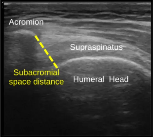
37
distance from the infero-lateral edge of the acromion to the humeral head (Leong, Tsui, Ying,
Leung, & Fu, 2012; Maenhout et al., 2012) (Figure 3). The participant was instructed to rest
between image trials with the arm at 0° abduction placing the hand and weight on his/her
thigh in order to prevent muscle fatigue during the testing session. Subacromial space
distance values were calculated as the average of three trials bilaterally. These values were
normalized to each participant’s height (subacromial space distance/height). We estabilished
intrasession reliability (ICC: 0.840), standard error of the measurement (SEM: 0.87mm), and
mean detectable difference (MDD: 2.41) through pilot testing.
FIGURE 2: Transducer Locations
FIGURE 3: Subacromial Space Distance
Humeral Head Supraspinatus
Subacromial space distance
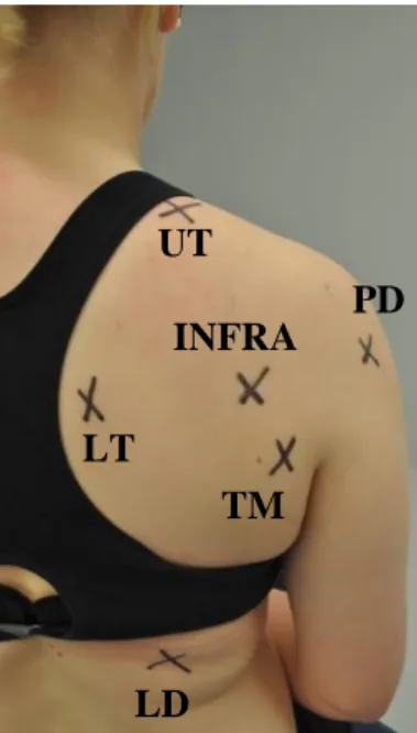
38 Muscle Stiffness
Muscle stiffness of the posterior deltoid, infraspinatus, teres minor, upper and lower
trapezius, and latissimus dorsi was recorded using a handheld myotonometer. Testing order
of the muscles was randomized for each participant. The participant was asked to remain
sitting with his/her feet resting flat on the floor and with the arm raised into 45° of shoulder
abduction. The same procedures previously outlined for subacromial space distance testing
were used in order to maintain the arm position at 45° of abduction. The participant was also
asked to hold a dumbbell, with the weight determined relative to body mass (1.4kg (3lbs) for
those weighing less than 68.1kg (150lbs) and 2.3kg (5lbs) for those weighing more than
68.1kg), in order to elicit activation of the muscles of interest (McClure et al., 2009). The
participant was instructed to rest the arm in 0° of shoulder abduction with the hand and
weight resting on the thigh between trials at each muscle in order to prevent excessive
muscle fatigue during the testing session. The following anatomical locations were used for
the placement of the myotonometer probe (Figure 4):
- Posterior deltoid - 2 fingerbreadths inferior to the posterior margin of the
acromion (Hung et al., 2010).
- Infraspinatus - 2 fingerbreadths below the medial portion of the spine of the
scapula (Hung et al., 2010).
- Teres minor - one-third of the distance between the acromion and inferior angle of
the scapula along the lateral border (Hung et al., 2010).
- Upper trapezius - midway between the spinous process of the seventh cervical
vertebra and the posterior margin of the acromion process (based on electrode
39
- Lower trapezius - obliquely upward and laterally along a linear pathway between
the intersection of the spine of the scapula with the vertebral border of the scapula
and seventh thoracic spinous process (based on electrode placement in
electromyography) (Cools et al., 2007).
- Latissimus dorsi - 5cm inferior to the inferior portion of the scapular border
(Laudner & Williams, 2013).
The mean of 5 trials at 8 different increments (0.25 – 2.00 kg) of 0.25kg of force
pressure was calculated during probe application at each muscle to determine tissue
displacement, which was used to calculate muscle stiffness. Procedures for tissue
displacement were completed bilaterally.
FIGURE 4: Myotonometer Probe Locations
** UT=Upper Trapezius, LT=Lower Trapezius, INFRA=Infraspinatus, PD=Posterior Deltoid, TM=Teres Minor, and LD=Latissimus Dorsi
UT
LT
INFRA PD
TM