THE ROLE OF HIPPOCAMPAL NEURAL IMMUNE SIGNALING IN STRESS-ENHANCED FEAR LEARNING: IMPLICATIONS FOR POST-TRAUMATIC STRESS
DISORDER
Meghan E. Jones
A dissertation submitted to the faculty of the University of North Carolina at Chapel Hill in partial fulfillment of the requirements for the degree of Doctor of Philosophy in the
Department of Psychology and Neuroscience.
Chapel Hill 2017
iii ABSTRACT
MEGHAN E. JONES: The role of hippocampal neural immune signaling in stress-enhanced fear learning: Implications for post-traumatic stress disorder.
(Under the direction of Donald T. Lysle)
Psychopathology and disease states involving depression and anxiety, including post-traumatic stress disorder (PTSD), have been associated with immune dysregulation. Preliminary data from our laboratory has suggested that severe stress induces a time-dependent increase in hippocampal IL-1β immunoreactivity and that centrally blocking IL-1 signaling prevented the development of stress-enhanced fear learning, a rodent PTSD-like phenotype. In parallel, astrocyte-derived cytokines and astroglial signaling have been linked to the development of PTSD-like phenotypes following severe stress. The goal of the current dissertation was to use the stress-enhanced fear learning paradigm to explore the role of neural immune signaling in the development of a PTSD-like phenotype. The experiments described in the current dissertation tested the overarching hypothesis that (1) severe stress induces changes in hippocampal astrocyte-derived IL-1β expression and the morphometric properties of hippocampal astrocytes and (2) that both blocking hippocampal IL-1 signaling and activating hippocampal astroglial Gi signaling prevent the development of SEFL.
iv
v
AKNOWLEDGEMENTS
I would first like to express my most sincere thanks to my advisor, Dr. Donald Lysle. I feel extremely lucky to have been able to complete my graduate work in the Lysle lab not only because of the excellent research environment that Don has created but also because his door was always open, no matter how silly the question. I do not think I could have completed as much working with any other advisor and I hope to know as much and be as great a researcher as Don is some day.
I would like to thank my lab mates, especially Christina Lebonville and Jackie Paniccia, for their help and support both in and out of lab throughout my time in graduate school. I would also like to thank all of the students and faculty of the Behavioral Neuroscience program and Psychology Department, especially my dissertation committee members. I have truly enjoyed the collaborative and supportive environment in our program and I know that it contributes to stronger science all around. I also especially want to thank Dr. Regina Carelli and Dr. Kathryn Reissner who have been important female role models for the ideal excellent researcher to me throughout graduate school.
vi
vii
TABLE OF CONTENTS
ACKNOWLEDGEMENTS………...v
LIST OF FIGURES………...………..xii
LIST OF ABBREVIATIONS………...….xiii
Chapter I. GENERAL INTRODUCTION………1
a. Post-traumatic stress disorder………1
b. Approaches to study PTSD using rodent models……….….3
c. Hippocampal function is important in PTSD and SEFL………...7
d. Role of cytokine signaling in stress response mechanisms………...8
e. Role of astroglial signaling in stress response mechanisms………....11
f. High resolution analysis of the morphometric properties of astrocytes……..13
g. Glial-expressing Designer Receptors Exclusively Activated by Designer Drugs………13
h. Specific Aims………...15
II. EXAMINATION OF STRESS-INDUCED HIPPOCAMPAL IL-1β: EFFECT OF HIPPOCAMPAL IL-1RA ON STRESS-ENHANCED FEAR LEARNING AND IDENTIFICATION OF THE CELLULAR SOURCE1………19
viii
a. Introduction………..19
b. Methods………23
i. Animals………....23
ii. Experiment 2.1: Effect of intra-dorsal hippocampal IL-1RA on the development of SEFL………..24
iii. Surgery………...24
iv. Stress-enhanced fear learning………..24
v. IL-1 receptor antagonist………...25
vi. Experiment 2.2: Immunofluorescence analysis of severe stress-induced changes in hippocampal GFAP, Iba-1, NeuN, and IL-1β………..…26
vii. Stress exposure……….26
viii. Immunohistochemistry………27
ix. Confocal microscopy, Bitplane Imaris colocalization analysis, and cell counting………..28
x. Statistical analyses………...29
c. Results………..30
i. Experiment 2.1: Intra-dorsal hippocampal IL-RA prevents SEFL………..30
ii. Experiment 2.2a: Stress-induced increase in hippocampal IL-1β is replicated………..32
iii. Experiment 2.2c: Stress-induced hippocampal IL-1β is colocalized primarily with GFAP in both stressed and non-stressed animals……….35
ix
III. EFFECT OF SEVERE STRESS ON THE MORPHOMETRIC
PROPERTIES OF HIPPOCAMPAL ASTROCYTES………..42
a. Introduction………..42
b. Methods………46
i. Animals………46
ii. Experiment 3.1 Verification of AAV5-GFAP-HA-hM3Dq-IRES-mCitrine as a membrane-dependent tag……….46
iii. Viruses……….46
iv. Surgery and Sacrifice………...46
v. Immunohistochemistry………47
vi. Confocal microscopy and Bitplane Imaris analysis……….48
vii. Experiment 3.2: Effect of stress on the morphometric properties of astrocytes………48
viii. Virus……….48
ix. Surgery……….48
x. Stress exposure and sacrifice………...49
xi. Confocal microscopy and Bitplane Imaris analysis……….50
xii. Image Acquisition………....50
xiii. Astrocyte volume, surface area, and colocalization with PSD95………..51
xiv. Quantification of hippocampal PSD95 immunoreactivity…………...52
xv. Statistical Analysis………...52
c. Results………..53
x
ii. Experiment 3.2: Effect of stress on the morphometric
properties of astrocytes………...56
iii. Stress exposure does not alter astrocyte volume, surface area or colocalization with PSD95………...56
iv. Stress exposure attenuates PSD95 Immunoreactivity………..59
d. Discussion………60
IV. EFFECT OF HIPPOCAMPAL ASTROGLIAL GI SIGNALING ON STRESS-ENHANCED FEAR LEARNING………..63
a. Introduction………..63
b. Methods………....66
i. Animals………....66
ii. Virus……….67
iii. Surgery……….67
iv. Experiment 4.1: Effect of hippocampal astroglial Gi activation on SEFL………...67
v. Stress-enhanced fear learning………..67
vi. Drug Administration………....68
vii. Sacrifice………...69
viii. Immunohistochemistry………....69
ix. Experiment 4.2: Effect of CNO on colocalization of mCherry-positive cells with cAMP………...70
x. Sacrifice………...70
xi. Immunohistochemistry………71
xii. Confocal microscopy and Bitplane Imaris colocalization analysis……….72
xi
xiv. Colocalization of mCherry and cAMP………72
xv. Statistical Analysis………...73
c. Results………..74
i. Experiment 4.1: Hippocampal astroglial Gi activation attenuates SEFL………...………74
ii. Experiment 4.2: CNO attenuated colocalization of mCherry-positive cells with cAMP……….76
d. Discussion………77
V. GENERAL DISCUSSION....………81
a. Summary of findings………81
b. Implications of the role of IL-1 signaling in SEFL………..82
c. Implications of the role of astrocytes, and in particular astroglial Gi activation, in SEFL………..84
d. Implications of the effect of severe stress on PSD95………..86
e. Role of additional neural immune signaling pathways in behavioral outcomes of severe stress………88
f. Summary of unanswered questions and future directions……….89
g. Concluding remarks……….92
xii
LIST OF FIGURES
Figure
1.1 Stress enhanced fear learning………..5
1.2 Stressor of the SFEL paradigm enhances general anxiety-like behavior seven days later………..6
1.3 Experiments described in Chapters 2 through 4 address three Specific Aims…………..18
2.1 Intra-dorsal hippocampal IL-1RA is sufficient to prevent SEFL………..33
2.2 Severe stress increases hippocampal IL-1β immunoreactivity………..34
2.3 Dorsal hippocampal Iba-1 immunoreactivity, but not GFAP immunoreactivity is attenuated 48 hours after severe stress……….36
2.4 IL-1β signal is colocalized with GFAP, and not with Iba-1 or NeuN, in the dorsal hippocampus in stressed and non-stressed animals……….38
2.5 IL-1β signal is colocalized with GFAP, and not with Iba-1 or NeuN, in the dorsal hippocampus in stressed and non-stressed animals……….39
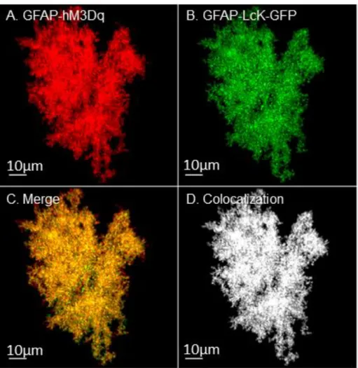
3.1 GFAP-Lck-GFP is expressed selectively in astrocytes……….57
3.2 GFAP-hM3Dq can be used to examine astrocyte morphology……….58
3.3 Foot shock does not alter the morphometric properties of astrocytes………...60
3.4 Cells included in morphology analyses were predominantly from CA1 and CA3………61
3.5 Foot shock attenuated PSD95 immunoreactivity………...62
4.1 Experimental timeline for Experiment 4.1……….…72
4.2 Experimental timelines for Experiment 4.2………...75
4.3 Astroglial Gi activation attenuates stress-enhanced fear learning………..79
xiii
LIST OF ABBREVIATIONS
AAV Adeno-associated virus
ANOVA Analysis of Variance
AP Anterior-posterior
ASR Acoustic Startle Response test
ATP Adenosine triphosphate
BDNF Brain-derived neurotrophic factor
CA1 Cornu Ammonis 1
CA3 Cornu Ammonis 3
cAMP Cyclic adenosine monophosphate
CNO Clozapine-n-oxide
CNS Central nervous system
DG Dentate gyrus
DH Dorsal Hippocampus
DREADD Designer receptors exclusively activated by designer drugs
DV Dorsal-ventral
EPM Elevated Plus Maze
GDNF Glial cell-derived neurotrophic factor
xiv
GFAP Glial fibrillary acidic protein
GPCR G protein coupled receptor
Iba-1 Ionized calcium-binding adaptor molecule-1
IL-1β Interleukin-1β
IL-1RA Interleukin-1 receptor antagonist
IL-6 Interleukin-6
LTP Long term potentiation
ML Medial-lateral
NeuN Neuronal Nuclear antigen
NIMH National Institute of Mental Health
PB Phosphate buffer
PTSD Post-traumatic stress disorder PSD95 Postsynaptic density 95
RDoC Research Domain Criteria Framework
ROI Region of Interest
SEFL Stress-enhanced fear learning
1 Chapter 1
GENERAL INTRODUCTION
Post-Traumatic Stress Disorder
2
Operations Iraqi Freedom and Enduring Freedom deployed about 2.5 million troops, approximately 13%-20% of which have developed PTSD (Hoge, Castro et al. 2004).
Most treatment options for individuals with PTSD involve cognitive behavioral therapy, re-exposure therapy, and/or the prescription of traditional antidepressants or antianxiety medications (De Jongh, Resick et al. 2016). There are currently no effective pharmacological treatments for PTSD specifically and the ones in current use in this context have small effects with unclear clinical relevance (Hoskins, Pearce et al. 2015). For example, while current pharmaceuticals used may improve mood or general anxiety temporarily, most do not improve symptoms of intrusions or dissociation (Byrne, Krystal et al. 2017). A better understanding of the neurobiological mechanisms driving PTSD is crucial for the development of more targeted pharmaceutical treatments.
3
Approaches to study Post-Traumatic Stress Disorder using rodent models
Psychiatric disorders, including PTSD, are multi-faceted and involve complex changes to cognition and mood. As such, PTSD can be difficult to thoroughly capture using rodent models. With the introduction of the Research Domain Criteria (RDoC) framework, the National Institute of Mental Health (NIMH) calls for researchers to approach mental health research by studying distinct behavioral constructs within mental disorders on a full continuum of adaptive to maladaptive behavior through multiple levels of analysis. In this way, researchers will uncover specific mechanisms involved in many aspects of a given disorder. The Research Domain Criteria framework defines behavioral constructs and subconstructs within five systems. Our approach to study PTSD is consistent with this organization in that the rodent paradigm used by our laboratory involves threat response constructs within the Negative Valence system defined by the NIMH, as described below.
4
excellently demonstrate hypervigilance or hyperreactivity to future fear learning, a key component of human PTSD, with impressive experimental control and measurement. The studies in the current dissertation take advantage of the SEFL model.
5
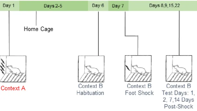
Figure 1.1 Stress-enhanced fear learning. In the SEFL paradigm in our laboratory, rats are exposed to 15 2 mA foot shocks in Context A. Six days later, animals are exposed to Context B without foot shocks to test for generalization of fear between the two contexts. On Day 7, rats are placed back into Context B where they receive a single 1 mA foot shock. Contextual fear learning to Context B is then analyzed. SEFL is used to study how the initial severe stressor in Context A enhances contextual fear learning to the single foot shock in Context B. This phenomenon reflects the hypervigilance or hyperreactivity to future stressors that is a key component of human PTSD.
6
brain following the severe stressor of SEFL will be important for understanding multiple behavioral outcomes. Converging mechanisms that drive depressive- and anxiety-like phenotypes that are part of the PTSD-like phenotype as a whole will be important in our overall understanding of behavioral outcomes following stress that lead to PTSD.
Figure 1.2 Stressor of the SEFL paradigm enhances general anxiety-like behavior seven days later. In order to compare the behavioral consequences of severe foot shock in SEFL to the behavioral consequences of stress in the predator scent stress and single prolonged stress models, rats were exposed to foot shock stress in Context A (15 2 mA scrambled foot shocks over 90 minutes) and then returned to their home cage for six days. Seven days after foot shock stress, animals were tested for anxiety-like behavior in the Elevated Plus Maze. Rats exposed to foot shock stress exhibited less time in the open arms of the maze (left panel) and a greater number of stretched attend postures (right panel, a measure of risk assessment behavior), both of which are reflective of enhanced anxiety in the model. * p < 0.05.
7
Thus, these results suggest that morphine treatment exerts its effect on behavioral outcomes following stress through a unique neural mechanism that drives enhanced fear learning.
While it is exciting that the use of morphine has shown promise to clinically prevent or alleviate PTSD, its use clinically in this context is severely limited by its abuse liability. Further, high co-morbidity between opioid abuse specifically and PTSD in a number of settings presents additional problems to its use as treatment (Fareed, Eilender et al. 2013; Dabbs, Watkins et al. 2014). The overarching goal of the current studies stems from an effort to elucidate the mechanisms through which morphine alters stress-enhanced fear learning in order to uncover more targets for the development of a prophylactic pharmaceutical to treat PTSD, without abuse liability.
Hippocampal function is important in PTSD and SEFL
non-8
traumatized controls (O'Doherty, Chitty et al. 2015) and an interesting recent report suggests that the dentate gyrus volume, specifically, is reduced (Hayes, Hayes et al. 2017). Furthermore, within PTSD populations, hippocampal volume is inversely related to treatment outcomes such that greater hippocampal volume predicts a more favorable treatment response (Rubin, Shvil et al. 2016).
A better understanding of the neurobiological mechanisms driving the acquisition of traumatic or stressful memories is an important step in understanding PTSD. The specific aims described herein make contributions to a greater effort to better understand the hippocampal mechanisms that influence memory acquisition and how such mechanisms might differ between traumatic or stressful vs. normal memories and experiences. How the current findings relate to potential mechanisms that have been hypothesized to be involved in memory acquisition and learning, including alterations in dendritic spine morphology/ synaptic remodeling (Yang, Pan et al. 2009; Lai, Franke et al. 2012; Giachero, Calfa et al. 2015) and secretion of glial-derived neurotrophic factors in the hippocampus (Bekinschtein, Cammarota et al. 2014; Rosas-Vidal, Do-Monte et al. 2014), is discussed throughout Chapters 3 through 5.
Role of cytokine signaling in stress response mechanisms
9
al. 2014; Hutchinson and Watkins 2014). Recent evidence also suggests that cytokines can directly influence learning and memory processes (Goshen, Kreisel et al. 2007; Goshen and Yirmiya 2009). The experiments described in the current dissertation are based on the conceptually innovative hypothesis that neuroimmunological mechanisms are involved in the development and expression of PTSD.
Interleukin-1β (IL-1β) is a proinflammatory cytokine thought to underlie many of the parallels between the body’s response to psychological stress and physical illness. Both conditions lead to a phenotype termed sickness behavior, which is characterized in part by enhanced anxiety (Dantzer 2009; Goshen and Yirmiya 2009). More recently, IL-1β has been shown to be critical to fear learning and memory related phenomena (Goshen, Kreisel et al. 2007). Interestingly, several groups have also reported upregulated circulating peripheral cytokines, including IL-1β, in PTSD patients (Gill, Saligan et al. 2009; Guo, Liu et al. 2012; Gola, Engler et al. 2013; Lindqvist, Wolkowitz et al. 2014; Passos, Vasconcelos-Moreno et al. 2015; Wang and Young 2016), even suggesting cytokine expression as a biomarker for affected individuals following trauma (Cohen, Meir et al. 2011).
10
immunoreactivity by hippocampal subregion, and the dentage gyrus exhibited the most dense IL-1β expression by far (relative to cornu ammonis (CA)1 and CA3). A subsequent experiment showed that centrally blocking IL-1 signaling through an intracerebroventricular (ICV) infusion of IL-1 receptor antagonist (IL-1RA) at 24 and 48 hours after Context A exposure prevented the development of SEFL (Jones, Lebonville et al. 2015). These data strongly suggest that IL-1 is causally related to SEFL in the hippocampus. Furthermore, the extremely dense IL-1β signal observed in the dentate gyrus is interesting given the recent report that the dentate gyrus, specifically, may be altered in human populations diagnosed with PTSD as well (Hayes, Hayes et al. 2017). Follow up studies found that the same systemic morphine treatment that prevented the development of SEFL (Szczytkowski-Thomson, Lebonville et al. 2013) also attenuated hippocampal stress-induced IL-1β (Jones, Lebonville et al. 2015). Collectively, our published work provides strong evidence that morphine exerts an anti-inflammatory effect on the hippocampus that has direct implications in fear learning behavior.
11
an important first step to this endeavor, the second goal of the current dissertation was to identify the cellular source of hippocampal stress-induced IL-1β.
Role of astroglial signaling in stress response mechanisms
While cytokines can be expressed by multiple cell types in the brain, astrocyte-derived cytokines, including IL-1β, have been increasingly implicated in stress response mechanisms (Goshen and Yirmiya 2009) (Sugama, Takenouchi et al. 2011). Data presented in Chapter 2 of the current dissertation provides even further support for this hypothesis in that that hippocampal astrocytes are the cellular source of stress-induced IL-1β, which we have shown to be causally related to the development of SEFL (See Figures 1.4 and 1.5). Traditionally viewed merely as neuronal “glue”, glial cells are now known to be critically involved in a diverse array of functions in development and disease of the central nervous system (CNS), however, the heterogeneity of glia remains underappreciated (Barres 2008). We argue that astrocyte function, especially in the context of severe stress, merits further scientific study.
Astrocytes are process-bearing cells that make direct contact with synapses (Blanco-Suarez, Caldwell et al. 2016). A “tripartite” synapse consists of a pre-synaptic cell, a post-synaptic cell, and an astrocyte that envelops the synapse. Converging evidence confirms that in this context, astrocytes engage in bidirectional communication with neurons through the release of gliotransmitters, such as ATP, d-serine, and/or glutamate, such that astrocytes can directly regulate synaptic transmission (Bernardinelli, Randall et al. 2014; Blanco-Suarez, Caldwell et al. 2016).
12
depressive and anxiety-like phenotypes that are observed in PTSD. Glial fibrillary acidic protein (GFAP) expression, a cytoskeletal protein selectively expressed by astrocytes and commonly employed as a marker of astrocyte activation or reactivity, has been reported to be altered in several brain regions, including the hippocampus, following a variety of different stress protocols (Colombo and Farina 2016) (Tynan, Beynon et al. 2013; Xia, Zhai et al. 2013; Choi, Ahn et al. 2016; Saur, Baptista et al. 2016). Impaired astrocyte glutamate transport, decreased release of astrocyte-derived neurotrophic factors, and altered astrocyte density have also been observed in the context of preclinical models of depression (Niciu, Henter et al. 2014). Furthermore, effective antidepressants, which are sometimes used to alleviate PTSD symptoms, have been associated with gliotrophic effects (Czeh, Muller-Keuker et al. 2007; Banasr, Chowdhury et al. 2010; Niciu, Henter et al. 2014). Given the significant role of the hippocampus in PTSD described above, it is important to note a recent report by Iwata and colleagues showed that the protective effect of imipramine in a model of learned helplessness was blocked by fluorocitrate, a reversible astrocyte inhibitor, directly into the hippocampus (Iwata, Shirayama et al. 2011). Similarly, Zhang and colleagues demonstrated that gastrodin, a compound shown to protect against depressive-like phenotypes, acts by enhancing astrocyte-derived brain-astrocyte-derived neurotrophic factor (BDNF) (Zhang, Peng et al. 2014).
13
allow researchers to thoroughly describe the morphometric properties of astrocytes and to directly manipulate astroglial G protein coupled receptor (GPCR) signaling in vivo.
High resolution analysis of the morphometric properties of astrocytes
Dr. Kathryn Reissner and colleagues have optimized a method to isolate and quantify astrocyte volume and synaptic contacts throughout a 3-dimensional reconstruction of an individual cell (Scofield, Li et al. 2016). With their method, an adeno-associated virus serotype 5 (AAV) is used to express Green Fluorescent Protein (GFP) in a membrane-dependent manner under a GFAP promoter such that entire astrocyte, including the most distal perisynaptic processes can be visualized and quantified. Double label fluorescence immunohistochemistry can be used to quantify the colocalization of GFP with synaptic markers, such as synapsin or postsynaptic density 95, within an individual cell. As such, high resolution confocal microscopy and Bitplane Imaris analysis can produce thorough measures of the volume, surface area, and synaptic colocalization of individual astrocytes. This technology has been employed in one peer-reviewed article thus far and is known to produce results that are robust and reliable (Scofield, Li et al. 2016). The goal of Chapter 3 of the current dissertation was to employ this technology to analyze how the severe stressor the SEFL paradigm alters the morphometric properties of hippocampal astrocytes.
Glial-expressing Designer Receptors Exclusively Activated by Designer Drugs
14
15
Specific Aims
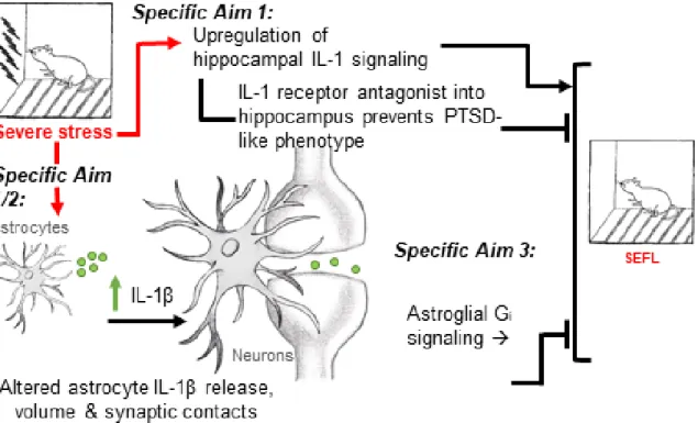
As reviewed above, PTSD is a debilitating condition that imposes severe fiscal and emotional costs on society. A better understanding of the neurobiological mechanisms driving PTSD is crucial to the development of more targeted treatments. Rodent paradigms that examine behavioral responses to severe stress can be used to elucidate mechanisms involved in PTSD-like behaviors. The goal of the current dissertation was to examine hippocampal neural immune signaling as a potentially important mechanism involved in stress-enhanced fear learning, a PTSD-like phenotype. To accomplish goal, the three specific aims described below examined stress-induced changes at the cellular level in the hippocampus following severe stress and tested the involvement of two specific signaling pathways in the development of SEFL.
16
of astrocytes and (2) that both blocking hippocampal IL-1 signaling and activating hippocampal astroglial Gi signaling prevent the development of SEFL (Figure 1.3).
Specific Aim 1 tested the hypothesis that blocking IL-1 signaling in the dorsal hippocampus prevents SEFL and isolated the specific cellular source of hippocampal stress-induced IL-1β. Strong preliminary data suggest that blocking IL-1 signaling directly in the dorsal hippocampus is sufficient to prevent the development of SEFL. However, the cellular source of hippocampal stress-induced IL-1 remains unknown. The experiments in this aim contribute to our understanding of the role of central IL-1 in SEFL in that we (1) directly tested whether dorsal hippocampal IL-RA is sufficient to attenuate SEFL and (2) identified the cellular source of stress-induced IL-1. Furthermore, analyses in this aim contribute to our knowledge of stress-induced changes of two common markers for astrocytes and microglia, glial fibrillary acidic protein (GFAP) and ionized calcium-binding adaptor molecule -1 (Iba-1). Specific Aim 1 is described in Chapter 2.
17
potential stress-induced reduction in postsynaptic density 95 (PSD95) are discussed. Specific Aim 2 is described in Chapter 3.
Specific Aim 3 tested the hypothesis that astroglial Gi activation attenuates SEFL
18
19 Chapter 2
EXAMINATION OF STRESS-INDUCED HIPPOCAMPAL IL-1β: EFFECT OF HIPPOCAMPAL IL-1RA ON STRESS-ENHANCED FEAR LEARNING AND
IDENTIFICATION OF THE CELLULAR SOURCE2 Introduction
Converging evidence from both human and animal studies has suggested that psychiatric disorders involving depression and anxiety, including post-traumatic stress disorder (PTSD), involve substantial immune system dysregulation (Silverman, Macdougall et al. 2007; Gill, Saligan et al. 2009; Koo and Duman 2009; Stepanichev, Dygalo et al. 2014; Jones, Lebonville et al. 2015). As mentioned previously, several published studies have reported that PTSD is associated with elevated peripheral cytokines, including interleukin-1β (IL-β), tumor necrosis factor-α (TNF-α), and interleukin-6 (IL-6) (Gill, Saligan et al. 2009; Guo, Liu et al. 2012; Gola, Engler et al. 2013; Passos, Vasconcelos-Moreno et al. 2015; Wang and Young 2016). Cohen and colleagues have even suggested IL-1 as a potential biomarker for susceptibility to PTSD (Cohen, Meir et al. 2011). Central IL-1 signaling is consistently shown to be upregulated by a variety of different stress protocols in rodents and to be critically
involved in stress response mechanisms that drive behavioral outcomes (Avital, Goshen et al. 2003; Goshen and Yirmiya 2009). For example, peripheral administration of IL-1β has been shown to lead to enhanced anxiety-like behavior in the elevated plus maze (EPM) (Swiergiel
20
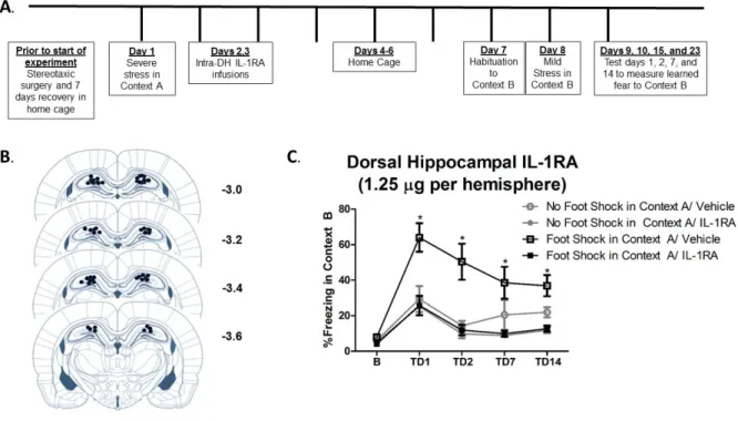
and Dunn 2007), and blocking IL-1 signaling centrally prevents stress-induced reductions in social interaction (Arakawa, Blandino et al. 2009). As mentioned in Chapter 1, we recently published the finding that stress-enhanced fear learning (SEFL), a preclinical animal model of PTSD developed by Rau and colleagues (Rau, DeCola et al. 2005), requires central IL-1 signaling. Our data demonstrated that the severe stressor of the SEFL paradigm (15 foot shocks) induces an increase in IL-1β in the dorsal hippocampus (DH) 24-48 hours after the stressor. Furthermore, blocking IL-1 signaling in the brain through an intracerebroventricular infusion of IL-1 receptor antagonist (IL-1RA) prevents the development of enhanced fear learning (Jones, Lebonville et al. 2015). Together these data suggest that central IL-1RA may be acting specifically in the hippocampus. Accordingly, the first goal of the current chapter was to test whether hippocampal IL-1 signaling 24-48 hours after severe stress is necessary for the expression of SEFL.
21
effect of stress on neuron-derived IL-1β. Kwon and colleagues reported an increase in IL-1β colocalized with neuronal nuclei following four days of restraint stress (Kwon, Seo et al. 2008). In contrast, there are several published studies to support the potential for microglia-derived or astrocyte-derived IL-1β, as described below.
22
immunoreactivity (MHC II, predominantly expressed by microglia) was increased in the hippocampus 24 hours after inescapable tail shock (Frank, Baratta et al. 2007), but they observed no change in either GFAP or Iba-1 immunoreactivity in the same tissue.
The final candidate for a potential source of stress-induced IL-1β is astrocytes. As mentioned in Chapter 1, though traditionally viewed merely as neuronal “glue”, astrocytes are now known to be critically involved in a diverse array of functions in development and disease (Barres 2008). Converging evidence from several laboratories using a variety of different severe stress procedures suggests that both GFAP expression and astrocyte process length are altered over time in the brain following stress (Tynan, Beynon et al. 2013; Xia, Zhai et al. 2013; Choi, Ahn et al. 2016; Saur, Baptista et al. 2016). Choi and colleagues observed an increase in the length and number of astrocyte processes but a decrease in GFAP in the DH one hour, but not 24 hours, after exposure to foot shock fear conditioning (Choi, Ahn et al. 2016). In contrast, others have observed decreases in the number of astrocyte processes either following chronic restraint stress (Tynan, Beynon et al. 2013) or 24-48 hours after foot shock exposure (Saur, Baptista et al. 2016). Of particular relevance here, Sugama and colleagues found that IL-1β expression was increased specifically in astrocytes, and not microglia, following cold stress (Sugama, Takenouchi et al. 2011).
23
whether IL-1 signaling in the DH is critical to the development of a PTSD-like phenotype in SEFL. Experiment 2.2 examined stress-induced changes in astrocytes and microglia in the DH and identified the cellular source of stress-induced IL-1β in this region. Analyses from Experiment 2.2a replicated our previous finding of stress-induced IL-1β in the dorsal hippocampus. Analyses from Experiment 2.2b quantified GFAP and Iba-1 immunoreactivity to examine stress-induced changes in astrocytes and microglia, respectively. Finally, analyses in Experiment 2.2c used Bitplane Imaris software in combination with confocal microscopy to visualize the colocalization of IL-1β with GFAP, Iba-1, and NeuN following foot shock to isolate and quantify astrocyte-derived, microglia-derived, and neuron-derived IL-1β, respectively. Collectively, these experiments tested the hypotheses that IL-1 signaling in the DH is critical for the development of SEFL and that astrocytes are the predominant cellular source of hippocampal IL-1β following stress in this context.
Methods
Animals
24
Experiment 2.1: Effect of intra-dorsal hippocampal IL-1RA on the development of SEFL
Surgery
Animals were anesthetized with a 1.0 mg/kg intraperitoneal injection of 9:1 (vol:vol) ketamine hydrochloride (100mg/ml) mixed with xylazine (100 mg/ml). Guide cannulae (26 Gauge, Plastics One, Roanoke, VA) were directed bilaterally at the DH (AP -3.4 mm, ML ± 3.1 mm, DV -2.2 mm, 15 degrees, relative to bregma). Animals were given one week for postoperative recovery prior to the start of any experimental procedures. Upon completion of the experiment, correct cannula placement was verified and any animals with incorrect placement were dropped from the analysis.
Stress-enhanced fear learning
25
cage. Context B was also associated with distinct textile, olfactory, and auditory characteristics from both Context A and the home cage. In addition, behavior in Context B was recorded using a video recording system (Sony Video Camera Model HDR-CX150). Similar to Rau and colleagues (Rau, DeCola et al. 2005), animals were exposed to Context B for 30 minutes without foot shocks being delivered to allow for habituation to the new context. On Day 8, animals were placed back into Context B where all animals received a single 1 mA scrambled foot shock, 3 minutes, 12 seconds after being placed into the context. Behavior during the three minutes prior to the single shock was recorded and analyzed to test for generalization of fear between the two contexts (these data are presented as ‘baseline’). On Days 9, 10, 15 and 22 (Test Days 1, 2, 7 and 14), animals were placed in Context B for 8 minutes, 32 seconds and behavior was recorded.
Ethovision XT video tracking software (Noldus Information Technology Inc.) was used to analyze freezing behavior, a measure of learned fear defined as a lack of all movement except that required for breathing. Specifically, the activity analysis feature (Activity Threshold = 10) was used to calculate the percent of time each animal was inactive during each contextual fear test and at baseline. No animals in any group demonstrated significant freezing behavior to Context B prior to the single foot shock, suggesting that there was no generalization of fear between contexts (Results 2.1). Thus, any differences observed between treatment groups presented here reflect altered learning to the single foot shock in Context B.
IL-1 receptor antagonist
26
from Context A, on Days 2 and 3, animals were microinfused with 1.25 µg of IL-1RA or sterile saline vehicle per hemisphere at a rate of 0.25 µl/min. Injectors had a 1mm projection and were left in place for 1 minute after the infusion to allow for diffusion. These time points were based on our earlier published findings that morphine administration and intracerebroventricular IL-1RA prevent the development of SEFL when administered 48 hours after Context A (Szczytkowski-Thomson, Lebonville et al. 2013; Jones, Lebonville et al. 2015).
Experiment 2.2: Immunofluorescence analysis of severe stress-induced changes in
hippocampal GFAP, Iba-1, NeuN, and IL-1β
Stress exposure
27 Immunohistochemistry
28
60 minutes. Sections were mounted onto SuperFrost Plus slides (Fisher Scientific, Pittsburgh, PA) using Vectashield hard set mounting medium (Vector Laboratories, Burlingame, CA). Tissue from poor perfusions that yielded high nonspecific background which interfered with thresholding and colocalization calculations was dropped from the analysis, and any such decision was made blind to treatment group.
Confocal microscopy, Bitplane Imaris colocalization analysis, and cell counting
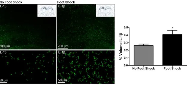
All image acquisition and analysis was completed by an experimenter blind to treatment group. Tissue was imaged using a Zeiss LSM800 confocal microscope with laser lines that excite at 405 nm, 488 nm, and 561 nm. Images were acquired using a 63X oil immersion lens. Z stacks of the dentate gyrus of the dorsal hippocampus (AP -3.12 mm through -3.84 mm from bregma) were acquired using a frame average of 4, 1024 by 1024 frame size, 12 bit image resolution, and 0.8 µm step size. We focused our analysis on the dentate gyrus based on our previous finding that IL-1β expression is most dense in this subregion of the DH (Jones, Lebonville et al. 2015).
29
signals. In addition to Imaris volume and colocalization analyses, the number of GFAP-positive and Iba-1-GFAP-positive cells in the dentate gyrus was counted in images acquired at 10X by an experimenter blind to treatment group.
Statistical analyses
30 Results
Experiment 2.1: Intra-dorsal hippocampal IL-RA prevents SEFL
31
32
Experiment 2.2a: Stress-induced increase in hippocampal IL-1β is replicated
We previously reported that the severe stressor of the SEFL paradigm induces an increase in hippocampal IL-1β immunoreactivity and mRNA that emerges at 6 hours and persists through 72 hours following stress exposure (Jones, Lebonville et al. 2015). Here, this effect is replicated in that exposure to the severe stressor of SEFL significantly enhanced hippocampal IL-1β immunoreactivity 48 hours later, t (10) = 2.083, p = 0.0319 (Figure 2.2).
33
Experiment 2.2b: Severe stress attenuates Iba-1, but not GFAP, in the dorsal hippocampus
34
35
Experiment 2.2c: Stress-induced hippocampal IL-1β is colocalized primarily with GFAP in
both stressed and non-stressed animals
Figures 2.4 and 2.5 show that there was an overwhelming amount of IL-1β colocalized with GFAP, 75% to 79% of the IL-1β signal, and only minimal colocalization with Iba-1 or NeuN, less than 5% of the IL-1β signal. Thus, there was over 15- fold greater colocalization of IL-1β with GFAP compared to the other two cell-type markers in both stressed and non-stressed animals. As such, there was a significant main effect of cell-type analyzed in both the % IL-1β signal colocalized, F (2, 28) = 2423.859, p < 0.001, and the Pearson’s correlation coefficient, F (2, 28) = 51.166, p < 0.001. Again, regarding both the % IL-1β signal colocalized and the Pearson’s correlation coefficient, post hoc comparisons confirmed significantly more colocalization with GFAP than Iba-1, p < 0.001, and with GFAP than NeuN, p < 0.001. There was no difference between colocalization of IL-1β with Iba-1 and with NeuN, p > 0.05.
36
37
38 Discussion
In the current chapter, we provide evidence that astrocytes are the cellular source of foot shock-induced hippocampal IL-1β, which plays a critical role in the development of SEFL, an animal model of PTSD. Experiment 2.1 supports our hypothesis that dorsal hippocampal IL-1 signaling is necessary for the development of SEFL in that intra-DH IL-1RA infused 24 and 48 hours following severe stress prevented the expression of SEFL. Further, Experiment 2.2 provides the first evidence that Iba-1 immunoreactivity is reduced 48 hours following foot shock stress and that the IL-1β signal at this critical time point for behavioral consequences of severe stress is almost exclusively colocalized with an astrocyte-specific marker, and not with microglia- or neuron-specific markers.
39
mechanisms involved in astrocyte regulation of stress and anxiety-related behavior remain unclear, our data and others’ reports converge to suggest a critical role for astrocytes in behavioral responses to stress.
40
We hypothesized that GFAP expression would increase following stress because the IL-1β signal is increased in the hippocampus at this time point following stress, and is highly colocalized with GFAP. While this hypothesis was not confirmed here, there are several potential explanations for our observed lack of effect. First, the amount of GFAP expression in the DH (1-1.5% of the region measured) is more than six times that of IL-1β (0.2-0.3% of the region measured). Thus, an increase in IL-1β in astrocytes can easily occur without a corresponding increase in GFAP. Second, while GFAP is one of the most canonical astrocyte markers relied on in the field, GFAP is a cytoskeletal protein that is only expressed within 15% of a given astrocyte’s area and even then, only by a subset of astrocytes (Benediktsson, Schachtele et al. 2005; Rajkowska and Stockmeier 2013). Measures of GFAP have also yielded results that have conflicted with other measures of astrocyte reactivity. Tynan and colleagues examined stress-induced changes in astrocyte activation and showed an increase in S100β, another astrocyte-specific marker, but a decrease in GFAP following the same stressor (Tynan, Beynon et al. 2013). Thus, our measure of GFAP immunoreactivity is an incomplete measure of astrocyte reactivity and experiments described in Chapter 3 take advantage of new technologies to study astrocyte morphology in greater detail.
41
implications of stress-induced alterations in additional immune signaling pathways would provide more information regarding the specificity of the IL-1 mechanism or could identify additional mechanisms that play a role.
42 Chapter 3
EFFECT OF SEVERE STRESS ON THE MORPHOMETRIC PROPERTIES OF HIPPOCAMPAL ASTROCYTES
Introduction
The goal of Chapter 3 was to conduct a more thorough analysis of severe stress-induced changes in hippocampal astrocyte morphology. Experiments described in Chapter 2 present a potential role for astrocytes in the development of SEFL. Specifically, our data provide evidence that the predominant source of hippocampal stress-induced IL-1β, which we have also shown to be causally related to SEFL, is astrocytes (Figures 2.4 and 2.5). Relatedly, astrocytes not only release IL-1 but also express IL-1 receptors, and IL-1β is thought to be a potent induction signal for astrocyte activation (Giulian, Woodward et al. 1988; Herx and Yong 2001; Proescholdt, Chakravarty et al. 2002; John, Lee et al. 2003). As such, stress-induced IL-1β may, over time, induce further changes in the morphometric properties of astrocytes. While we did not observe a stress-induced change in hippocampal GFAP immunoreactivity, as described in Chapter 2, others have after a variety of stress protocols (Tynan, Beynon et al. 2013; Xia, Zhai et al. 2013; Choi, Ahn et al. 2016; Saur, Baptista et al. 2016) and inconsistencies in our understanding of hippocampal astrocyte function in the context of behavioral responses to stress remain.
Changes in astrocyte morphology are important to investigate because astrocyte morphology and synaptic contact can directly influence astrocyte and neuronal function
43
glutamate homeostasis, synaptic remodeling, secretion of neurotrophic factors, and synaptic strength (Ben Menachem-Zidon, Avital et al. 2011; Scofield and Kalivas 2014; Blanco-Suarez, Caldwell et al. 2016). In the context of our data, if stress induces an increase in the volume, surface area, or synaptic contacts of hippocampal astrocytes, then more IL-1β might reach the synapse.
Current studies that have examined astrocyte morphology following stress have been limited by the reliance on GFAP or S100β immunoassays (Tynan, Beynon et al. 2013; Xia, Zhai et al. 2013; Choi, Ahn et al. 2016; Saur, Baptista et al. 2016). As mentioned in Chapter 2, GFAP constitutes only about 15% of the total volume of an astrocyte and is limited to a subset of astrocytes (Benediktsson, Schachtele et al. 2005; Rajkowska and Stockmeier 2013). Thus, while these studies do provide support to our hypotheses, they do not provide full information about how hippocampal signaling might be influenced by astrocyte changes. For example, how fine processes of glial cells that make synaptic contacts are altered following stress is unclear from immunoassays of GFAP alone (Scofield, Li et al. 2016).
44
based thresholding to quantify astrocyte volume, surface area, and colocalization with synaptic contacts. Here, we apply this technology in the context of the severe stressor within the SEFL paradigm.
45
can induce a reduction in PSD95 in cortex that is associated with poor performance in the rotarod and Morris water maze tests (Mir, Sen et al. 2014). Collectively, while a hypothesis regarding the specific memory mechanism involved in mild vs. stress-enhanced fear learning would be premature, we hypothesize that the severe stressor in the context of SEFL may be associated with a reduction in PSD95. Thus, measuring the levels of PSD95 in the acquired Z stacks used for analysis of the morphometric properties of astrocytes is both an important control for any implications in terms of astrocyte morphology as well as an interesting experimental question.
46 Methods
Animals
Male Sprague Dawley rats (225-250 g, Charles River Laboratories, Raleigh, NC) were housed individually under a reversed 12 hour light-dark cycle. They were given ad libitum access to food and water and were handled regularly throughout all experiments. All procedures were conducted in accordance with and approval by the UNC Institutional Animal Care and Use Committee.
Experiment 3.1 Verification of AAV5-GFAP-HA-hM3Dq-IRES-mCitrine as a
membrane-dependent tag
Viruses
AAV5-GFAP-HA-hM3Dq(Gq)-IRES-mCitrine was obtained directly from the UNC Gene Therapy and Vector Core (Chapel Hill, NC). AAV5-GFAP-Lck-GFP was provided by Dr. Kathryn Reissner. Purified viruses were obtained pre-dialyzed (350mM NaCl, 5% D-sorbitol in phosphate buffered saline), and combined to a single stock containing both viruses prior to surgery.
Surgery and Sacrifice
47
minute. Injectors were left in place for 15 minutes to allow for diffusion of the virus away from the injector site. Three weeks later, all animals were sacrificed via transcardial perfusion. Briefly, rats were deeply anesthetized with a 1 ml intraperitoneal injection of 9:1 (vol:vol) ketamine hydrochloride (100 mg/ml) mixed with xylazine (100 mg/ml). Animals were transcardially perfused with cold 0.1 M phosphate buffer (pH = 7.4) for three minutes at a rate of 15 mls/ minute and then with cold 4% paraformaldehyde in 0.1 M phosphate buffered saline (pH = 7.4) for seven minutes at a rate of 15 mls/ minute. Brains were extracted and post-fixed in 4% paraformaldehyde in 0.1 M phosphate buffered saline for 4–6 hours and then sliced into 100 µm sections on vibratome.
Immunohistochemistry
48
times for 10 minutes, mounted onto SuperFrost Plus slides (Fisher Scientific, Pittsburgh, PA) and cover slipped using Vectashield hard set mounting medium (Vector Labs, Burlingame, CA). For all antibodies, control experiments verified that our staining for all target antigens was visible and specific using this method.
Confocal microscopy and Bitplane Imaris analysis
A Zeiss LSM800 confocal microscope was used to acquire Z stacks of individual cells in the dorsal hippocampus that were transduced by both GFAP-hM3Dq and GFAP-Lck-GFP. Z stacks were acquired using a 63X oil immersion lens, 1024 x 1024 frame size, frame average of 4, and step size of 0.8 µm. Laser lines that excite at 405nm, 488nm, 561nm were used to visualize GFP, and the Alexa Fluor tags used to label GFAP-hM3Dq, GFAP, and PSD95. Images were deconvolved using Bitplane AutoQuant X3 (10 iterations, (Lee, Wee et al. 2014)) and exported to Bitplane Imaris software (Zurich, Switzerland).
Experiment 3.2: Effect of stress on the morphometric properties of astrocytes.
Virus
AAV5-GFAP-HA-hM3Dq(Gq)-IRES-mCitrine was obtained directly from the UNC Gene Therapy and Vector Core (Chapel Hill, NC). Purified virus was obtained pre-dialyzed (350mM NaCl, 5% D-sorbitol in phosphate buffered saline) and microinjected at 3.7 x 10¹² particles/ml.
Surgery
49
were infused with AAV5-GFAP-HA-hM3Dq-IRES-mCitrine. Injectors (26 Gauge, Plastics One, Roanoke, VA) were directed bilaterally at the dorsal hippocampus (AP -3.4 mm, ML ± 3.1 mm, DV -3.2 mm, 15 degrees, relative to bregma). Virus was injected in a volume of 0.7 µl per hemisphere at a rate of 0.1 µl per minute. Injectors were left in place for 15 minutes to allow for diffusion of the virus away from the injector site. Experimental procedures began three weeks later.
Stress exposure and sacrifice
Animals (N = 16, n = 8) were randomly assigned to either a Foot Shock (in Context A) or No Foot Shock (in Context A) treatment and exposed to only the initial severe stressor of the SEFL paradigm described in Experiment 2.1. Thus, animals assigned to receive foot shocks were exposed to 15 2 mA scrambled foot shocks in Context A, an environment distinct from the home cage, while control animals were exposed to the same context without foot shocks being delivered. Forty-eight hours after removal from Context A, rats were deeply anesthetized with a 1 ml intraperitoneal injection of 9:1 (vol:vol) ketamine hydrochloride (100 mg/ml) mixed with xylazine (100 mg/ml). Animals were transcardially perfused with cold 0.1 M phosphate buffer (pH = 7.4) for three minutes at a rate of 15 mls/minute and then with cold 4% paraformaldehyde in 0.1 M phosphate buffered saline (pH = 7.4) for seven minutes at a rate of 15 mls/minute. Brains were extracted and post-fixed in 4% paraformaldehyde in 0.1 M phosphate buffered saline for 4–6 hours and then sliced into 100 µm sections on vibratome. Immunohistochemistry
50
primary control stain was used to analyze nonspecific background and ensure analyzed signal was specific to the antigens. Brain sections were washed three times for 10 minutes in 0.1 M phosphate buffer (PB, pH = 7.4) and incubated in 10% Normal Goat Serum and 2% TritonX100 for 60 minutes. Tissue was then incubated in primary antibody for three nights at 4°C in 10% Normal Goat Serum, 2% TritonX100, and rabbit anti-HA (Cell Signaling, Danvers, MA, Cat # mAb3724, 1:500) and mouse-anti-PSD95 (1:500, Thermo Fisher Scientific, Cat # MA1-045). The following day, tissue was washed three times for 10 minutes in 0.1 M PB and incubated in 10% Normal Goat Serum, 2% TritonX100, and secondary antibody for three nights at 4°C. Secondary antibodies conjugated with Alexa Fluor dyes (Thermo Fisher Scientific, Waltham, MA, 1:1000) were be used for visualization. Tissue was then washed 3 times for 10 minutes, mounted onto SuperFrost Plus slides (Fisher Scientific, Pittsburgh, PA) and cover slipped using Vectashield Hard Set mounting medium (Vector Labs, Burlingame, CA). For all antibodies, control experiments verified that our staining for all target antigens was visible and specific using this method.
Confocal microscopy and Bitplane Imaris analysis
Image Acquisition
51
at 405nm, 488nm, 561nm were used to visualize GFP, and the Alexa Fluor tags used to label GFAP-hM3Dq, GFAP, and PSD95. Acquisition parameters including Master Gain, Digital Offset and Laser Power were kept the same throughout all acquisition. Images were deconvolved using Bitplane AutoQuant X3 (10 iterations, (Lee, Wee et al. 2014)) and exported to Bitplane Imaris software (Zurich, Switzerland).
Astrocyte volume, surface area, and colocalization with PSD95
52
the dorsal hippocampus was dropped from the analysis, and any such decision was made blind to treatment group.
Quantification of hippocampal PSD95 immunoreactivity
PSD95 immunoreactivity in each of the acquired Z stacks was quantified. A no primary antibody control stain revealed that the secondary incubation was associated with a non-specific band of signal towards the edge of the tissue. This portion of the Z stack was cut from the analysis manually by an experimenter blind to treatment group. Ten random samples of the absolute intensity of the PSD95 signal through the Z stack were recorded and the mean intensity was used to set the absolute intensity threshold. Again, care was taken to ensure that the PSD95 signal was at least 6-fold greater than background detected in the no primary antibody control stain in all Z stacks included in the analysis. The ROI was defined as the whole Z stack and the % ROI with voxels above the absolute intensity threshold determined was recorded.
Statistical Analysis
53 Results
Experiment 3.1 AAV5-GFAP-HA-hM3Dq-IRES- -mCitrine is expressed in a
membrane-dependent manner
54
55
56
Experiment 3.2: Effect of stress on the morphometric properties of astrocytes.
Stress exposure does not alter astrocyte volume, surface area or colocalization with PSD95
There was no effect of foot shock on astrocyte volume, t (11) = 0.8686, p = 0.8686, or surface area, t (11) = 0.05384, p = 0.9580. There was also no effect of foot shock on the %ROI colocalized with PSD95, t (8) = 1.394, p = 0.2008. These data as well as representative images of a 3-D reconstruction of an astrocyte co-labeled with PSD95 from each treatment group are presented in Figure 3.3.
As we have previously published data to suggest that stress-induced IL-1β is most dense in the dentate gyrus of the dorsal hippocampus (Jones, Lebonville et al. 2015), it is important to note that all of the cells that met the criteria for analysis were acquired from CA1 and CA3 (Figure 3.4). Because virus expression was very dense in this region (Figure 3.4), there were no cells that were non-overlapping and extended fully through the x, y, and z planes in this region.
57
58
59 Stress exposure attenuates PSD95 Immunoreactivity
As shown in Figure 3.5, foot shock attenuated PSD95 immunoreactivity, t (8) = 1.883, p = 0.482. Thus, the slight mean difference observed suggesting a decrease in astrocyte colocalization with PSD95 in Figure 3.3 is confounded by the fact that foot shock induced a decrease in the PSD95 signal.
60 Discussion
The hypothesis that severe stress induces changes in the morphometric properties of astrocytes 48 hours later was not supported here. There was no change induced by foot shock exposure in Context A in hippocampal astrocyte volume, surface area, or colocalization with PSD95. However, we observed a significant decrease in PSD95 immunoreactivity. The interesting implications of a stress-induced decrease in hippocampal PSD95 are discussed below.
It is important to note that dynamic stress-induced changes in astrocyte morphology may make critical time points for this variable difficult to pinpoint. While important to examine, 48 hours post-stress is not the only time point at which changes in astrocyte morphology might be important to SEFL and may represent a very short time point relative to the initial manipulation. Reissner and colleagues have reported changes in nucleus accumbens astrocyte morphology that were measured following 14-16 days of extinction after self-administration of cocaine (Scofield, Li et al. 2016). A later time point in the SEFL model might allow us to detect changes in the morphometric properties of astrocytes that occur on a slower timeline. For example, examining astrocyte morphology seven days after Context A exposure would give us a measure of astrocyte morphology at the time at which conditioning in Context B would normally take place, and thus the time at which stress-induced changes in plasticity are important for processing future stressors.
61
that PSD95 is important for the stabilization of new synaptic contacts, a reduction in PSD95 could be involved in a stress-induced change in dendritic morphology or the rate of spine turnover as another mechanism that is important for this effect (Berry and Nedivi 2017). Interestingly, Qiao and colleagues recently reported that an injection of Brain Derived Neurotrophic Factor (BDNF) rescued chronic mild unpredictable stress-induced deficits in hippocampal dendritic spine density and PSD95 (Qiao, An et al. 2017). Indeed, BDNF function has been implicated in both rodent models of PTSD (Ji, Peng et al. 2017; Lee, Shim et al. 2017) and human PTSD (Rakofsky, Ressler et al. 2012; Green, Corsi-Travali et al. 2013), further supporting the hypothesis that a better understanding of synaptic remodeling in the hippocampus in this context is important. Thus, while the stress-induced reduction in PSD95 should be replicated using multiple methods of measurement and a larger region of tissue sample given that the area we measured was limited to Z stacks of 8-10 cells per rat, our data suggest that attention to synaptic remodeling and spine turnover in the context of SEFL could lead to promising discoveries.
62
63 Chapter 4
EFFECT OF HIPPOCAMPAL ASTROGLIAL GI SIGNALING ON STRESS-ENHANCED FEAR LEARNING
Introduction
64
65
Impressively, these advances in chemogenetics have allowed researchers to manipulate astroglial GPCR signaling in vivo, and four groups have shown that manipulating astrocytes in the CNS directly influences behavioral outcomes (Agulhon, Boyt et al. 2013; Bull, Freitas et al. 2014; Scofield, Boger et al. 2015; Yang, Qi et al. 2015). Experiments described in the current chapter took advantage of this technology to selectively activate Gi signaling specifically in astrocytes within the dorsal hippocampus in the context of SEFL.
It is important to note that glial-expressing DREADD constructs are still new and, to our knowledge, only one effect has been reported with the AAV8-GFAP-hM4Di-mCherry to date (Yang, Qi et al. 2015). Yang and colleagues reported that hypothalamic astroglial Gi activation enhanced ghrelin-evoked food intake. In the same report, they also showed that hypothalamic astroglial Gq activation attenuated ghrelin-evoked food intake. In an effort to verify both of the glial DREADD constructs the group used, they also reported that CNO administration enhanced GFAP colocalization with cFos, an immediate early gene, only in AAV-GFAP-hM3Dq-mCherry-transduced rats and not in AAV-GFAP-hM4Di-mCherry transduced- rats (Yang, Qi et al. 2015). Thus, while their virus-specific and CNO-specific enhancement of feeding and GFAP colocalization with cFos are supportive of the validity of GFAP-hM4Di, there are no published data reported to directly confirm that CNO activates Gi -coupled signaling when used with this construct.
66
is possible that a floor effect may confound attempts to verify the hM4Di construct in naïve animals. To account for this, animals selected for this assay were also injected with lipopolysaccharide (LPS, derived from E. coli) to induce a state of central neuroinflammation in which we might be better able to detect CNO-induced changes in Gi-dependent messengers, such as cAMP, in astrocytes (Tarassishin, Suh et al. 2014).
Collectively, the goal of experiments described in the current chapter was to test the hypothesis that hippocampal astroglial Gi signaling is sufficient to attenuate SEFL. Experiment 4.1 used AAV8-GFAP-hM4Di-mCherry to selectively activate astroglial Gi signaling in the hippocampus at 1 mg/kg or 3 mg/kg CNO in SEFL. Experiment 4.2 used fluorescence immunohistochemistry, confocal microscopy, and Bitplane Imaris colocalization analysis to examine cAMP expression in GFAP-hM4Di-mCherry-positive astrocytes following LPS and CNO or Vehicle injection.
Methods
Animals
67 Virus
AAV8-GFAP-hM4Di(Gi)-mCherry was obtained directly from the UNC Gene Therapy and Vector Core (Chapel Hill, NC). Purified virus was obtained pre-dialyzed (350mM NaCl, 5% D-sorbitol in phosphate buffered saline) and microinjected at 2.0 x 10¹² particles/ml. Surgery
Animals (N = 64, n = 8) were anesthetized with a 1.0 mg/kg intraperitoneal injection of 9:1 (vol:vol) ketamine hydrochloride (100mg/ml) mixed with xylazine (100 mg/ml). All animals were infused with AAV8-GFAP-hM4Di-mCherry. Injectors (26 Gauge, Plastics One, Roanoke, VA) were directed bilaterally at the dorsal hippocampus (AP -3.4 mm, ML ± 3.1 mm, DV -3.2 mm, 15 degrees, relative to bregma). Virus was injected in a volume of 0.7 µl per hemisphere at a rate of 0.1 µl per minute. Injectors were left in place for 15 minutes to allow for diffusion of the virus away from the injector site. Experimental procedures started three weeks later.
Experiment 4.1: Effect of hippocampal astroglial Gi activation on SEFL
Stress-enhanced fear learning
68
vehicle immediately, 24, and 48 hours after removal from Context A. Contextual fear to Context B was measured by analyzing freezing behavior at baseline and on test days 1, 2, 7, and 14. Behavior was recorded as described in Chapter 2 and analyzed manually by a rater blind to the treatment group. Similar to Experiment 2.1, there was no significant generalization of fear between contexts in any treatment group (Results of Experiment 4.1), thus any differences observed reflect altered learning to the single shock in Context B.
Figure 4.1 Experimental timeline for Experiment 4.1. Rats were infused with AAV8-GFAP-hM4Di-mCherry bilaterally into the dorsal hippocampus, given three weeks to recover, and exposed to the SEFL paradigm. Rats were injected with CNO or vehicle (1 or 3mg/kg) immediately, 24, and 48 hours after Context A exposure.
Drug Administration
69 Sacrifice
All animals were sacrificed via transcardial perfusion. Briefly, rats were deeply anesthetized with a 1 ml intraperitoneal injection of 9:1 (vol:vol) ketamine hydrochloride (100 mg/ml) mixed with xylazine (100 mg/ml). Animals were transcardially perfused with cold 0.1 M phosphate buffer (pH = 7.4) for three minutes at a rate of 15 mls/minute and then with cold 4% paraformaldehyde in 0.1 M phosphate buffered saline (pH = 7.4) for seven minutes at a rate of 15 mls/minute. Brains were extracted and post-fixed in 4% paraformaldehyde in 0.1 M phosphate buffered saline for 4–6 hours, cryoprotected in 30% sucrose in 0.1 M phosphate buffer (pH = 7.4) for at least 48 hours, and sliced into 40 µm sections on freezing microtome. Brains were stored in 0.1 M phosphate buffer with 0.1% sodium azide (pH = 7.4) at 4°C until the time of assay.
Immunohistochemistry
70
Scientific, Waltham, MA) and cover slipped using Vectashield hard set mounting medium (Vector Labs, Burlingame, CA). For all antibodies, control experiments verified that our staining for all target antigens was visible and specific using this method. Slides were stored at 4°C until the time of microscopy. A Zeiss LSM800 confocal microscope was used to ensure that mCherry colocalized with GFAP, and not with NeuN, and thus was specific to astrocytes. Only data from animals with virus expression that was both hippocampus- and astrocyte- specific were included in the final analyses.
Experiment 4.2: Effect of CNO on colocalization of mCherry-positive cells with cAMP
Sacrifice
71
Figure 4.2 Experimental timeline for Experiment 4.2. A subset of animals from Experiment 4.1, that had been bilaterally infused with AAV8-GFAP-hM4Di-mCherry into the dorsal hippocampus, were injected with CNO or vehicle (3 mg/kg, s.c.) and 30 minutes later were injected with LPS (1mg/kg, s.c.). Three hours later, animals were sacrificed by transcardial perfusion and brains were extracted and processed for immunohistochemistry.
Immunohistochemistry
72
-20°C until the time of assay. In addition, as for all other antibodies used, no primary control experiments verified the specificity of the anti-cAMP signal.
Confocal microscopy and Bitplane Imaris colocalization analysis
Image Acquisition
A Zeiss LSM800 confocal microscope was used to acquire Z stacks of individual cells in the dorsal hippocampus that were transduced by AAV8-GFAP-hM4Di-mCherry. Acquisition and analyses were completed by an experimenter blind to treatment group. Z stacks were acquired using a 63X oil immersion lens, 1024 x 1024 frame size, 12 bit image resolution, frame average of 4, and step size of 0.8 µm. Laser lines that excite at 488nm, 561nm were used to visualize Alexa Fluor 488 and the mCherry signal. Acquisition parameters including Master Gain, Digital Offset and Laser Power were kept the same throughout all acquisition. Images were deconvolved using Bitplane AutoQuant X3 (10 iterations, (Lee, Wee et al. 2014)) and exported to Bitplane Imaris software (Zurich, Switzerland).
Colocalization of mCherry and cAMP
73
colocalized was recorded. In addition, to avoid a potential confounding floor effect given that we expected CNO to reduce cAMP in mCherry-positive cells, cells from both treatment groups that exhibited less than 10% colocalization were dropped from the analysis. Seven cells from the CNO-treated group and eight cells from the vehicle-treated group were dropped for this reason.
Statistical Analysis