CALCIUM-INDUCED STRUCTURAL REARRANGEMENTS RELEASE AUTOINHIBITION IN THE RAP-GEF, CALDAG-GEFI.
Aaron A. Cook
A dissertation submitted to the faculty at the University of North Carolina at Chapel Hill in partial fulfillment of the requirements for the degree of Doctor of Philosophy in the Department
of Biochemistry and Biophysics in the School of Medicine.
Chapel Hill 2018
ABSTRACT
Aaron A. Cook: Calcium-induced structural rearrangements release autoinhibition in the Rap-GEF, CalDAG-GEFI
(Under the direction of Wolfgang Bergmeier and John Sondek)
When blood vessels are injured, extracellular matrix is exposed leading to the recruitment and activation of platelets where they aggregate to staunch bleeding. To aggregate, platelets require the release of intracellular calcium linked to the activation of Rap1B. Rap1B is a small GTPase that is activated by guanine nucleotide exchange factors (GEFs) related to Cdc25. CalDAG-GEFI is a Cdc25-related GEF likely to coordinate increased calcium levels with Rap1B activation in platelets. However, the molecular details for how CalDAG-GEFI activates Rap1B are unknown. Here we show that calcium directly regulates CalDAG-GEFI to activate Rap1B. In particular, purified CalDAG-GEFI robustly activated Rap1B using a fluorescence-based
nucleotide exchange assay and exchange was lost when CalDAG-GEFI was treated to chelate calcium. Conversely, exchange was fully restored by reintroduction of calcium. Furthermore, substitution within the calcium binding loop of either EF hand crippled the exchange capacity of CalDAG-GEFI. Indeed, substitution in both EF hands abrogated calcium-dependent nucleotide exchange on Rap1B. Many EF hands undergo calcium-dependent conformational
rearrangements. To better understand potential conformational rearrangements that regulate CalDAG-GEFI, the differential uptake in deuterium between active CalDAG-GEFI and the form with both EF hands disabled was measured. These studies highlighted calcium-induced
ACKNOWLEDGEMENTS
I would like to thank both John and Wolfgang for the encouragement, advice, and support throughout my time here. I am very grateful for their scientific mentorship, financial support, and compassion for my personal and scientific growth. This has been a remarkable journey for me and I am indebted to you both for changing the course my life and for the new lens for which to see the world around me. It was not always easy having two advisors but because of your
different styles and unique approaches to science, in the end I got twice the training than I would have without this collaboration.
Although I had two mentors, I did all of my experiments in John's lab and will cherish the experiences I had there. John's passion for rigorous science and his attention to detail is both inspiring and contagious. He has inspired me to always improve and instilled in me the will to be better. He has solidified my beliefs in the scientific process which gives me the courage and confidence to move forward in my career knowing I can find an answer to any question so long as I work smarter and not harder.
Dr. David Paul for his support, encouragement while I navigated graduate school as a husband and dad. He is a wonderful example of how to maintain high achievement and work-life balance.
I want to acknowledge to my collaborators in Spain, Atlanta, and at UNC for providing the opportunities and resources to push my science forward and to reach a broader audience. I especially thank my committee for their encouragement and honest assessment of my progress. I was consistently held to a very high standard and appreciate the opportunity to fall down and get back up. This experience has shaped me in ways I had never imagined. Thank you.
I also want to thank my parents, sister, and her family who have always encouraged to be my best self. I want to thank Dr. Webb for her care and compassion and for whom I relied on to navigate me through some of the rough patches and for her reminder to always seek Joy over all else. I also thank Dr. Bob for his support. Without using his simple tools, I would not be where I am today!
TABLE OF CONTENTS
LIST OF FIGURES ... ix
LIST OF ABBREVIATIONS AND FIGURES ... x
CHAPTER 1: INTRODUCTION ... 1
CHAPTER 2: PLATELET-RECEPTOR SIGNALING, CALCIUM REGULATION, AND EF HAND BIOLOGY ... 5
Platelet receptors signaling ... 5
Mechanisms for regulating calcium in platelets ... 10
The function and calcium-binding properties of EF hand domains ... 13
CHAPTER 3: MECHANISMS FOR REGULATING SMALL GTPASES BY CDC25 DOMAIN NUCLEOTIDE EXCHANGE FACTORS ... 17
CalDAG-GEF (RasGRP) subfamily ... 20
Ral GEF subfamily ... 22
Sos subfamily ... 24
Epac subfamily ... 25
CHAPTER 4: A NEGATIVE-FEEDBACK LOOP REGULATING ERK1/2 ACTIVATION AND MEDIATED BY RASGRP2 PHOSPHORYLATION ... 27
Introduction ... 27
Results ... 28
Discussion ... 37
Introduction ... 38
Discussion ... 48
Experimental procedures ... 54
CHAPTER 6: GENERAL CONCLUSIONS AND FUTURE DIRECTIONS ... 58
LIST OF FIGURES
FIGURE 1. THE PENTAGONAL BIPYRAMIDAL GEOMETRY OF CALCIUM COORDINATION OF THE EF
HAND DOMAINS ... 16
FIGURE 2. CDC25 DOMAIN GUANINE NUCLEOTIDE EXCHANGE FACTORS... 19
FIGURE 3. ERK2 PHOSPHORYLATES RASGRP2 AT SER394 IN VITRO ... 29
FIGURE 4. RASGRP2 IS PHOSPHORYLATED IN RESPONSE TO EPIDERMAL GROWTH FACTOR (EGF) STIMULATION IN HEK293T CELLS. ... 31
FIGURE. 5. PHOSPHORYLATION OF RASGRP2 IMPAIRS ITS NUCLEOTIDE EXCHANGE ACTIVITY ... 33
FIGURE 6. PHOSPHORYLATION OF RASGRP2 NEGATIVELY REGULATES ERK1/2 ... 35
FIGURE 7. CALDAG-GEFI REQUIRES CALCIUM TO ACTIVATE RAP1B. ... 41
FIGURE 8. MUTATIONS IN THE CALCIUM-BINDING EF HANDS REDUCE THE CAPACITY OF CALDAG-GEFI TO ACTIVE RAP1B. ... 43
FIGURE 9. DIFFERENTIAL HYDROGEN-DEUTERIUM EXCHANGE BETWEEN CALDAG-GEFI WT AND EF1+2. ... 45
FIGURE 10. SUBSTITUTION OF A CONSERVED VALINE FULLY RESTORES THE EXCHANGE ACTIVITY OF EF1+2. ... 47
LIST OF ABBREVIATIONS AND FIGURES
Å Angstrom
aC1 Atypical C1 domain with secondary structure that does not bind DAG
ADP Adenosine diphosphate
BEH Ethyl-bridged hybrid
BODIPY 4,4-difluoro-4-bora-3a,4a-diaza-s-indacene B-Raf v-Raf murine sarcoma viral oncogene homolog B
C1 PKC DAG-binding domain
C18 HPLC Octadecyl carbon chain (C18)-bonded silica High performance liquid chromatography
CalDAG-GEF Calcium and diacylglycerol-regulated nucleotide exchange factor containing residues 1-551
CalDAG-GEFI EF1 N-terminal EF hand mutant CalDAG-GEFI with substitution E450A CalDAG-GEFI EF1+2 EF hand mutant CalDAG-GEFI with substitutions E450A and E479A CalDAG-GEFI EF1+2+V406E EF hand mutant CalDAG-GEFI with substitutions E450A and E479A
and glutamate substitution for valine at position 406
CalDAG-GEFI EF2 C-terminal EF hand mutant CalDAG-GEFI with substitution E479A CalDAG-GEFI+V406E CalDAG-GEFI with substitution V406E
Cdc25 Catalytic domain in Ras/Rap GEFs
cDNA Complimentary deoxyribonucleic acid
CNB domain Cyclic nucleotide binding domain
Da Dalton
DAG Diacylglycerol
DEP domain Dishevelled, Egl-10, and Pleckstrin domain
DH domain Dbl-homology domain
DTS Dense tubule system
DTT Dithiothreitol
ECM Extracellular matrix
EF hand Canonical calcium binding domain
EGF Epidermal growth factor
EGTA Ethylene glycol-bis (β-aminoethyl ether)-N, N, N', N'-tetraacetic acid
Em Emission wavelength
EPAC Exchange protein directly activated by cyclic adenosine monophosphate
ERK1/2 Extracellular-signal-regulated kinase
Ex Excitation wavelength
FBS Fetal bovine serum
FLAG Peptide tag with sequence DYKDDDDK used for isolating proteins
GAP GTPase activating protein
GDP Guanosine diphosphate
GEF Guanine nucleotide exchange factor
Glu (E) Glutamate
GP Glycoprotein
GPCR G-protein coupled receptor
GRB2 Growth factor receptor-bound protein 2
GST Glutathione S-transferase
GTP Guanosine triphosphate
GTPase Enzyme that hydrolyzes guanosine triphosphate
H-Ras Harvey-Rat sarcoma small GTPase
HEK293T Human embryonic kidney cells 293 with SV40 large T antigen His-tag Six histidine residues added to proteins for purification
HisTrap HP Nickel Sepharose High Performance and designed for simple, high-resolution purification of histidine-tagged proteins
HLH Helix-loop-helix as in EF hands
HRP Horseradish peroxidase
IP3 Inositol 1,4,5- triphosphate
IPTG Isopropyl β-D-1-thiogalactopyranoside
ITAM Immuno-tyrosine based-activated motif
K Kelvin
K-Ras Kirsten-Rat sarcoma small GTPase
Kd Dissociation constant
kDa Kilodalton
KO Knock-out (refers to a gene that is no longer expressed)
L Leucine
LB Lysogeny broth
M Molar
M-Ras Muscle Rat sarcoma small GTPase
MCF2 MCF2 cell line derived transforming sequence
MEF Mouse embryonic fibroblasts
MEK Mitogen activated protein kinase
mg Milligram
MgCl2 Magnesium chloride
ml Milliliter
NaCl Sodium chloride
Ni-NTA Nickel-nitrilotriacetic acid
nm Nanometer
nM Nanomolar
NMR Nuclear magnetic resonance
NP40 Nonyl phenoxypolyethoxylethanol
ORF Open reading frame
P loop Phosphate binding loop in small GTPases p15LIC2 pET15b ligation-independent cloning vector
PAR Protease activated receptor
PBS Phosphate-buffered saline
PCR Polymerase chain reaction
PDGF Platelet-derived growth factor
PDZ domain PSD-95, Dlg, ZO domain
pH Potential of hydrogen
PH domain Pleckstrin homology domain
Phe (F) Phenylalanine
PI(3)K phosphatidylinositol-3-OH kinase
PIP2 Phosphatidylinositol 4,5-diphosphate PIP3 Phosphatidylinositol 3,4,5-trisphosphate PIPES Piperazine-N,N′-bis(2-ethanesulfonic acid)
PKA Protein kinase A
PKC Protein kinase C
PLC Phospholipase C
Q-Tof Quad Time of flight
R-Ras Ras viral related-Rat sarcoma small GTPase
RA domain Ras associated domain
Ral Ras-like small GTPase
RalGDS-RBD Ral guanine disassociation stimulator Rap binding domain
RanBP2 Ran-binding protein 2
Rap1B Ras-related protein, isoform 1B
Ras Rat sarcoma small GTPase
RASA3 Ras p21 protein activator 3
RasGRP Ras guanyl-releasing proteins
REM Ras-exchange motif
RPM Revolutions per minute
SDS-PAGE Sodium dodecyl sulphate-polyacrylamide gel electrophoresis
SH-2/3 Src-homology domain 2 and 3
Sos1 Son-of-sevenless Ras GEF
Src Sarcoma (as in Src homology domain)
SWI Switch one region in small GTPases
SWII Switch two region in small GTPases
TC21 Small GTPase encoded by Rras2
TCR T-cell receptor
TEV Tobacco etch virus
Tris Tris (hydroxymethyl) aminomethane
Trp (W) Tryptophan
v/v Volume/volume
WT Wild type. Also, refers to CalDAG-GEFI (1-551)
ZnSO4 Zinc sulfate
µl Microliter
CHAPTER 1: INTRODUCTION
Blood is comprised of three main types of cells: i) red blood cells that carry oxygen to tissue, ii) white blood cells that fight off infection, and iii) platelets that prevent blood loss at sites of vascular injury. The data gathered from my studies and described in this dissertation pertain only to platelet function and principally address the mechanisms for how calcium
regulates the activity of calcium and diacylglycerol-regulated guanine nucleotide exchange factor (CalDAG-GEFI), and the small GTPase Ras-associated protein (Rap) 1B in platelets. These two proteins mediate essential platelet functions necessary for proper hemostatic plug formation at the site of vascular injury (1, 2).
When the endothelial lining of blood vessels is injured, the extracellular matrix (ECM), becomes exposed, which serves as a substrate activating platelets as they flow out of the compromised blood vessel (3). One way platelet activation can be quantified is by measuring their change in morphology, from a discoid shape to a star-like shape with numerous filopodia, and by measuring the amount of activated adhesion receptors, integrinaIIbb3 on their plasma membrane, activated platelets adhere tightly to each other albeit, indirectly. Fibrinogen, a protein secreted in the plasma in blood binds to the activated integrins on adjacent platelets to form platelet aggregates that continue to accumulate, eventually recruiting enough platelets to form a stable hemostatic plug that prevents further bleeding (4).
and Rap isozymes in response to an increase in calcium concentration in the cytosol (5). Subsequently, the IAC binds to the cytoplasmic tail of the integrins causing them to undergo conformational rearrangements that are transmitted back through the membrane necessary for integrin activation. Increases in the intracellular calcium concentration coincide with high levels of activated integrin on the platelet surface. Improper regulation of calcium cripples the platelets’ ability to function normally because they cannot activate aIIbb3 on their surface (6, 7). For example, integrin activation is dramatically reduced in platelets that have defective calcium channels (8, 9). These platelets are dysfunctional because calcium cannot be released from the dense tubular system (DTS). Clearly, calcium is important for platelet function, but the
mechanism linking calcium to integrin aIIbb3 activation is unknown (10).
The IAC is critical for inside-out activation of integrins. Rap1B is a molecular switch, cycling between active and inactive state. Rap1B is 'on' or active when bound to GTP and 'off' when bound to GDP. Platelets express all Rap isozymes. However, Rap1B is the most abundant isozyme, and comprises nearly 0.1% of total platelet proteins (11). The abundance of Rap1B emphasizes its importance in platelet function. Platelets from genetically modified mice lacking Rap1B, have many defects including a marked reduction in integrin activation in response to agonist stimulation. These findings are important because they provide a link between integrin activation and Rap1B and delayed aggregation (12).
surface that interacts specifically with Ras-family proteins (13-16). Interestingly, the most abundant GEF in platelets is a Rap-specific GEF, CalDAG-GEFI (17, 18). Platelets from mice lacking CalDAG-GEFI have normal calcium regulation but showed a dramatic reduction in Rap signaling and integrin activation. These findings suggest a link between CalDAG-GEFI, Rap1B, and integrin aIIbb3 signaling pathways. However, to determine the molecular mechanisms regulating CalDAG-GEFI, my work focused to determining how CalDAG-GEFI is regulated and define whether calcium is directly activating CalDAG-GEFI, required for proper platelet
function.
Platelets lacking CalDAG-GEFI have normal calcium mobilization but a marked delay in Rap1B activation and aIIbB3 integrin activation. However, these platelets eventually do form a hemostatic plug. These data suggest two facts about Rap1B activation: 1) There is more than one phase of Rap1B activation, and 2) the early phase of Rap1B activation requires CalDAG-GEFI activity. Because there are multiple phases to Rap1B activation, we set out to determine the biochemical function of calcium in the first phase of Rap1B activity whether calcium interacts directly with CalDAG-GEFI (2). To answer this question, we developed a functional assay to measure nucleotide exchange on Rap1B; we use purified CalDAG-GEFI and Rap1B to determine whether calcium directly regulates CalDAG-GEFI activity. Thus, my thesis work addressed the following aims:
1) Determine whether CalDAG-GEFI activity is calcium-dependent.
2) Determine the conformational rearrangements in CalDAG-GEFI caused by calcium binding.
CHAPTER 2: PLATELET-RECEPTOR SIGNALING, CALCIUM
REGULATION, AND EF HAND BIOLOGY
Platelets are shed into the vascular system from megakaryocytes that reside in bone marrow, spleen and lungs after they mature and undergo apoptosis, releasing platelets. Each megakaryocyte is capable of producing 2000-5000 platelets that are vital for the development and maintenance of our vasculature (19). In the developing fetus, platelets are required for the proper segregation of the lymphatic and blood vascular systems, while in adults, the major functions of platelets are to prevent blood loss and promote wound healing.
Platelets in circulation have a discoid shape and are the smallest cells circulating in the blood stream. Platelets detect an injured vessel when the ECM, lining the outside of the vessel, becomes exposed (20). The exposed ECM then serves as substrates for receptors on the platelet surface (21). When these receptors are activated they transmit signals across the membrane, initiating a cascade of signaling events that result in dramatic changes in platelet morphology and their adhesive properties (22, 23). In this section, I will discuss platelets in the context of three phases of hemostatic plug formation: i) platelet activation and firm adhesion to the ECM, ii) platelet-platelet cohesion, and iii) clot retraction.
Platelet receptors signaling
on/off rate of GPIb/V/IX binding to vWF allows platelets to be tethered and detached over a lawn of vWF, required to recruit platelets to sites of injury where the shear forces inhibit the interaction of other platelet receptors that engage the ECM but have slower binding rates. For example, platelets have receptors that directly bind collagen however, without tethering to collagen through vWF, platelets would have a lower incidence of binding to the ECM directly because of the shear forces (20). As a result, platelets would accumulate more slowly at the site of injury, hindering hemostatic plug formation. However, once GP Ib/V/IX engages vWF, signals are transmitted across the membrane to activate proteins at the inner leaflet to produce new signals being transmitted back across the membrane, activating two platelet receptors that directly bind collagen (25).
GP VI and a2b1 integrins are receptors on the plasma membrane, bind collagen and enhance the calcium signals propagated by GP Ib/IX/V necessary for firm platelet adhesion (26). GPVI is an immunoreceptor tyrosine-based activation motif (ITAM) receptor activated when its cytoplasmic tail is phosphorylated by Fyn/Lyn kinases (27). These kinases are recruited to the Fc gamma chain (ITAM)-containing intracellular tail of GPVI. The interaction, between the
cytoplasmic tails, enhances Fyn/Lyn kinase activity and enhances clustering of GPVI within the membrane. Collagen binding also leads to GPVI clustering, and transmits a signal back across the membrane, amplifying the intracellular Fyn/Lyn kinase activity (27). These inside-out signals propagated by GVI clustering, activates another collagen receptor on the surface of platelets, a2b1 integrin.
formation of the integrin activation complex (IAC) (5, 28-30). The IAC includes proteins such as talin, kindlin3 and the small GTPase, Ras-associated protein (Rap) 1. Assembly of the IAC at the cytoplasmic tail of the b subunit leads to a conformational change in a2b1, shifting it from a low affinity state to a high affinity state. Clustering of a2b1 receptors enhances the avidity for
collagen (4, 31, 32). Platelets tethered and adhered to collagen form a ring that encircles the injury site and recruit additional platelets (24). These newly recruited platelets adhere to the platelets in the ring, and to one another, forming a monolayer, adhering in concentric circles, eventually closing the wound. An important intracellular event downstream of activated
receptors and platelet adhesion is the release of stored calcium into the cytosol (7, 33). Previous work demonstrated a close correlation between calcium mobilization and integrin activation on the platelet surface. Importantly, the kinetics of the activation of Rap1, a crucial member of the IAC, correlates with calcium mobilization and integrin activation; both calcium and Rap1 are the major focus of my studies and in chapter 5, I will discuss the precise mechanism for how Rap isozymes become activated downstream of calcium release.
N-terminus of the PAR receptor, between arginine 41 and serine 42, revealing a new N-N-terminus that becomes the activating ligand for the receptor itself. All PAR receptors are activated by thrombin that is sustained by high concentrations of intracellular calcium.
Sustained signals through PARs cause platelets to secrete the contents of their granules into the extracellular space. Adenosine diphosphate (ADP), is one of the molecules released from granules and is an important substrate for autocrine and paracrine signaling.
Autocrine and paracrine signals positively regulate platelet activation insuring that platelet signaling, in and around the hemostatic plug, is robust and sustained. (2, 21). Cohesion between platelets, when platelets adhere to one another, builds layers of platelets, stabilizing hemostatic plugs over holes in vessels. Platelet cohesion provides a means for relaying both, outside-in and inside-out signals from surface receptors, unifying the activation state of all the platelets in these layers by promoting granule secretion (22). Interestingly, platelets within the hemostatic plug secrete two types of granules; dense granules, containing ligands such small molecules, and alpha granules containing peptides and proteins (36, 37). The granule releasates activate receptors both on platelets from which they are released and receptors on platelets in close proximity, mechanisms known as autocrine or paracrine signaling, respectively. Although, many receptors are important for autocrine and paracrine signaling, I will only discuss two important receptors on the platelet surface: the P2Y receptors for Adenosine diphosphate (ADP), and aIIbb3 integrin receptors for fibrinogen and fibrin.
signal through calcium, but provide robust signals through Phosphatidylinositol-3-kinase (PI3K) which sustains the activity of Rap1 isozymes and subsequently, aIIbb3 integrin (22, 39, 40).
aIIbb3 integrins are platelet-specific adhesion receptors and are the most abundant receptors on their surface (11). These integrins are activated through inside-out signaling pathways and form a high-affinity conformation, by the identical mechanism as a2b1 integrins, as previously discussed. Activated aIIbb3 in-turn, transmits signals back across the membrane, providing outside-in signals. The aIIbb3 integrins are important for all phases of platelet adhesion and are thus crucial for hemostatic plug formation.
Fibrinogen, a protein produced in the liver, circulates within the bloodstream. Within the hemostatic plug, fibrinogen is the ‘glue’ that holds platelets together. Like vWF, fibrinogen acts like a tether between platelets, linking them together through binding to activated aIIbb3
integrins.
The final phase of platelet plug formation requires both, the proteolytic cleavage of fibrinogen to form fibrin, and the contraction of the actin cytoskeleton within each platelet, forming a stable and dense hemostatic plug that can withstand the shear forces exerted by blood flow and has the flexibility to withstand contractions and dilations by the blood vessel (41). To form a more stable hemostatic plug, fibrinogen is cleaved by thrombin to form fibrin. Fibrin self assembles into longer fibrin strands and the platelet- fibrin interaction provides the strength and resilience to the hemostatic plug to withstand the contraction of platelets as part of the final phase of plug formation, clot retraction (37).
their adhesions to collagen, and the cohesion between one another. Platelet retraction stabilizes the hemostatic plug, promotes the recruitment of more immune cells, and seals off the wound to prevent further blood loss. Once the platelets are in their final contracted state, they will
eventually become necrotic, but will remain in the hemostatic plug throughout the healing process; bound to collagen in the ECM and linked together by fibrin. In the end, the hemostatic plug is broken down by proteases and removed by phagocytic immune cells (22).
As outlined above, a critical event during platelet activation is the release of calcium from the intracellular stores of platelets and the influx of calcium through ion channels in membranes. The concentration of calcium in platelets fluctuates throughout their lifetime in circulation. The release of stored calcium induced by cellular activation, dramatically increases its concentration in the cytoplasm of. High calcium concentrations are sustained throughout all phases of hemostatic plug formation because of autocrine and paracrine signals required for aIIb3 integrins in platelets. Molecular mechanisms of calcium release and calcium regulation downstream of these will be discussed in greater detail in the following section.
Mechanisms for regulating calcium in platelets
In this section I will discuss two main aspects of calcium regulation in platelets, calcium mobilization from inner stores, and calcium transport from the extracellular space into the cytosol. Critical for the generation of the calcium signal is the activation of phospholipase (PLC) isozymes, which are located at the inner leaflet of the plasma membrane (42). Once activated downstream of GPCR or ITAM receptors, PLC beta (b) and gamma (g) isozymes hydrolyze phosphatidylinositol (4,5) phosphate (PIP2), on the glycerol side of the phosphodiester bond.
PIP2 hydrolysis produces two important second messengers, diacylglycerol, (DAG), a lipophilic
compound, that diffuses throughout the cytosol (38). DAG is an important substrate for
activating many downstream signaling networks, most notably, protein kinase signaling. DAG also activates some CalDAG-GEF isozymes, but not CalDAG-GEFI. CalDAG-GEFI is the focus of my work thus, activation of other CalDAG-GEF isozymes by DAG is only pertinent because it contrasts the activation mechanism of CalDAG-GEFI. Instead, I will focus on the more pertinent second messenger IP3, because it binds to inositol triphosphate receptors (IP3R) in the
dense tubule system (DTS), causing a dramatic increase in cytosolic calcium concentrations required for CalDAG-GEFI activation (43).
The DTS is an organelle similar to the endoplasmic reticulum of other cell types. It is unique to platelets and is their main storage compartment for calcium. The IP3R is the main
calcium channel in the DTS, which remains closed in the absence of receptor signaling, keeping calcium sequestered from the cytosol. Once receptors become activated, the activity of PLC increases producing a bolus of IP3 that binds to and opens IP3R channels in the DTS, allowing
calcium to flow into the cytoplasmic space. The result of this is a significant increase in the levels of calcium in the cytoplasm, from ~60 nM to nearly 1 µM (7). This dramatic increase in calcium concentration triggers further downstream signals, including the activation of CalDAG-GEFI. Although, calcium released from the DTS produces a rapid increase in calcium
concentration in the cytosol, the DTS is only a finite source of free calcium. As previously discussed, there are high concentrations of calcium sustained throughout hemostatic plug
and clusters it in the plasma membrane forming a channel. Calcium is actively transported back into the cytoplasm through the Orai1 channel; a mechanism called store-operated calcium entry (SOCE) (48).
STIM isozymes (1 and 2) are multi-domain, transmembrane proteins, which form dimers in the DTS membrane. STIM proteins sense calcium in the DTS with their N-terminal EF hand domain, which is localized to the inner leaflet of the DTS membrane. In the absence of agonist stimulation, the concentration of calcium in the DTS remains high. Therefore, the EF hand in STIM isozymes are bound to calcium However, the EF hand has a low affinity for calcium so, once calcium is mobilized from the DTS, it is displaced from the EF hand causing STIM
isozymes to cluster (43). Orai isozymes are transmembrane proteins that form dimers when they are in the inactive state and bind to calcium channels in the plasma membrane to prevent calcium transport from the extracellular space into the platelet cytosol. Once STIM isozymes are
clustered they recruit Orai dimers away from the calcium channels, allowing them to form tetramers in the plasma membrane which allows for SOCE. SOCE serves two roles: maintain high calcium concentrations in the cytosol and restore calcium to the DTS (46). Once the calcium concentration in the DTS is sufficiently high, calcium binding to the EF hands is restored, the STIM-Orai complex is dissolved.
The function and calcium-binding properties of EF hand domains
Calcium ions serve diverse functions in proteins and bind these proteins to either, enhance protein stability, or regulate enzyme catalysis (49). Proteins that bind calcium ions to enhance their stability, typically, have calcium binding pockets formed by multiple domains within close proximity to each other, and calcium binding is necessary to maintain their secondary structure (50, 51). In contrast, proteins that bind calcium ions to regulate their catalytic activity, have unique calcium binding sites, made up of a single domain, the EF hand.
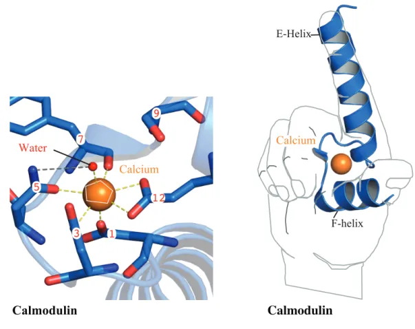
The EF hand nomenclature comes from the relationship between their two helices connected by a loop, (helix-loop-helix, HLH). Classically, the N- and C-terminal helices are designated by ‘E’ and ‘F’ helix, respectively (52). The tertiary structure of the HLH when superimposed on the forefinger and thumb of a person's right hand, is modeled with the linker region represented by the curvature of skin, between the thumb (F helix) and forefinger (E helix) (53). While not a rule, EF hands typically come in pairs but, both EF hands within the pair do not always bind calcium; an example of this is CalDAG-GEFII protein. Using isothermal titration calorimetry, (ITC), Iwig et al., showed that in the isolated pair of EF hands, calcium only bound to the N-terminal EF hand (54).
calcium binding within the loop region induces conformational rearrangements between the E and F helices that are transmitted throughout the protein, often regulating enzymatic function allosterically (56).
When calcium binds to the loop region of an EF hand, the E helix undergoes
conformational rearrangements relative to the F helix. Figure 1,illustrates the arrangement of a calcium-free HLH, overlaid with the arrangement of an HLH bound to a calcium ion. Calcium binding to EF hands imposes a pentagonal- bi-pyramidal geometric arrangement within the loop that is important for providing the thermodynamic energy required to rearrange the E and F helices, making it possible to unpack previously solvent inaccessible, hydrophobic regions.
EF hands typically pack tightly to the core of the protein through hydrophobic
interactions between the EF hands and the protein core. Thus, conformational changes induced by calcium exposes hydrophobic regions in EF hand proteins that are otherwise solvent
inaccessible. Exposing these hydrophobic regions promotes protein-protein interactions (57). However, substrate binding is not limited to only the proximity of the hydrophobic regions exposed by the EF hand movement. Often, the movement of EF hands are translated throughout the protein and thus, calcium binding can modulate the tertiary structure of proteins, promoting allosteric binding of substrates or its translocation to the hydrophobic membrane. In addition, the movement of the helices can also displace regions of a protein that are upstream of the E helix or downstream of the F helix. Such is the case for CalDAG-GEFI as proposed by the data presented in chapter 5.
directly activates CalDAG-GEFI, but the question remained, what was calcium doing to
Figure 1. The pentagonal bipyramidal geometry of calcium coordination of the EF hand domains
A calcium ion is coordinated by residues 1, 3, 5, 7, 9, 12, and a water molecule. loop region. Once calcium is bound within the loop, the adjoining helices are perpendicular to one another as in the shape of the forefinger and thumb of the right hand.
E-Helix
F-helix Water
1 3
5
7
12 9
Calcium
Calcium
CHAPTER 3: MECHANISMS FOR REGULATING SMALL GTPASES BY
CDC25 DOMAIN NUCLEOTIDE EXCHANGE FACTORS
There are approximately 150 small GTPases in eukaryotes. The major isozymes discussed here is Ras associated protein (Rap). Rap is a member of the Rat Sarcoma (Ras) family. However, the most abundant GTPase expressed in platelets are the five Rap isozymes, Rap1A, Rap1B, and Rap2A-C. In particular, Rap1B is expressed 10-fold higher than the other Rap isozymes, accounting for 0.1% of total platelet proteins (58). GTPases function like
molecular switches that are switched off when they are bound by guanosine diphosphate (GDP) and switched on when GDP is exchanged for guanosine triphosphate (GTP), a reaction catalyzed by a guanine nucleotide exchange factor (GEF) (59). The rate of nucleotide exchange in small GTPases is intrinsically slow due to the structural flexibility within SWI and SWII, which intermittently displaces the magnesium ion, allowing GTP loading. The concentration of GTP in a cell is nearly 10 times that of GDP thus once GDP is released GTP preferentially binds (60).
Inactive GTPases have a GDP molecule in their nucleotide binding pocket that is
is released, GTP is able to bind, switching the GTPase on. Once the GTPase is turned on, the GEF dissociates which allows the magnesium ion to re-coordinate within SWII, stabilizing GTP within the nucleotide-binding pocket. (PDB: 3X1W, 3X1X).
Figure 2. Cdc25 domain guanine nucleotide exchange factors
A,Domain architecture which include: Ras exchange motif (REM), cell-division-cycle 25 (Cdc25) domain, EF hand (EF), C1 domain, Ras-associated (RA) domain, Dbl homology (DH) domain, Pleckstrin homology (PH) domain, Rho GEF domain, Dishevelled, Egl-10, and
Pleckstrin (DEP) domain, cyclic-nucleotide binding (CNB) domain.
PH
DH/RhoGEF REM Cdc25
CNB
DEP REM RA Cdc25
CNB
CNB DEP REM RA Cdc25
EF EF C1
REM Cdc25
RA
REM Cdc25
SOS 1/2
Epac1
Epac2
CalDAG-GEF I-IV Ral-GEFs
The chapter will conclude by detailing the mechanism for how these small GTPases are switched off.
CalDAG-GEF (RasGRP) subfamily
The calcium and diacylglycerol-regulated guanine nucleotide exchange factor (CalDAG-GEF) family consists of four members (I-IV), also referred to in the literature as Rat sarcoma (Ras) guanyl-nucleotide exchange-releasing protein (RasGRP) (RasGRP1-4). CalDAG-GEF isozymes catalyze nucleotide exchange on both Ras and Rap small GTPases and are composed of an N-terminal Ras exchange motif (REM) domain, a catalytic Cdc25 domain, two EF hands, and a C1 domain.
The CalDAG-GEFII-IV isozymes are primarily regulated by DAG and activate Ras and Rap isozymes. Interestingly, overexpression of these CalDAG-GEF isozymes transforms certain cell types from normal cells to a more cancer-like cell; phenotypes that are associated with aberrant signaling downstream of Ras and/or Rap proteins (61). Indicative of this transformation are changes in cell morphology, an increased rate of cell division, and prolonged cell survival (62). However, the exact regulatory mechanisms for each CalDAG-GEF isozyme attributing to these pathological cellular transformations are not well understood. In contrast, some of the mechanisms regulating each isozyme under physiological conditions are better understood, beginning with CalDAG-GEFII.
The CalDAG-GEFII is a selective nucleotide exchange factor for Ras isozymes, H-, M-, R-Ras1 and R-Ras2, which is highly expressed in distinct portions of the brain and a subset of immune cells (16). Although the role of CalDAG-GEFII in neurons is unclear, CalDAG-GEFII is required for developing and maintaining a healthy immune system, promoting T cell
Recent work published by Iwig et al. provided structural and functional data on the molecular mechanisms underlying CalDAG-GEFII function. Data from a crystal structure of the truncated form of CalDAG-GEFII, lacking the C1 domain, suggests that in the absence of calcium CalDAG-GEFII is autoinhibited (PDB: 4L9M). Furthermore, it was shown that calcium binds to the N-terminal EF hand in CalDAG-GEFII, and that calcium binding leads to
conformational changes that perturb an autoinhibitory linker region between the Cdc25 domain and EF hand 1.
Data from a second crystal structure of the isolated C1 domain of CalDAG-GEFII revealed another mechanism regulating autoinhibition, suggested by two subunits of the C1 domains that form coiled-coiled homodimers within the unit cell of these crystals. The C1
domain of CalDAG-GEFII shares significant sequence homology with the C1 domains in protein kinase C (PKC) isozymes, for which DAG binding is best understood. The C1 domain of
CalDAG-GEFII isozymes binds DAG with nanomolar affinity (Kd»1.5 nM) (64). Functional studies using full-length CalDAG-GEFII with substitutions in residues that prevent dimer formation, revealed an enhanced nucleotide exchange activity, supporting not only homodimer formation in the full-length protein, but suggested that the dimers may be disrupted by DAG binding.
CalDAG-GEFIII is a nucleotide exchange factor for the same repertoire of small
data provides evidence for how the localization of CalDAG-GEFIII is regulated, but also suggests that DAG binding may release an autoinhibited state, allowing for its translocation to membranes and subsequent activity.
The CalDAG-GEFIV isozymes is a selective nucleotide exchange factor for H-Ras (16, 68). CalDAG-GEFIV has high expression in fetal tissues, specifically the lungs, liver, and spleen, whereas in adult tissue, it is expressed predominantly in leukocytes. CalDAG-GEFIV is the least studied isozyme in the CalDAG-GEF family. However, data has shown that fibroblast-like cells overexpressing CalDAG-GEFIV and treated with phorbol esters had enhanced
recruitment of CalDAG-GEFIV to the membrane, had increased activity of H-Ras isozymes, and increased levels of phosphorylated ERK1/ERK2. These data were corroborated by studies using another cell type overexpressing CalDAG-GEFIV, which were also transformed, by a Ras-dependent mechanism, suggesting a possible role for CalDAG-GEFIV as an oncogene (62).
Ral GEF subfamily
All members of the Ras-like (Ral) GEF family activate both RalA and RalB isozymes of Ral GTPases and is comprised of Ral guanine-dissociation stimulator (RalGDS), Ral releasing factor (Rlf), and RalGEF-like (RGL) isozymes (69). RalGEF isozymes are implicated as part of the signaling cascades required for endocytosis, gene transcription, cell proliferation, cell survival, and the production of second messengers. However, it has been difficult to ascertain their true function in these processes because of overlap between their activity and the activity of other Ras and Rap nucleotide exchange factors that are implicated in these same processes (70).
isozymes, and the RA domain is a binding site for activated Ras isozymes. Activated Ras
isozymes bind to RalGEFs inducing conformational rearrangements that release an autoinhibited state necessary for the normal function of Ral isozymes. Not surprising, oncogenic Ras
dramatically increases the activation of RalGEFs, leading to enhanced expression of transcription factors associated with tumorigenesis (71). Given the effect of oncogenic Ras has on the activity of Ral isozymes, Ral has become the target of small molecules for cancer therapy (72).
Unfortunately, little is known about the mechanism for how RalGEFs are regulated by Ras. Data from a crystal structure of the isolated RA domain of one RalGEF, Rlf reveals very little
information about the effect of Ras on the RA domain in the context of the full-length protein (PDB:1RLF) (73).
Commonly, small GTPases are localized to the membrane and therefore GEFs must be recruited to the membrane to activate them. A proline-rich region at the N-terminus of Rlf, upstream of the REM domain, is thought to serve as a ‘flagpole’ to recruit Src-homology (SH) 3 domain proteins (PDB:4JGW) (75). This SH3 binding site of Rlf may be a docking site for interactions with a well-known adaptor protein, growth factor receptor-bound protein (Grb) 2, shown to regulate other GEFs by recruiting them to the plasma membrane (76).
Sos subfamily
The Son of Sevenless (Sos) family of GEFs consists of the Sos1 and Sos2 isozymes whose complex regulatory mechanisms are attributed to their elaborate domain architecture. The Sos isozymes contain an N-terminal, histone binding domain, a DH/PH domain, PH/REM helical linker, REM domain, Cdc25 domain, and C-terminal Grb2/E3b binding domain. A wealth of data about the function of some of these regulatory domains has been compiled from
biochemical and crystallographic studies defining key aspects for how individual domains contribute to the activity of Sos isozymes such as their cellular localization, their dual specificity for Rho and Ras isozymes, and positive-feedback mechanisms (77).
Inactive Sos isozymes are primarily localized to the cytosol but are recruited to the inner leaflet of the plasma membrane with the help of adaptor proteins that sense changes in the composition of phospholipid composition of the membrane- namely an increase in the concentration of PIP2. The DH domain of SOS binds PIP2, but sterically occludes by the PH
domain, preventing the ability of Sos to activate Rac isozymes until it interacts with PIP2. Upon
exchange nucleotide on Ras isozymes, suggesting the specificity of Sos for both Rac and Ras isozymes is allosterically regulated.
Data from a crystal structure of a truncated version of Sos1, containing the DH-PH, REM, and Cdc25 domains corroborates the biochemical data for the allosteric regulation of Sos isozymes. These data revealed that in the absence of PIP2, the DH-PH domain is in a bent
conformation creating contacts between the REM and Cdc25 domain (79). This structural arrangement of the DH-PH domain is thought to be an autoinhibitory mechanism as determined by the reduced capacity for these truncated Sos proteins to exchange nucleotide on Rac and Ras isozymes. Concomitantly, biochemical data showed that Sos1, lacking the DH-PH domain has an enhanced capacity for nucleotide exchange on Rac (76). In addition, data from another crystal structure of Sos1, containing just the REM and Cdc25 in complex with Ras, defines a helical hairpin in the Cdc25 domain that undergoes conformational rearrangement when Ras proteins bind to the Cdc25 domain. These rearrangements break bonding contacts between the REM and Cdc25 domain revealing two hydrophobic patches, one in each domain, creating an allosteric binding site for Ras-GTP. Thus, Sos proteins bind Ras with a 2:1 stoichiometry. Importantly, when Ras-GTP binds at the allosteric site, nucleotide exchange activity by Sos1 at the primary site in the Cdc25 domain is dramatically enhanced. This allosteric mechanism dramatically increases the effective concentrations of active Ras as it clusters around receptors at the cell surface (PDB: 1NVV, 1NVW, 1XD2, 1XD4, 1XDV).
Epac subfamily
architecture. The N-terminus of Epac1 has a Dishevelled, Egl-10, Pleckstrin (DEP) domain, followed by a cyclic nucleotide-binding domain (CNBD), a REM domain, a Ras-associated domain (RA), and catalytic Cdc25 domain. Epac 2 isozymes contain an N-terminal high affinity-CNBD domain, a DEP domain, a second, low affinity-affinity-CNBD, a REM domain, a Ras-associated domain (RA), and catalytic Cdc25 domain (80).
Epac isozymes were first discovered in a database search for unique proteins in cells that could activate Rap isozymes, downstream of increased concentrations of cAMP. Epac1 and Epac2 isozymes are differentially expressed; Epac1 isozymes are most abundant in the heart, liver, blood vessels, and adipose tissue. In the heart, Epac1 signals through b1-adrenergic
receptors and is necessary for cardiac contractility, relaxation, and establishing a consistent heart rate (81). However, sustained, elevated levels of cAMP in cardiac myocytes can lead to cardiac hypertrophy, and/or heart failure. Epac2 isozymes are expressed in the adrenal glands and pancreas (126). Rap1 activation by Epac2 triggers insulin and glucagon secretion in the pancreas without which increases the risk of obesity (82, 83).
Data from crystal structures of Epac2 show that these isozymes remain in an
autoinhibited state that is released by conformational rearrangements induced by cAMP binding. The details about the rearrangements have been described using data from crystal structures comparing the conformations of active Epac2 to inactive Epac2 isozymes. Autoinhibition is released when cAMP binds to the CNBD and phenylalanine 435 is displaced exposing the
CHAPTER 4: A NEGATIVE-FEEDBACK LOOP REGULATING ERK1/2
ACTIVATION AND MEDIATED BY RASGRP2 PHOSPHORYLATION
Introduction
The Ras-Raf-MEK-ERK signaling pathway is essential for many cellular processes, including growth, cell-cycle progression, differentiation, and apoptosis. ERK1/2 phosphorylates hundreds of substrates in vivo, but these activities are restricted by cell type and subcellular compartment (87). Furthermore, the complexities and dynamics of ERK1/2 signaling are modified by both positive and negative-feedback loops (88). For example, Sos1 is a GEF that actives Ras isoforms. In turn, active Ras allosterically enhances the exchange capacity of Sos1 and this positive feedback loop ultimately impacts ERK1/2 activation (76, 89). Conversely, ERK1/2 phosphorylates both Sos1 and Raf to suppress further activation of ERK1/2 in a negative-feedback loop (90-92).
ERK1/2 is also activated downstream of the small GTPase, Rap1, in platelets. (10, 16, 92, 94). However, in contrast to activation by Ras isoforms, very little is known about potential feedback regulation by ERK1/2 on Rap1 signaling. RasGRP2 (alternative name CalDAG-GEFI) is the predominant GEF that activates Rap1 in platelets. RasGRP2 specifically activates Rap1 but not Ras isoforms both in vitro and in vivo (17, 18). In this study, using purified proteins and cellular studies, we show that ERK1/2 phosphorylates RasGRP2 to limit its exchange activity. In addition, we show that this connection establishes a negative-feedback loop that ultimately controls the dynamics of active ERK1/2.
93Ren, J., Cook, A. A., Bergmeier, W., and Sondek, J. (2016) A negative-feedback loop regulating ERK1/2
Results
ERK2 phosphorylates RasGRP2 at Ser394 in vitro
RasGRP family members have four conserved functional domains: i) an N-terminal Ras-exchange motif (REM), ii) a catalytic CDC25 domain, and a C-terminal regulatory region comprised of iii) two EF hands and iv) an atypical C1 domain (Fig. 3A). Post-translational modifications to RasGRP2 have previously been identified and are available from several
databases that provide mass spectral data (95, 96). From these databases, we noted that RasGRP2 is phosphorylated at Ser394 within the linker between the CDC25 domain and EF hands. In particular, this region occludes the Ras-binding region of RasGRP1 in its autoinhibited state and is conserved in other RasGRP isoforms (54). Furthermore, sequence adjacent to Ser394 matches a consensus motif of many ERK1/2 substrates defined by P-X-S/T-P, where “X” indicates any amino acid and S/T is the site of phosphorylation (Fig. 3A). Thus, we hypothesized that ERK1/2 phosphorylates RasGRP2 at Ser394 to potentially regulate its capacity to activate Rap1.
Figure 3. ERK2 phosphorylates RasGRP2 at Ser394 in vitro
A. Domain architecture of human RasGRP2. Sequence alignment for the individual RasGRP family members highlights the conserved ERK1/2 phosphorylation motif “PXSP” (red) in the linker between the Cdc25 domain and the EF hands. Asterisk identifies Ser 394 in
RasGRP2. B. ERK2 phosphorylates RasGRP2 in vitro. C. ERK2 phosphorylates RasGRP2 at Ser394. For panels B and C, bacterially purified proteins (5 µg) were incubated with
recombinant, constitutively active ERK2 in the presence of (32P) γ-ATP for 10 min prior to
SDS-PAGE and autoradiography
CalDAG-GEFII RNHRAPPLTPSKP---PVV-VDWASGVSPKP CalDAG-GEFI R-SKSSPTSPTSCTPPPRPPVLEEWTSAAKPKL CalDAG-GEFIII RNSKSQPTSPTTPN---KPVVPLEWALGVMPKP CalDAG-GEFIV RCPKSLPPSPFNA---PLV-VEWAPGVTPKP
Figure 3.
A. B.
72 55
28
17
GSTH -RasRap1BCalDAG-GEFIGST H-RasRap1BCalDAG-GEFI
coomassie blue autoradiography
autoradiography
coomassie blue
WT S394A
C.
CalDAG-GEFI
CDC25-EF linker
CalDAG-GEFI S394
126 150
4 388 430 499 551 609
*
55
55
The ERK1/2 pathway is required for phosphorylation of RasGRP2 in cells
HEK239T cells are competent to activate ERK1/2 downstream of EGF activation.
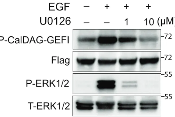
Figure 4. RasGRP2 is phosphorylated in response to epidermal growth factor (EGF) stimulation in HEK293T cells.
A. RasGRP2 is basally phosphorylated at Ser394 in HEK293T cells. Plasmids encoding FLAG-tagged RasGRP2 wild-type (WT) or S394A were transfected into HEK293T cells prior to immunoprecipitation of the expressed protein and western blotting as indicated. The parental vector was used as a control and treated similarly. B Phosphorylation of RasGRP2 in response to EGF is eliminated by the MEK inhibitor, U0126. Cells were pretreated with the indicated
amount of U0126 or DMSO for 1 hr prior to stimulation with 50 ng/ml EGF for 5 min. FLAG-RasGRP2 was immunoprecipitated from cell lysates and followed by western blotting as indicated. Cell lysates were also blotted for total ERK1/2 (T-ERK1/2) and phos-ERK1/2 (P-ERK1/2) 72 72 pcDN A WT S394A
A.
B.
Flag P-CalDAG-GEFI P-ERK1/2 T-ERK1/2EGF
U0126
+ + +_ 1 (µM)
However, RasGRP2(S394A) is poorly phosphorylated under these conditions. Upon stimulation with EGF, phosphorylation of RasGRP2 is increased and conversely, this phosphorylation is dramatically decreased upon inhibition of MEK1 with the small molecule, U0126 (Fig. 4B).
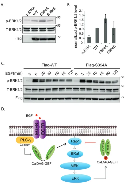
Phospho-mimetic mutation of RasGRP2 impairs nucleotide exchange activity
A fluorescence-based, guanine nucleotide exchange assay was used to assess the capacities of purified RasGRP2 proteins to activate Rap1b (Fig. 5A-B). The intrinsic rate of nucleotide exchange of Rap1b is low and this rate was increased dramatically by the addition of RasGRP2. A similar increase in exchange activity was seen upon addition of RasGRP2(S394A). However, the phospho-mimetic mutant, RasGRP(S394E), was unable to activate Rap1b to the same degree. Consistent with these in vitro findings, the expression of RasGRP2(S394E) in cells led to
Figure. 5. Phosphorylation of RasGRP2 impairs its nucleotide exchange activity
A. Purified proteins (3 µg) were subjected to SDS-PAGE and stained with Coomassie Brilliant Blue. B. In vitro nucleotide exchange assay. Equivalent amounts of Rap1b and BODIPY-FL-GDP were preincubated prior to the addition (arrow) of the indicated forms of RasGRP2. C. GST-RalGDS was used to precipitate GTP-Rap1 from HEK293T cells expressing the indicated forms of RasGRP2 prior to western blotting for proteins as indicated. D. GTP-Rap1 levels as shown in C were normalized to that of WT (average ± SEM).
0.9 1 1.1 1.2 1.3 1.4 1.5
0 10 20 30
Figure 5.
A. C. WT S394E B. D.RasGRP2 phosphorylation negatively regulates ERK1/2 activation
ERK1/2 activation by Rap1 requires B-Raf (10). The previous experiments indicate that ERK1/2 phosphorylates and inhibits the Rap1 GEF, RasGRP2. Thus, it is possible that these proteins participate in a negative-feedback loop whereby active ERK1/2 leads to decreased active Rap1 through RasGRP2. Since HEK293T cells also express B-Raf in addition to being competent to activate ERK1/2 as shown above, we used this cell line to delineate potential linkages between RasGRP2 and ERK1/2. Consistent with a negative-feedback loop, expression of RasGRP2(S394A), which is not able to be phosphorylated, increased levels of phos-ERK1/2. (Fig. 6A-B). Expression of the phospho-mimetic mutant, RasGPR2(S394E), did not substantially reduce phos-ERK1/2 levels, but this situation might arise due to the already high basal levels of phosphorylated WT RasGRP2 in these cells (Fig.6).
Studies show disruption of other ERK1/2 negative-feedback loops increase the amplitude and duration of ERK1/2 activation upon stimulation (3). If phosphorylation of RasGRP2 at Ser394 is critical for a negative-feedback loop involving active ERK1/2, then we might expect to see an increase in the amplitude and duration of ERK1/2 activation upon expression of
Figure 6. Phosphorylation of RasGRP2 negatively regulates ERK1/2
A. P-ERK1/2 levels in HEK293T cells transfected with the indicated forms of RasGRP2. Cells were lysed 24 hrs after transfection and analyzed by western blotting as indicated. B. Levels of P-ERK1/2 as shown in A for three independent experiments (average ± SEM), analyzed with Image J and normalized to that of the cells transfected with WT RasGRP2. C. RasGRP2(S394A) increases the magnitude and duration of ERK1/2 phosphorylation in response
WT S394A pcDNA S394E p-ERK1/2
Flag T-ERK1/2
normalized p-ERK1/2 level
A. B.
C.
p-ERK1/2
T-ERK1/2
Flag-WT Flag-S394A
0 5 20 60 90 120
0 0.3 0.6 0.9 1.2 1.5 1.8 2.1 WT S394A pcDNA S394E D.
0 5 20 40 60 90 120
Discussion
The equivalent of Ser394 and surrounding residues of RasGRP2 are conserved in all members of the RasGRP family suggesting that the negative-feedback loop described here for RasGRP2 is also relevant for signaling by other RasGRP family members. Consistent with this hypothesis, phosphorylation of human RasGRP3 at Ser391, the equivalent of Ser394 in
RasGRP2, was identified in two independent phosphoproteomics studies (97, 98).
In addition to ERK1/2, PKA has also been shown to phosphorylate RasGRP2 at multiple sites (99-101). However, these studies have produced conflicting results. In the original studies, phosphorylation by PKA resulted in the inhibition of RasGRP2 in HEK293T cells, while the most recent study showed PKA-mediated activation of RasGRP2 in Cos-7 cells. It is difficult to reconcile these results and perhaps future studies using purified proteins will clarify this issue. It seems likely that the RasGRP proteins are directly targeted by diverse kinases and that the regulation of RasGRP2 by phosphorylation is crucial for a range of cellular processes.
Signaling cascades leading to the activation of ERK1/2 are subject to stringent
homoeostatic control through both positive and negative-feedback loops. A number of inhibitors that target these cascades are used to treat cancer. However, drug-resistant tumors frequently emerge leading to disease progression. Alteration in the feedback regulation of ERK1/2 pathway is one potential cause of acquired drug resistance. Our study has defined a new
CHAPTER 5: CALCIUM-INDUCED STRUCTURAL
REARRANGEMENTS RELEASE AUTOINHIBITION IN THE RAP-GEF,
CALDAG-GEFI
Introduction
Human survival depends on our ability to prevent blood loss at sites of vascular injury. Upon damage to the endothelial lining, blood platelets detect exposed extracellular matrix and locally produced thrombin, become activated, and form a hemostatic plug. Critical to plug formation is the engagement of the aIIbb3 integrins on the cell surface, a process that depends on the inside-out signaling to these receptors (4).
Given the unique high shear environment found in blood vessels, the signal transduction in platelets leading to integrin inside-out activation has been optimized for sensitivity and speed. Upon receptor stimulation, the second messenger calcium, is rapidly released from the dense tubule system (DTS). Calcium also enters through channels in the plasma membrane, effectively increasing intracellular calcium concentrations 50-fold. Calcium plays an important role in the activation of aIIbb3. Integrin activation also depends strongly on the focal adhesion proteins, talin-1 and kindlin-3, as well as the small GTPase Ras-related protein (Rap) 1 (2, 4). The Rap1 isozymes, Rap1A and Rap1B, are guanine nucleotide binding proteins and members of the large superfamily of Ras small GTPases. Small GTPases act like molecular switches, cycling between an inactive, GDP-bound, and an active, GTP-bound, state. The biological role of Rap1B in platelets has been well studied. Genetically modified mice lacking the Rap1B isozyme have a
102 Cook, A. A., Deng, W., Ren, J., Li, R., Sondek, J., and Bergmeier, W. (2018) Calcium-
number of platelet defects, including a marked reduction in integrin activation in response to agonist stimulation (1).
Small GTPase activity is modulated by guanine nucleotide exchange factors (GEFs) and GTPase-activating proteins (GAPs). The most abundant Rap-GEF and Rap-GAP in platelets are calcium- and diacylglycerol-regulated guanine nucleotide exchange factor I, (CalDAG-GEFI, RasGRP2) and Ras p21 protein activator 3, (RASA3, GAP1IP4bp), respectively (17, 40).
CalDAG-GEFI contains a Ras exchange motif (REM) domain with no known function, a
catalytic Cdc25 domain, a pair of calcium binding EF hands, and an atypical C1 domain with no known function. Studies in mice, dogs, and humans lacking functional CalDAG-GEFI,
demonstrate that CalDAG-GEFI/Rap1B signaling is crucial for rapid activation of aIIbb3 required for platelet adhesion at sites of vascular injury (17, 103-105).
EF hands are composed of pairs of helices that typically bind calcium using acid residues within the intervening loop. These residues are defined by their relative positions (1, 3, 5, and 12) and in particular position 12 is a highly conserved glutamate that coordinates calcium through bidentate interactions. Mutating this glutamate cripples calcium binding (51, 56). Calcium binding generally leads to major conformational changes in the EF hands and other regions of the protein (53).
Our functional studies in platelets suggested calcium is involved the regulation of CalDAG-GEFI activity (10). In this work, we present biochemical evidence that both EF hands contribute to CalDAG-GEFI activation and that activity require calcium. Hydrogen-deuterium exchange mass spectrometry studies further suggest that calcium binding induces global
mutant forms of GEFI confirms the linker is important for autoinhibiting GEFI in the absence of calcium. Thus, our work provides the first evidence that (1) CalDAG-GEFI activity is directly regulated by calcium and (2) release of autoinhibition as the molecular mechanism underlying CalDAG-GEFI activation.
Results
CalDAG-GEFI activation is calcium-dependent.
Previous studies using isothermal titration calorimetry determined the isolated EF hands of CalDAG-GEFI (amino acid residues 417-495) bind calcium with very high affinity (Kd~80 nM) (54). To determine if calcium affects catalytic activity in full length CalDAG-GEFI, we established a cell-free nucleotide exchange assay using purified human CalDAG-GEFI and Rap1B proteins. Purified CalDAG-GEFI (amino acid residues 1-551, WT) was slightly truncated at the C-terminus to increase protein stability. Rap1B (amino acid residues 1-181) was purified with a substitution, cysteine 181 to serine, to increase solubility (Fig. 7A). Purification was performed using affinity and size exclusion chromatography. Proteins were >95% pure as confirmed by SDS-PAGE (Fig. 1B, inset). Proteins were stable at 4˚C for up to 15 hours. Compared to Rap1B alone, exchange activity increased by ~16-fold in samples containing Rap1B and CalDAG-GEFI. Of note, exchange activity was not altered by the addition of free calcium (not shown). However, treatment of CalDAG-GEFI with a 20mM ethylene glycol-bis (β-aminoethyl ether)-N, N, N', N'-tetraacetic acid (EGTA), a calcium-selective chelator, dramatically reduced catalytic activity. Importantly, exchange activity was restored upon
Figure 7. CalDAG-GEFI requires calcium to activate Rap1B.
A.Domain architecture. Rap1B is primarily composed of a Ras-like domain (red) while CalDAG-GEFI is a multi-domain protein consisting of a Ras exchange motif (REM) domain (salmon), catalytic Cdc25 domain (green), a putative autoinhibitory linker (red), two calcium-binding EF hands (blue), and an atypical C1 domain (yellow). CalDAG-GEFI was truncated (dotted lines) at residue 551 for purification. Truncated residues are not conserved and do not impact the capacity of CalDAG-GEFI to activate Rap1B. Substitutions in CalDAG-GEFI used in this paper are marked (red circles) below its domain architecture. B. Activation of Rap1B by CalDAG-GEFI monitored by the increased fluorescence of BODIPY FL GDP loaded onto Rap1B. Nucleotide (100 nM) and GTPase (1 µM) were incubated in four wells monitored simultaneously (lex/em = 480/520). Select reactions also included 10 mM EGTA as indicated. At
12 min (left arrow), 400 nM CalDAG-GEFI was added to all reactions except the one marked “Rap1B alone”. Addition of 10 mM free Ca2+ (right arrow) reconstituted exchange activity.
Inset: purified CalDAG-GEFI and Rap1B (2 µg) used in nucleotide exchange reactions; stained gel after SDS-PAGE.
0 10 20 30 40 50 60 70 80 1.0 1.5 2.0 2.5 Time (min) Rap1B alone
EGTA10 mM Ca 2+ + + + -- --
-+ Ca lDAG
-GEFI
+ Ca 2+ CalDA G-GEFI Rap1B 72 kDa 55 36 28 17
REM CDC25 EF EF C1 4 126 150 388 430 499 551 609
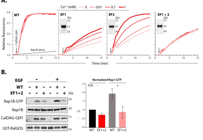
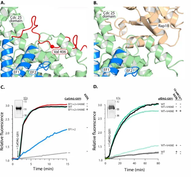
Both EF hands are critical for regulating CalDAG-GEFI exchange activity.
To determine if and how the individual EF hands regulate CalDAG-GEFI activity, we mutated a key residue in the calcium binding loops (Fig. 8A). Substitutions of glutamic acid at position 12 in the calcium binding loop of EF hands has been shown to reduce the binding affinity for calcium by over 100-fold (106, 107). Glutamic acid substitution for alanine at position 450 in the N-terminal EF hand (EF1) or position 479 in the C-terminal EF hand (EF2) markedly decreased nucleotide exchange towards Rap1B. Catalytic activity was restored by adding increasing concentrations of calcium in EF2, while only a partial recovery was observed in the EF1. Catalytic activity in the double EF hand mutant (EF1+2) could not be restored (Fig. 8A). These data provide strong evidence that calcium binding to EF1 is essential for exchange activity in CalDAG-GEFI, while EF2 is important but not essential in this process. To validate our in vitro findings in a cellular context, we studied agonist-induced Rap1 activation in
HEK293T cells expressing WT or EF1+2 CalDAG-GEFI (Figure 8B). Cellular stimulation with epidermal growth factor (EGF) led to a significant increase in Rap1-GTP levels in cells
Figure 8. Mutations in the calcium-binding EF hands reduce the capacity of CalDAG-GEFI to active Rap1B.
A. Nucleotide exchange was monitored as described in Figure 1. EF1 and EF2 indicate mutant forms of CalDAG-GEFI substituted (E450A and E479A) at equivalent positions within the N- and C-terminal EF hands, respectively; EF1+2 indicates the double mutant. Insets: stained gels of purified, mutants of CalDAG-GEFI (2 µg) after SDS-PAGE. B., left panel: stained gel for GST-RBD pull down of active Rap1B from HEK-293T cells expressing WT or EF1+2 proteins after SDS-PAGE and Western blot analysis. Right panel (representative of three independent experiments). Cells were incubated for 5 minutes in the presence and absence of 100ng/ml epidermal growth factor (EGF) before lysis. Right panel: Quantification of Rap1-GTP levels (mean± SD).
28
28 kDa
55 72
Calcium binding to CalDAG-GEFI induces conformational rearrangements required for
its activity.
We next performed hydrogen-deuterium exchange mass spectrometry experiments (HDX-MS) to determine the differences in deuterium uptake between WT and EF1+2 proteins. Measuring the rate of deuterium uptake defines the stability of hydrogen bond networks between residues stabilizing secondary structure in proteins as well as residues that are more solvent accessible (108, 109). The most stable hydrogen bonds have the slowest exchange while more dynamic regions exchange faster. WT and EF1+2 proteins were exposed to deuterated water for a designated amount of time and then quenched and digested for mass spectrometric analysis. We recovered 326 peptides from the WT sample and 329 from the EF1+2 sample, with complete coverage of both samples.
Figure 9. Differential hydrogen-deuterium exchange between CalDAG-GEFI WT and EF1+2.
A. Heat maps of deuterium uptake for CalDAG-GEFI and EF1+2. Proteins were incubated in deuterated water for indicated times prior to measurements of deuterium uptake using mass spectrometry. Largest differences in uptake between the two proteins span the putative autoinhibitory linker and EF hands shown for individual peptides below the heat maps. B. Homology model of CalDAG-GEFI. Blue regions map enhanced deuterium uptake (> 12%; excluding the linker) of CalDAG-GEFI relative to EF1+2. The majority of these regions cluster within the binding site for Rap1B and EF hands shown in expanded views
deuterium uptake at 166 min
EF hands
C1 domain 100
EF hands
4 126 150 388 430
363 431 499 551 Cdc25 domain REM domain 0
REM Cdc25 Linker EF1 EF2 C1
Exposu
re time (min)
WT EF1+2 0.15 1.6 16 166 0.15 1.6 16 166 0.01 0.01
Relative percent uptake
0.001 0.01 0.1 1 10 100 1000 Exchange time (min)
0.001 0.01 0.1 1 10 100 1000 0.0010 0.01 0.1 1 10 100 1000
10 20 30 40 50 % u pta ke WT EF1+2
Residues 363-376 Residues 378-407
Valine 406 contributes to maintain CalDAG-GEFI in an autoinhibited state.
We next determined critical residues within the putative autoinhibitory linker region using a homology model of inactive CalDAG-GEFI, based on predictions by the Iterative Threading ASSembly Refinement (I-TASSER) server (for more details see Material and Methods section) (https://zhanglab.ccmb.med.umich.edu/I-TASSER/). We identified amino acids 406-410 (VLEEW) as the residues that insert directly into the Rap1B binding groove (Fig. 10A, B). This region of the linker is fully conserved in all mammals. To test whether
Figure 10. Substitution of a conserved valine fully restores the exchange activity of EF1+2.
Discussion
The four members of the CalDAG-GEF (RasGRP) family are critical for the proper function of different blood cell types (94). They all possess a characteristic domain structure with an N-terminal REM/Cdc25 catalytic domain and a C-terminal regulatory domain consistent of a pair of EF hands and a C1 domain. In this work, we investigated the mechanistic details by which calcium affects nucleotide exchange activity in CalDAG-GEFI, a key regulator of Rap1 signaling in platelets. Compared to CalDAG-GEFII (RasGRP1), the best studied family member, CalDAG-GEFI shows significant differences in the Cdc25 catalytic domain and the EF hand and C1 regulatory domains. CalDAG-GEFII is a RasGEF that exists as a dimer in solution. Binding of diacylglycerol (DAG) to its C1 domain is critical for dimer release and thus CalDAG-GEFII function. In contrast, CalDAG-GEFI is primarily a Rap-GEF, does not dimerize, and does not contain a typical, DAG binding C1 domain. There are also significant differences with regard to the EF hand regulatory domain; while CalDAG-GEFII contains only one active, low affinity (KD > 1uM) EF hand, CalDAG-GEFI contains two fully functional EF hands with high affinity for calcium (KD < 100nM) (54). The high affinity for calcium is consistent with the documented role of CalDAG-GEFI in the rapid, calcium-dependent activation of Rap1 and integrin aIIbb3 that is required for platelet adhesion under shear stress conditions (10). Using biochemical and biophysical approaches, we demonstrate that both EF hands are critical for CalDAG-GEFI function and we provide evidence that calcium binding induces global conformational changes in CalDAG-GEFI, most prominently in an autoinhibitory linker region that prevents Rap1 binding to the Cdc25 domain in absence of calcium.
Figure 11. Model for calcium-dependent rearrangements within CalDAG-GEFI required for its engagement and activation of Rap1B
At low concentrations of intracellular calcium (left), the linker encompassing valine 406 blocks the surface of the Cdc25 domain that engages Rap1B. As calcium levels rise (right), the EF hands bind calcium and change conformation. This rearrangement moves the linker,
liberating the surface of CalDAG-GEFI needed to bind Rap1B.
Figure 11.
A. B.
Rap1B
Rap1B
C1 C1
Cdc25 Cdc25
REM
REM Rap1 binding pocket 406 Rap1 binding pocket 406
EF hands EF hands
Calcium